Effect of Different pH Media on Fluoride-releasing of Silver Diamine Fluoride in Primary Teeth: An in Vitro Study
ABSTRACT
Objective: To investigate the effect of different pH media on the concentration of fluoride-released ions from silver diamine fluoride (SDF) after its application on the enamel surface of primary molars.
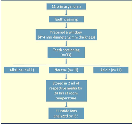
Material and Methods: A total of 33 specimens were prepared in sectioned parts of primary molar teeth with the following measurements (4×4 mm in height and width). Each specimen was immersed in an airtight polyethylene bottle containing 2 mL of pH media (neutral, alkaline, and acidic). The fluoride release pattern of each sample was taken on days 1, 2, 7, 15, and 30 using an Orion fluoride ion-selective electrode.
Results: On day 1, acidic media showed the least mean fluoride release (8.83 ppm) compared to neutral (23.17 ppm) and alkaline media (18.92 ppm). There was apersistent decrease in the mean fluoride release at all time intervals for each media. On day 30, the fluoride release of acidic, neutral, and alkaline media was 0.477 ppm, 3.325 ppm, and 4.183 ppm, respectively. The acidic media showed a significant decrease in fluoride release (p=0.00l) from day 1 to day 30 compared to neutral and alkaline media. However, non-significant differences were observed between neutral vs. alkaline media at all time intervals.
Conclusion: The neutral group showed the highest fluoride release compared to other groups, where the different pH media strongly affect fluoride release from SDF material.
Keywords:
Dental Caries; Tooth; Deciduous; Hydrogen-Ion Concentration
Introduction
Silver nitrate was first introduced in Japan by Nishino, Yamaga, and others in the 19th century to arrest dental caries, followed by rapid development during the 20th century to create a more effective formulation [1]. The formulation started with Howe’s ammoniacal silver nitrate, followed by silver fluoride and silver diamine fluoride (SDF) [2, 3]. At the beginning of the 21st century, the SDF was used in China as a caries arresting agent for school children [4]. From 2005 to 2009 in Australia, a series of in vitro studies conducted by Knight et al. [5, 6] and their group proved its effectiveness as a caries-arresting and antimicrobial agent. Later, it was used as a caries arresting agent in other parts of the world [7, 8]. The topical application of SDF as a non-invasive dental treatment is currently gaining popularity worldwide [9].
The US Food and Drug Administration (FDA) approved the use of SDF as a desensitizing agent in 2014. In 2015, 38% SDF solution was the first commercial product available in the United States as a preventive and caries arresting agent [10]. In January 2016, a new Code on Dental Procedures and Nomenclature (CDT) D1354 allowed billing claims for off-label use of SDF as an interim caries arresting medicament [11, 12]. A solution of 38% SDF (Ag (NH3)2F) is alkaline (pH=10) in nature, having a colorless solution containing 24-27% silver (Ag), 8.5-10.5% ammonia (NH3), and 5.0-6.0%fluoride (F) [13]. It is reported that the annual application of 38% SDF could arrest active root caries by 90% [14].
After three decades of infrequent studies, clinical trials with more rigorous experimental designs evaluated the effectiveness of the SDF in the management of dental caries [3]. A study focused on the incidence of new lesions reported that the prevented fractions were 77% and 73% after 2.5 years [15]. The effects of SDF are primarily the prevention of caries at the enamel and dentin levels since it contains fluoride (F), which mainly affects enamel, while the effect on dentin is predominantly by silver [2, 16, 17]. The literature reported that the application of SDF increased the bond strength of resin-based composite to carious-affected dentin [18]. SDF has been used as an inhibitor of caries in deciduous and permanent teeth because of the presence of silver compounds as a sterilizing agent. Moreover, it is used to promote the remineralization of enamel and dentin, reducing caries incidence because of its fluoride containment [10].
Studies were conducted to determine the SDF’s initial contents, besides the short-term stability of fluoride and silver concentrations over 28 days, an inverse relationship was found between fluoride and silver contents by increasing fluoride ions and reducing silver ions in contrast [19, 20]. Furthermore, a study that investigated the concentration of fluoride ions in saliva at three different intervals, which are before, immediately after, and one hour after 38% SDF application on enamel, concluded that fluoride concentration reached its peak immediately, and after one hour it had returned to the baseline [21]. However, the major limitation of these studies is that they were only experimented on human saliva, not on different pH media. The anti-cariogenic effect of fluoride-releasing materials depends on the amount and sustainability of fluoride release [22]. The pattern of release is typically characterized by an initial rapid release, followed by a significant reduction in the release rate after only a few days of immersion [23].
Several studies have been reported on the effect of pH on the fluoride release from various types of restorative materials [24, 25, 27]. However, the effect of pH on the release of fluoride from SDF has not been well documented. Considering the usefulness of SDF in caries prevention, the present laboratory study aimed to investigate the effect of different pH media on the concentration of fluoride-released ions from 38% silver diamine fluoride (SDF) after applying it to primary teeth at different time intervals. It was hypothesized that the concentration of fluoride released from SDF would be higher in an acidic pH medium.
Material and Methods
This was a randomized, controlled, single-blind (at the investigator level) laboratory study. Ethical permission was taken from the institutional review board (IRB-2021-02-483).
Teeth Selection and Sample Size
Using the effect size of Cohen criteria (G Power version 3.1.9.2, Heinrich-Heine-Universität Düsseldorf), taking alpha= 0.05 and 80% and based on the previous study [28], a minimum sample size of 30 (10/pH group) was calculated. The tested teeth (n = 33) were collected from pediatric dental clinics at Imam Abdulrahman Bin Faisal University, Dammam, Saudi Arabia. The teeth were carefully chosen to meet the inclusion criteria, i.e., primary posterior teeth with no carious or superficial enamel caries, free from cracks or developmental defects, and not restored previously. The selected teeth were sectioned from the cemento-enamel junction using an ortho-stripping disk (DynaFlex Corp., Chennai, India), and the sectioned coronal parts of teeth were stored in normal saline.
Specimen Preparation
Fluoride-free pumice and low-speed handpiece (KaVo Dental, Biberach, Germany) were used to clean all surfaces of the teeth included in this study. An occlusal cavity (4 mm × 4 mm height and width) was prepared in enamel and dentin part in each tooth; then the crown was cut at CEJ level and cutting the crown to 3 slides horizontally using a standard # 330 diamond bur (Brasseler USA, Savannah, GA, USA) with high-speed handpiece (KaVo Dental, Biberach, Germany) (Figure 1). Then 38% SDF - Advantage Arrest Silver Diamine Fluoride 38% (Elevate Oral Care LLC, West Palm Beach, FL USA) was applied on the prepared wall according to the manufacturer’s instructions. Each sectioned specimen was immersed in an airtight polyethylene bottle containing 2 mL of respective pH media.

Preparation of Different pH Media
In this study, one liter of alkaline, neutral, and acidic media was prepared. The alkaline pH media was prepared by mixing deionized water (Hach Lange GmbH, Düsseldorf, Germany) with 1.5 mM calcium chloride (CaCl2) (PanReac AppliCham, ITW Reagents, Darmstadt, Germany), 0.9 mM sodium dihydrogen phosphate(NaH2PO4) (Sigma Aldrich Chemie GmbH, Steinheim, Germany), and 150 mM potassium chloride (KCl) (Techno Pharmchem, New Delhi, India), Tris buffer 20 Mm (Sigma-Aldrich Inc., St. Louis, USA), and 0.02% sodium nitrate (NaN3) (Research-Lab Fine Chem Industries, Mumbai, India) were added, and pH was adjusted to 8-9 by the addition of dilute hydrochloric acid (HQ) (Honeywell Specialty Chemicals Seelze GmbH, Seelze, Germany).
The neutral pH media was prepared like the alkaline pH media, and the pH was adjusted to 6-7 by adding diluted HQ. The acidic pH media was prepared by mixing 2.0 πiM CaCl2, 2.0 πiM NaH2PO4, 75 mM acetate buffer (NaCH3COO) (Techno Pharmchem, New Delhi, India), and 0.02% NaN3 .Drops of diluted HC1 and diluted sodium hydroxide (NaOH) (Emsure; Merck KGaA, Darmstadt, Germany) were added to adjust pH to 4-5 [29].
The pH of the solutions was monitored using a pH test strip, immersing it directly into the solution and keeping it in contact with the solution for 5 seconds. The strip was visually compared to the chart provided with the stripes. The samples in each pH were incubated at 37°C in each media for days 1, 2, 7, 15, and 30. At each time interval, pH media was collected for the fluoride release analysis and replaced with fresh pH media in each tube (Figure 2).

Fluoride Release Analysis
The fluoride ion selective electrode (ISE) (Orion Dual Star pH; Thermo Fisher Scientific Inc., Waltham, MA, USA) was connected to an ISE meter to measure fluoride ions at days 1, 2, 7, 15, and 30 under normal atmospheric conditions. The fluoride ISE was calibrated using three standard fluoride solutions (Hach Lange GmbH, Düsseldorf, Germany) at 0.1, 1.0, 10.0, and 100.0 mgL-1 concentration. After each specific interval, the specimens were removed from stored pH media and stirred continuously with lmL of deionized water mixed with an equal amount of total ionic strength adjustment buffer (TISAB III) (Thermo Fisher Scientific Inc., Waltham, MA, USA). The buffer solution was used for washing the discs, which were later dried with absorbent paper and then re-incubated in a new 2 rπL of fresh pH media. A magnetic stirrer was placed in a plastic vial with the previous respective media. The dipped electrode into the solution recorded the fluoride concentration in parts per million (ppm).
Statistical Analysis
Data analyses were performed using SPSS-20.0 (IBM Corp., Chicago, IL, USA). Numerical data based on measurements of fluoride release under the exposure to the three-pH media were presented as means ± standard deviations (SD). These numeric variables were explored for the test of normality by using the Kolmogorov-Smirnov test, which revealed a non-Gaussian (non-normal) distribution. A non-parametric Kruskal Wallis test was performed to compare mean fluoride release among three media at various time intervals. A post-hoc Mann-Whitney U test was used to compare mean fluoride release between each of the two media types. A non-parametric Wilcoxon sign rank test was used to compare mean fluoride release at various time intervals within each media. P-value ≤0.05 was considered a statistically significant difference of means.
Results
The mean fluoride release under the exposure of an acidic medium showed a significantly persistent decrease in fluoride release from day 1 to day 30 (p=0.00l) compared to neutral and alkaline media. However, a non-significant difference was observed in mean fluoride release between neutral vs. alkaline media at all time intervals (p>0.05).
The mean fluoride release within each media on days 1, 2, 7, 15, and 30 was significant in neutral and acidic media (p<0.00l). In contrast, a non-significant difference in mean fluoride release on days 2 and 7 was observed in alkaline media (p=0.875). However, significant decreases were seen on days 15 and 30 (p<0.00l) using the Wilcoxon sign rank test, as presented in Table 1 and Figure 3.
Fluoride release (ppm) at various time slots under the exposure to the three different media.

Using the Kruskal-Wallis test, fluoride release was significantly different at various time durations among the three media types (p=0.001). There was a significant difference in mean fluoride release between acidic vs. neutral media and acidic vs. alkaline media (p=0.001), with non-significant differences between neutral vs. alkaline media at all time intervals (p>0.05) (Tables 2 and 3 and Figures 3 and 4). In addition, a significantly persistent lower mean fluoride release under exposure of all three media from day 1 to 30, except alkaline media non-significant from day 2 to 7 (p=0.875). Therefore, the results of our experiment showcase SDF’s significant in-vitro efficacy in its potential preventive characteristics against various bacteria, especially in acidic media.
Comparison of cumulative fluoride release (ppm) at various time slots under the exposure to the three-pH media.

Comparison of cumulative fluoride release (ppm) at various time slots under different pH media.
Discussion
SDF has been used for many years to prevent caries and arrest material in primary and permanent dentition [30, 31]. In this in vitro study, the effect of different pH media on the concentration of fluoride release from SDF has been investigated. The maximum amount of fluoride release was observed in an alkaline medium on day 30. Thus, this result is comparable with the null hypothesis.
The formation of a high plaque index and a reduction in the pH or acidogenic ability causes decay. The influence of pH in demineralization indicates that decreasing the pH may favor the cariogenic process. Reversion of this tooth demineralization can be induced when the pH of the oral cavity is found to be at 5.5 [32]. SDF is known to prevent and control dental caries effectively and is used in different concentrations in children with high caries risk. The topical application of SDF is a cost-effective, user-friendly, and non-invasive approach in caries management [33]. Fluoride is an essential component of SDF to facilitate remineralization and hardening of the tooth structure. It is reported that the SDF is associated with a definite increase in fluoride levels [34].
The present study evaluated the effect of different pH media on the concentration of F release of 38% silver diamine fluoride for one month. Fluoride release was the highest on day one and started to decrease gradually, with this reduction being the least in acidic media. These data indicate that the product delivers the advertised concentration of fluoride when first opened; however, the concentrations decrease after opening [29]. Previous research has shown that SDF application results in the formation of CaF2, resulting in the formation of fluorapatite crystals, which are more resistant to acidic dissolution [35].
The highest fluoride concentration of SDF in all storage media occurred 24 hours post-application and is known as the “burst effect”. These findings are similar to various restorative materials such as glass ionomer cement, resin-modified glass ionomer cement, compomer, and composite resin, where the “burst effect” occurred 24 hours after post storing in different pH media [29, 36, 37]. In this study, the amount of fluoride released was influenced by the storage medium. The mean fluoride release value showed a significant difference between acidic and alkaline media at all intervals. In acidic media, a minimum fluoride release value was observed, whereas maximum fluoride release was observed in alkaline media. The possible reasons for the minimum fluoride release value in the acidic media could be the partial demineralization of the crystals that might attract and absorb fluoride to the crystal surface and the crystal behaving like fluorapatite. Meanwhile, in alkaline, the crystals are not affected, and there is no need for fluoride absorption; therefore, the fluoride content is high in the alkaline media [38, 39]. Also taken into consideration, the reduction of fluoride release occurred in acidic media as a result of using competitor anions in preparing acidic media like chloride, nitrate, sulfate, and phosphate. In addition, phosphate and sulfate have larger ionic radii and higher surface charger densities than fluoride, resulting in a slight decrease in fluoride release (40). In contrast, previous investigations of fluoride release from glass-ionomer cement and resin-based composites showed more fluoride release with acidic media [29, 36].
It is reported that annual application of either SDF solution or high fluoride-releasing glass ionomer can arrest active dentine caries with a rate of 79% and 82%, respectively. Increasing the frequency of application every six months can increase the caries arrest rate of SDF applications by 91% [41]. In the present study, the mean fluoride release at days 1, 2, 7, 15, and 30 was significant in neutral (p<0.001). This result is comparable to a study conducted to analyze the amount of fluoride released from SDF in artificial saliva at a pH of 7 on days 1, 7, and 14 [42].
A significantly persistent decrease in mean fluoride release was observed under exposure to all three media from day 1 to 30, except alkaline media, which was non-significant from day 2 to 7 (p=0.875). In contrast, a study reported that the percentage of fluoride ions released following the application of SDF within 60 days was increased [43]. However, the results of this study contradict previous studies, which found that ammonia ions bond with silver ions, forming a stable complex ion called the silver diamine ion [Ag(NH3)2]+ [44].
Based on previous studies, fluoride in low concentration (up to 1 ppm) in a solution significantly protects against demineralization of teeth structure [45, 46]. The same was obtained in this study, as SDF in all tested media during the 30 days of trial effectively reduced the dissolution of tooth structure.
One of the limitations of this study is that it was conducted under in vitro conditions, which cannot replicate the oral cavity’s complexity regarding temperature and other factors. However, it is felt that the result from the present investigation will enable pediatric dentists and other dental professionals to know and critically analyze the materials based on their fluoride-releasing properties.
Conclusion
The fluoride release concentration of 38% SDF is under the influence of pH storage media. All the specimens released fluoride ions during the entire experimental period. Abrupt fluoride release was found in all groups; however, a continuous release pattern was observed in all media. Overall, the neutral group showed the highest fluoride release compared to others. The release concentration from all groups is sufficient to restrict the caries.
-
Financial Support
None.
Data Availability
The data used to support the findings of this study can be made available upon request to the corresponding author.
References
-
Kumar C. The synergism of silver and fluoride: A new arsenal in caries management. Macromolecular Symposia 2024; 413(2). https://doi.org/10.1002/masy.202300129
» https://doi.org/10.1002/masy.202300129 -
Xu GY, Zhao IS, Lung CYK, Yin IX, Lo ECM, Chu CH. Silver compounds for caries management. Int Dent J 2024; 74(2):179-186. https://doi.org/10.1016/j.identj.2023.10.013
» https://doi.org/10.1016/j.identj.2023.10.013 - Nishino M. Studies on the topical application of ammoniacal silver fluoride for the arrest of dental caries. Osaka Daigaku Shigaku Zasshi 1969; 14(1):1-14. [In Japanese].
-
Vishwanathaiah S, Maganur PC, Syed AA, Kakti A, Hussain Jaafari AH, Albar DH, et al. Effectiveness of silver diamine fluoride (SDF) in arresting coronal dental caries in children and adolescents: A systematic review. J Clin Pediatr Dent 2024; 48(5):27-40. https://doi.org/10.22514/jocpd.2024.101
» https://doi.org/10.22514/jocpd.2024.101 -
Knight GM, McIntyre JM, Craig GG, Mulyani, Zilm PS, Gully NJ. An in vitro model to measure the effect of a silver fluoride and potassium iodide treatment on the permeability of demineralized dentine to Streptococcus mutans. Aust Dent J 2005; 50(4):242-5. https://doi.org/10.1834-7819.2005.tb00367.x
» https://doi.org/10.1834-7819.2005.tb00367.x - Knight GM, McIntyre JM, Craig GG, Mulyani, Zilm PS, Gully NJ. Inability to form a biofilm of Streptococcus mutans on silver fluoride- and potassium iodide-treated demineralized dentin. Quintessence Int 2009; 40(2):155-161.
- Phan D, Wen ZT, Fidel PL, Chapple AG, Collins A, Johnson JT. Silver Diammine fluoride usage in general dentistry offices in Louisiana. J Dent Child 2024; 91(1):3-9.
-
Timms L, Choi S, Marshman Z, Rodd H, Wilson AR, Tiwari T. Parental acceptability of silver diamine fluoride: The UK and US experiences. Int J Paediatr Dent 2025; 35(1):13-21. https://doi.org/10.1111/ipd.13195
» https://doi.org/10.1111/ipd.13195 -
Kaewkamchai S, Thanyasrisung P, Sukarawan W, Samaranayake L, Tuygunov N, Songsiripradubboon S. Efficacy of silver diamine fluoride (SDF) in arresting dentin caries against inter-kingdom biofilms of Streptococcus mutans and Candida albicans. PLoS One 2024; 19(9):e0308656. https://doi.org/10.1371/journal.pone.0308656
» https://doi.org/10.1371/journal.pone.0308656 -
Crystal YO, Niederman R. Evidence-based dentistry update on silver diamine fluoride. Dent Clin North Am 2019; 63(1):45-68. https://doi.org/10.1016/j.cden.2018.08.011
» https://doi.org/10.1016/j.cden.2018.08.011 -
U.S. Food and Drug Administration. FDA Product Classification. Silver Spring, MD. U.S. Department of Health & Human Services. Available from: https://www.accessdata.fda.gov/scripts/cdrh/cfdocs/cfpcd/classification.cfm [Accessed on December 14, 2020].
» https://www.accessdata.fda.gov/scripts/cdrh/cfdocs/cfpcd/classification.cfm -
American Dental Association. American Dental Association Releases CDT 2016 Dental Procedure Codes. ADA News Releases. 168. Available from: https://commons.ada.org/newsreleases/168 [Accessed on December 14, 2020].
» https://commons.ada.org/newsreleases/168 -
Elevate Oral Care. Safety Data Sheet. Advantage Arrest Silver Diamine Fluoride 38%. West Palm Beach, FL. Elevate Oral Care. Available from: https://www.elevateoralcare.com/site/images/AASDS082415.pdf. [Accessed on August 29, 2016].
» https://www.elevateoralcare.com/site/images/AASDS082415.pdf. -
Asghar M, Omar RA, Yahya R, Yap AU, Shaikh MS. Approaches to minimize tooth staining associated with silver diamine fluoride: A systematic review. J Esthet Restor Dent 2023; 35(2):322-332. https://doi.org/10.1111/jerd.13013
» https://doi.org/10.1111/jerd.13013 - Horst JA, Heima M. Prevention of dental caries by silver diamine fluoride. Compend Contin Educ Dent 2019; 40(3):158-163.
-
Liu BY, Lo ECM, Li CMT. Effect of silver and fluoride ions on enamel demineralization: a quantitative study using micro-computed tomography. Aust Dent J 2012; 57:65-70. https://doi.org/10.1111/j.1834-7819.2011.01641.x
» https://doi.org/10.1111/j.1834-7819.2011.01641.x -
Zhi QH, Lo ECM, Kwok ACY. An in vitro study of silver and fluoride ions on remineralization of demineralized enamel and dentine. Aust Dent J 2013; 58:50-56. https://doi.org/10.1111/adj.12033
» https://doi.org/10.1111/adj.12033 -
Firouzmandi M, Mohaghegh M, Jafarpisheh M. Effect of silver diamine fluoride on the bond durability of normal and carious dentin. J Clin Exp Dent 2020; 12(5):e468-e473. https://doi.org/10.4317/jced.56303
» https://doi.org/10.4317/jced.56303 -
Crystal YO, Rabieh S, Janal MN, Rasamimari S, Bromage TG. Silver and fluoride content and short-term stability of 38% silver diamine fluoride. J Am Dent Assoc 2019; 150(2):140-146. https://doi.org/10.1016/j.adaj.2018.10.016
» https://doi.org/10.1016/j.adaj.2018.10.016 -
Mei ML, Chu CH, Lo EC, Samaranayake LP. Fluoride and silver concentrations of silver diammine fluoride solutions for dental use. Int J Paediatr Dent 2013; 23(4):279-85. https://doi.org/10.1111/ipd.12005
» https://doi.org/10.1111/ipd.12005 -
Widianti TA, Bahar A, Maharani DA, Tumen EC, Yavuz I. Effect of silver diamine fluoride application on fluoride concentration in saliva. J Phys Conf Ser 2018; 1073:052001. https://doi.org/10.1088/1742-6596/1073/5/052001.
» https://doi.org/10.1088/1742-6596/1073/5/052001. -
Tyas MJ. Clinical evaluation of glass-ionomer cement restorations. J Appl Oral Sci 2006; 14:10-13. https://doi.org/10.1590/S1678-77572006000700003
» https://doi.org/10.1590/S1678-77572006000700003 -
Itota T, Carrick TE, Yoshiyama M, McCabe JF. Fluoride release and recharge in giomer, compomer and resin composite. Dent Mater 2004; 20(9):789-795. https://doi.org/10.1016/j.dental.2003.11.009
» https://doi.org/10.1016/j.dental.2003.11.009 -
Carey CM, Spencer M, Gove RJ, Eichmiller FC. Fluoride release from a resin-modified glass-ionomer cement in a continuous-flow system. Effect of pH. J Dent Res 2003; 82(10):829-832. https://doi.org/10.1177/154405910308201013
» https://doi.org/10.1177/154405910308201013 -
Koga H, Kameyama A, Matsukubo T, Hirai Y, Takaesu Y. Comparison of short-term in vitro fluoride release and recharge from four different types of pit-and-fissure sealants. Bull Tokyo Dent Coll 2004; 45(3):173-179. https://doi.org/10.2209/tdcpublication.45.173
» https://doi.org/10.2209/tdcpublication.45.173 -
Behrend B, Geurtsen W. Long-term effects of four extraction media on the fluoride release from four polyacid-modified composite resins (compomers) and one resin-modified glass-ionomer cement. J Biomed Mater Res 2001; 58(6):631-637. https://doi.org/10.1002/jbm.1062
» https://doi.org/10.1002/jbm.1062 -
Anusavice KJ, Zhang NZ, Shen C. Effect of CaF2 content on rate of fluoride release from filled resins. J Dent Res 2005; 84(5):440-444. https://doi.org/10.1177/154405910508400508
» https://doi.org/10.1177/154405910508400508 - Upadhyay S, Rao A, Shenoy R. Comparison of the amount of fluoride release from nanofilled resin modified glass ionomer, conventional and resin modified glass ionomer cements. J Dent 2013; 10(2): 134-140.
-
Nigam A, Jaiswal J, Murthy R, Pandey R. Estimation of fluoride release from various dental materials in different media-an in vitro study. Int J Clin Pediatr Dent 2009; 2(1):1-8. https://doi.org/10.5005/jp-journals-10005-1033
» https://doi.org/10.5005/jp-journals-10005-1033 -
Worthington HV, Lewis SR, Glenny AM, Huang SS, Innes NP, O’Malley L, et al. Topical silver diamine fluoride (SDF) for preventing and managing dental caries in children and adults. Cochrane Database Syst Rev 2024; 11(11):CD012718. https://doi.org/10.1002/14651858.cd012718.pub2
» https://doi.org/10.1002/14651858.cd012718.pub2 -
Alowid AM, Hebbal M, Almufarji FS, Almutairi GG, Alkait SS, Jodalli P. Antimicrobial effect of silver diamine fluoride (SDF) in arresting dentine caries of permanent teeth: A Systematic review. F1000 Res 2024; 13:1495. https://doi.org/10.12688/f1000research.158864.1
» https://doi.org/10.12688/f1000research.158864.1 -
Yip HK, Lam WT, Smales RJ. Fluoride release, weight loss and erosive wear of modern aesthetic restoratives. Br Dent J 1999; 187(5):265-270. https://doi.org/10.1038/sj.bdj.4800256
» https://doi.org/10.1038/sj.bdj.4800256 -
Zhao IS, Gao SS, Hiraishi N, Burrow MF, Duangthip D, Mei ML, et al. Mechanisms of silver diamine fluoride on arresting caries: a literature review. Int Dent J 2018; 68(2):67-76. https://doi.org/10.1111/idj.12320
» https://doi.org/10.1111/idj.12320 -
Delbem AC, Bergamaschi M, Sassaki KT, Cunha RF. Effect of fluoridated varnish and silver diamine fluoride solution on enamel demineralization: pH-cycling study. J Appl Oral Sci 2006; 14(2):88-92. https://doi.org/10.1590/S1678-77572006000200005
» https://doi.org/10.1590/S1678-77572006000200005 - Yamaga R, Nishino M, Yoshida S, Yokomizo I. Diammine silver fluoride and its clinical application. J Osaka Univ Dent Sch 1972; 12:1-20.
-
MarkovicDL, Petrovic BB, Peric TO. Fluoride content and recharge ability of five glass ionomer dental materials. BMC Oral Health 2008; 8:21. https://doi.org/10.1186/1472-6831-8-21
» https://doi.org/10.1186/1472-6831-8-21 -
Yoda A, Nikaido T, Ikeda M, Sonoda H, Foxton RM, Tagami J. Effect of curing method and storage condition on fluoride ion release from a fluoride-releasing resin cement. Dent Mater J 2006; 25(2):261-266. https://doi.org/10.4012/dmj.25.261
» https://doi.org/10.4012/dmj.25.261 -
Chawhuaveang DD, Yu OY, Yin IX, Lam WYH, Chu CH. Topical agents for nonrestorative management of dental erosion: A narrative review. Healthcare 2022; 10(8): 1413. https://doi.org/10.3390/healthcare10081413
» https://doi.org/10.3390/healthcare10081413 -
Amaechi BT, van Loveren C. Fluorides and non-fluoride remineralization systems. Monogr Oral Sci 2013; 23:15-26. https://doi.org/10.1159/isbn.978-3-318-02207-0
» https://doi.org/10.1159/isbn.978-3-318-02207-0 -
Dehghani MH, Farhang M, Alimohammadi M, Afsharnia M, Mckay G. Adsorptive removal of fluoride from water by activated carbon derived from CaQ2 -modified Crocus sativus leaves: Equilibrium adsorption isotherms, optimization, and influence of anions. Chemical Engineering Communications 2018; 205(7):955—965. https://doi.org/10.1080/00986445.2018.1423969
» https://doi.org/10.1080/00986445.2018.1423969 -
Zhi QH, Lo EC, Lin HC. Randomized clinical trial on effectiveness of silver diamine fluoride and glass ionomer in arresting dentine caries in preschool children. J Dent 2012; 40(11):962-967. https://doi.org/10.1016/jjdent.2012.08.002
» https://doi.org/10.1016/jjdent.2012.08.002 -
Thakur T, Lahiri PK, Karmakar M, Sarvaiya B, Datta P, Saha R. Comparison of the fluoride release of silver diamine fluoride, fluoride varnish, acidulated phosphate fluoride gel on extracted teeth over various time intervals in artificial saliva. J Pharm Res Int 2021; 33(26A):55-63. https://doi.org/10.9734/jpri/2021/v33i26A31471
» https://doi.org/10.9734/jpri/2021/v33i26A31471 -
Soekanto SA, Dayanara P, Daviq H, Sarwono AT, Sahlan M. Effectiveness of silver diamine fluoride and propolis fluoride varnish application on dentin within 60 days. Int J App Pharm 2019; 11(1):190-193. https://doi.org/10.22159/ijap.2019.v11s1.17138
» https://doi.org/10.22159/ijap.2019.v11s1.17138 - Chu CH, Lo EC. Promoting caries arrest in children with silver diamine fluoride: A review. Oral Health Prev Dent 2008; 6(4):315-321.
-
Alsaadawi A, Felemban O, Nassar HM, Abdelbaki M. Shear bond strength and fluoride release of a universal adhesive: An in-vitro study on primary teeth. Materials 2023; 16(7):2573. https://doi.org/10.3390/ma16072573
» https://doi.org/10.3390/ma16072573 -
Rošin-Grget K, Peroš K, Sutej I, Bašić K. The cariostatic mechanisms of fluoride. Acta Med Acad 2013; 42(2):179-188. 10.5644/ama2006-124.85
» https://doi.org/10.5644/ama2006-124.85
Edited by
-
Academic Editor:
Ana Maria Gondim Valença