Comparison of Edge Testing Between Indirect Composite-Layered Zirconia Crowns and Hand-Layered All-Ceramic Crowns without Aging
ABSTRACT
Objective: To compare the edge strength of hand-layered all-ceramic crowns and indirect composite veneers on zirconia frameworks to evaluate their durability.
Material and Methods: Edge strength of hand-layered ceramic on zirconia coping (Group 1) and indirect composite veneered on zirconia coping (Group 2) were compared. 20 samples, 10 in each group, underwent tooth preparation on a typodont model, followed by digital scanning, coping fabrication using digital software, and zirconia milling. These copings were either layered with veneering ceramic (Group 1) or indirect composite (Group 2). Edge testing , performed with an Instron universal testing machine, assessed the force required for chipping at 0.5 mm from the cusp edge Statistical analysis employed the unpaired t-test.
Results:: The mean force required to edge-chip an indirect composite veneered on a zirconia framework crown was 305 N, and to edge-chip a hand-layered all-ceramic layered crown was 231.50 N. A significant difference between the groups was observed (p < 0.05).
Conclusion: Indirect composite offers a unique blend of esthetics and strength and can be used as an alternative veneering material in routine clinical practice.
Keywords:
Zirconium Oxide; Crowns; Ceramics.
Introduction
For more than a hundred years, dentists have been trying to find the best material to replace damaged or missing teeth. Over time, many different options have been used - composites, PMMA, metals, ceramics, PEEK, and several newer biomaterials [1-3]. Among these, ceramics have become especially popular because they are biocompatible, chemically stable, maintain their color well, and are abrasion-resistant for everyday wear [4,5]. Despite the widespread use of materials, a consensus on the preferred restorative material - be it composite or ceramic - remains elusive in the current literature. In esthetic regions, layered restorations, recommended for various indications, provide a conservative therapeutic approach [6]. Current literature shows that there is still no clear agreement on which restorative material is ideal. Ceramic-veneered zirconia crowns have reported survival rates between 82% and 96% over periods of 10 to 21 years. Most failures occur from veneer fractures, which account for about 12-20%, and from marginal defects, which range from 5.6-11% [7]. In this context, it should be understood that ceramics and even the layers of their glaze can be subject to degradation due to acid or mechanical stress associated with use, and this might affect their appearance and tendency to form biofilms.
In general, chipping is considered one of the major reasons for restoration failure [8]. However, many clinical studies do not clearly describe the extent or specific characteristics of these chipping events. In veneered zirconia restorations, the most common cause of failure is chipping of the porcelain veneer, often triggered by wear on the biting surfaces [9]. Such damage typically starts from the contact areas in the teeth, where small internal cracks begin to grow until they become large enough to crack through the veneering surface. Evidence shows that stress induced by contact together with residual thermal stress plays an important role in causing this common phenomenon [10]. To address these issues, alternative materials such as indirect composites and monolithic zirconia have been introduced and refined over time [11]. As a result, numerous investigations have been conducted to assess the fracture behavior and susceptibility of various dental ceramics using a variety of testing methods. Although the concept of edge strength has long been known, a clear measurement method for this property was only recently established [12]. Edge strength specifically refers to a material's ability to resist fracture along a thin edge, measured by the force needed to cause a fracture 0.5 mm from the sample's edge [13]. This property depends on the ability of the restoration to resist any loading without losing its integrity. The magnitude of the force needed to induce a fracture of the edge is dependent on several aspects, such as the shape of the object used for loading, the location at which the force is exerted, the angle at which loading occurs, the geometry of the edge and its fracture toughness.
There has been quite an extensive examination of the edge strength of ceramic veneering materials. On the contrary, there is not enough research regarding the edge strength of composites as veneering materials. According to the current literature, no investigation has ever focused on the edge strength of indirect composites before. Hence, there appears to be a gap in the literature with regard to the edge strength of the aforementioned materials. Bridging the identified gap would help to get better insight into the properties of those materials and the need for further investigations on that matter. Therefore, this study would be the first study to focus on the edge strength of indirect composite and hand-layered ceramic restorations on zirconia cores. Previous studies focused on the fracture characteristics of ceramics as veneering material, while this study aims at evaluating the edge chipping resistance. By doing so, it shows how material compositions and layers influence the edge strength and hence the lifetime of restorations.
The aim of this study was to investigate and compare the edge strength of hand-layered all-ceramic crowns and indirect composite veneers fabricated on zirconia copings. The results of this investigation could contribute to the development of future restorations by providing information on the edge chipping resistance
Material and Methods
Study Design and Ethical Clearance
The present in vitro study was conducted in the Department of Prosthodontics, Saveetha Dental College and Hospitals, Chennai, India, after approval from the Institutional Systematic Review Board (Approval No. SRB/SDC/PROSTHO-2105/22/168).
Sample Size and Preparation
Sample size estimation was carried out using statistical parameters derived from a previous study [14] and performed by using G*Power 3.1.9.3 software on mac OS®. In total, there were 20 specimens, with 10 specimens assigned to hand-layered ceramic onto zirconia coping (Group 1) and 10 specimens assigned to indirect composite veneer onto zirconia coping (Group 2).
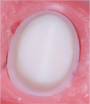
Tooth preparation was done on a single first premolar tooth typodont (Nissin Manufacturing Co., Ltd., Kyoto, Japan) (Figure 1). Then, the typodont was scanned with an extraoral laboratory scanner (3Shape E series, 3Shape, Copenhagen, Denmark). The first premolar tooth was chosen for testing because of its importance from both functional and esthetic point of views. Following scanning and obtaining the STL file, the copings were designed using the 3Shape Dental Designer 2021 software (3Shape A/S, Copenhagen, Denmark). The milling of the zirconia copings (Upcera HT White; Upcera Dental America Inc., Cerritos, CA, USA) was done with a 5-axis milling machine (Imes iCore 350i, Im Leibolzgraben 16, Eiterfeld). Following milling, sintering of the copings was performed as per the manufacturer’s instructions, using the high-temperature furnace (MIHM-VOGT GmbH & Co. KG, Stutensee, Germany). The sintering process comprised of a controlled heating up to 1,500 °C (range of 1,450 - 1,600 °C), with the dwelling stage being held to allow for densification of the material with no porosity left. Overall, the sintering process took about 7-8 hours to finish and was controlled in order to produce the best zirconia coping properties.

Layering of the Samples
The zirconia copings were fabricated before veneering ceramic (Emax Ceram, Ivoclar Vivadent AG, Schaan, Liechtenstein) application on these copings, which resulted in defining the first group of samples during the research. The technique of veneering layer buildup was employed, consisting of baking the veneering ceramic in the muffle furnace using standard conditions of the manufacturer. Such conditions involved the temperature within the range of 650-1000 °C, increasing the temperature by 55 °C per minute and the peak temperature maintaining for 5 minutes under atmospheric pressure. The preparation of precisely measured paste of 2 g of powdered porcelain in 0.7 ml of distilled water occurred after the process. After layering and contouring, glaze firing was performed at 770 °C per the manufacturer’s instructions to achieve a uniform surface finish. Similarly, indirect composite (Group 2) samples were layered onto the zirconia copings (Ceramage, Shofu, Kyoto, Japan) and cured with a light-cure unit (Solidilite V, Shofu Dental India PVT. LTD, New Delhi, India) for 1 min (Figure 2). Before indirect composite layering, a priming agent containing functional monomer MDP (Clearfil photo bond with Clearfil porcelain bond activator) was used. The samples were then polished with a 400-grit silicon carbide abrasive paper under running water for 10 seconds by a single operator and then used for further analysis.

Hand-layered ceramic on zirconia coping and indirect composite veneered on zirconia coping samples.
Edge Testing
The edge chipping test was conducted using a universal testing machine (Instron 4501, Instron Corp., Canton, MA) in accordance with the ISO 23146:2012 standard [15]. To ensure precision, an XY stage was affixed, and a designated slot securely positioned the Vickers indenter (Shanghai Toyo Diamond Tools Co., Ltd., Shanghai, China). The use of a USB 40x digital microscope ensured accurate observations. Specimens were sectioned vertically before any tests. As the cusp edges are more susceptible to force concentrations, the cusp tips were chosen for the test. The cutting process was done using a 0.3 mm diamond disc at a speed range of 200 to 300 rpm using continuous water irrigation for heat avoidance and prevention of microcracks formation. Specimen calibration was achieved through the use of a calibration system to properly align the specimen, setting up of the XY stage and camera settings, and indentations formation on an aluminum surface (Figure 3).

The force needed to create chipping at a particular distance of 0.5 mm from the cusp edge was assessed. Load application of 1 kgf for 30 seconds was used to create indentations and the edge chipping patterns observed at 40x magnification. Kc (toughness) values were computed through the use of fracture mechanics equation: ; Where Kc = toughness; fc = critical chipping load; β (a dimensionless coefficient) = 9.3; and d = edge distance.
Statistical Analysis
Data collection and tabulation were performed using Google Forms. Mean edge chipping values were analyzed using an unpaired t-test in SPSS version 26.0 (IBM SPSS, Chicago, IL, USA) after confirming the dataset's normality. The level of statistical significance was set at 0.05.
Results
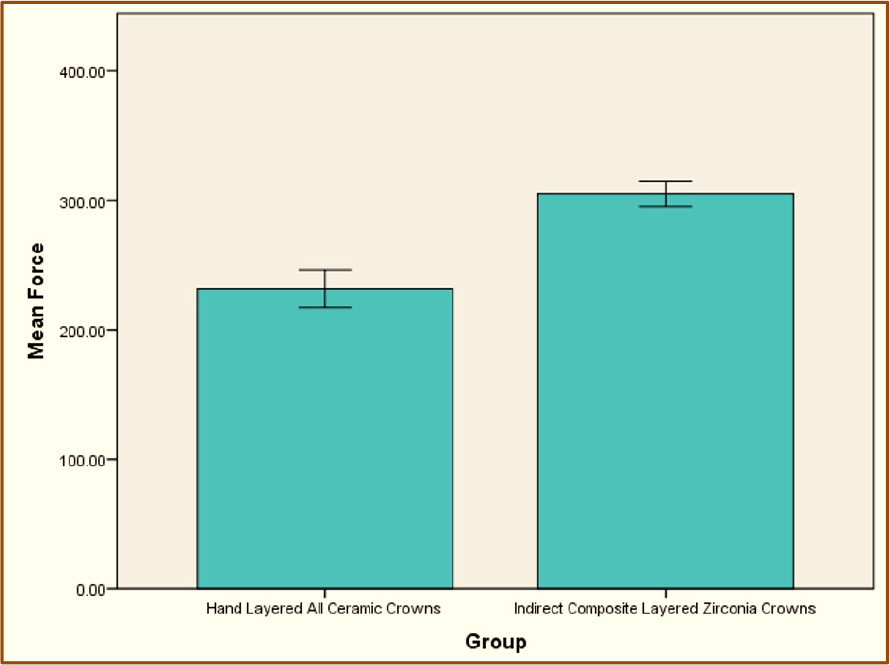
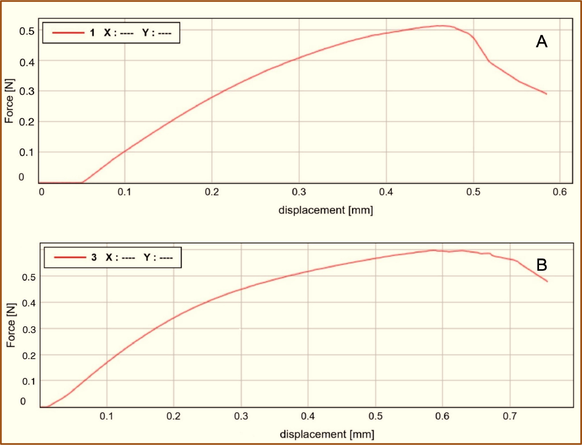
The Vickers test for edge chipping forces revealed distinct resistance characteristics between the two groups. Hand-layered all-ceramic crowns (Group 1) required a force of 231.50 ± 20.145 N, while indirect composite veneered on zirconia (Group 2) needed 305 ± 13.944 N to induce edge chipping (Figure 4 and Table 1). A statistically significant difference in the edge-chipping forces between the groups was observed (p < 0.05). Further analysis of the failure mechanisms in hand-layered ceramic samples showed different outcomes: (a) simultaneous failure of the zirconia coping and veneer, (b) complete debonding of the veneering material, and (c) under the microscope (Figure 5), the fracture line in the ceramic samples was sharp and well-defined. In contrast, the fracture line in the indirect composite samples appeared more diffuse, with cracks spaced apart or showing a larger damaged area (Figure 6).

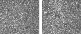
40X magnification images of edge chipped samples (A) Hand-layered all-ceramic sample. (B) Indirect composite veneered sample.
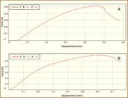
Edge test results. (A) Hand-layered ceramic on zirconia coping. (B) Indirect composite veneered on zirconia coping.
Discussion
The primary focus of this study was the meticulous, comparative analysis of the edge strength of hand-layered all-ceramic crowns and indirect composite veneers on zirconia copings. The average fracture strength recorded at the junction of the hand layered ceramic against the zirconia coping test specimens was found to be statistically lower than that of the indirect composite veneered zirconia coping. This made it possible to reject the null hypothesis. The failure characteristics associated with the hand layered ceramic sample showed a distinct crack line, contrary to indirect composite, whose failure characteristics revealed a dispersed crack line, appearing more like a drift line [16]. The brittle nature of layered ceramics, along with localized stress concentrations, often leads to sharp, well-defined fractures. In contrast, the denser structure of indirect composite tends to produce a more diffuse, drift-like fracture pattern [17].
Several studies have also used devices integrated with universal testing machines to assess the edge-fracture strength of different materials [18]. Notwithstanding the assortment of equipment deployed, the fundamental principle underpinning edge chipping evaluations remains unswerving: the methodology entails applying force with an indenter at predetermined distances from the material's periphery, thereby documenting the requisite force required to initiate chipping.
Chipping can be considered the main cause behind the failures of ceramic restorations, which may explain the main reason (45%) for focusing on edge chipping research among ceramic materials. The multi-layered structures of ceramics tend to have more predisposition to chipping regardless of their composition or manufacturing process compared with monolithic structures of ceramics, which usually offer better toughness properties. Chipping of the veneering ceramics becomes the main cause of ceramic restoration failure [17,19].
In posterior dental prostheses, chipping fractures typically result from contact-induced damage that begins at a wear facet on the occlusal surface [20]. Hence, minute cracks starting below the point of contact join together to create one big crack in the substrate of the veneer. When there is considerable chipping in the ceramic veneer, the cracks usually run into the border line, changing direction. However, tiny chips will still occur in the veneer, although the underlying substrate acts as a barrier for the development of cracks [21].
Given that disparities in the thermal-mechanical properties of ceramics accentuate the propensity for chipping, the proposition of monolithic restorations has been advanced as a potential mitigation strategy, as they inherently exhibit chipping resilience that can be up to 4 times greater than that of their bilayer counterparts [17].
The layered ceramic materials can be described as highly advanced composite materials, whereby leucite crystal particles are incorporated into the oxide glass matrix in an amount that ranges from 1% to 30%. While often described as a monolithic glass, the inclusion of leucite results in the thermal expansion property being altered from the one characteristic of ordinary glass material. The presence of leucite particles in the glass matrix causes formation of stress within it that may serve as a point for initiation of a fracture.
On the other hand, the indirect composite materials have a more compact structure, thus making them more resilient to edge stresses [22]. Moreover, the cooling process influences the coefficient of thermal expansion, a pivotal determinant that diminishes the resilience of ceramics - an effect not observed in Indirect composites. The propensity for chipping observed in Indirect Composites may be ascribed to the interfacial adhesion dynamics between the zirconia framework and the overlaid indirect composite [23].
Many factors, including the type of composite, the nature of the polymer matrix, the filler content, and the size, shape, and distribution of the filler particles, influence fracture toughness. It is also affected by the surface treatment of the fillers and by polymerization carried out under heat and pressure [24]. Earlier studies have shown that variations in fracture toughness largely stem from differences in material composition and microstructure [25,26]. The size and distribution of the crystalline phase further contribute to the distinct behavior seen among materials. The higher edge strength observed in indirect composites can be especially advantageous in clinical situations where restorations must withstand substantial occlusal forces. In these cases, the composite's resistance to chipping can enhance the longevity of restorations by reducing the need for frequent repairs or replacements [27]. Additionally, in areas requiring minimal tooth reduction, indirect composites provide a conservative option, combining esthetic resilience with structural stability, making them ideal for restorations in esthetic zones with thin margins or where preservation of natural tooth structure is prioritized. Dental composites experience changes in their material properties as they degrade and age under different environmental conditions. This degradation can lead to the development of microcracks that do not always align with the direction of the applied load [20,27]. Fractures in indirect composites usually occur due to cracks that form on the surface or subsurface plane at an angle of about 20 to 30 degrees to the direction of the force exerted. Due to the heterogeneous nature of composites, the cracks formed can often take a curved path. This feature is more realistic in terms of modeling fractures and fatigue in dentistry, as cracks in teeth usually form at an angle to the direction of force.
Conversely, the results of this study can be used to demonstrate clinically significant benefits of indirect composites over ceramics. These include increased fracture resistance, improved performance in acidic or high-pressure environments, and lowered chances of chipping. All of the aforementioned qualities can contribute to reduced maintenance needs, longevity of esthetics, and increased patient satisfaction with their dental restoration procedures, which means that indirect composites may represent a promising veneering alternative in terms of edge chipping resistance; however, comprehensive clinical evaluation encompassing long-term durability, bond strength, color stability, and aging behavior is required before definitive clinical recommendations can be made.
Despite the fact that the results are clinically significant in many respects, there are also certain limitations to the applicability of these materials based on the study. First and foremost, there is inconsistency in the testing environment. This is seen in differences in the design of the indenters used, edge-distances, sample sizes, and even the surface roughness of the materials. The lack of a unified testing protocol is one of the main limitations to the clinical application of these materials, and future studies should aim at examining the performance of these composites over longer periods in real-world applications with the presence of occlusal forces, temperature shifts, and acidic conditions. It will be important to consider patient-related variables in these assessments to obtain more conclusive data.
Conclusion
This variation in strength at the edges suggests that indirect composites exhibit greater longevity compared to ceramics hand-laid by a dentist. This conclusion justifies their employment as an effective means in cases when chip resistance is a major concern since it will help reduce problems and improve patient results. At the same time, the presence of both esthetics and high strength makes these materials suitable to meet both cosmetic and functional needs, making them a potentially suitable material for selected dental procedures where chip resistance and esthetics are primary considerations, pending further clinical validation.
-
Financial Support
None.
Data Availability
The data used to support the findings of this study can be made available upon request to the corresponding author.
ν References
-
[1] Narde J, Ahmed N, Keskar V, Pandurangan KK. Evaluation of the colour stability and surface roughness of polymethylmethacrylate and indirect composites with and without ageing: An in-vitro study. Cureus 2024; 16(8):e68073. https://doi.org/10.7759/cureus.68073
» https://doi.org/10.7759/cureus.68073 -
[2] Elbasty RS, Nabil O, Elgohary MS, Taymour M. Comparison of vertical marginal gap and internal fit of chairside laminate veneers fabricated from advanced versus conventional lithium disilicate ceramics: An in vitro study. J Prosthet Dent 2025; 134(3):748.e1-748.e6. https://doi.org/10.1016/j.prosdent.2025.02.044
» https://doi.org/10.1016/j.prosdent.2025.02.044 -
[3] Aboushelib MN, Feilzer AJ, Kleverlaan CJ. Bridging the gap between clinical failure and laboratory fracture strength tests using a fractographic approach. Dent Mater 2009; 25(3):383-391. https://doi.org/10.1016/j.dental.2008.09.001
» https://doi.org/10.1016/j.dental.2008.09.001 -
[4] Vohra M, Pandurangan KK, Shenoy A, Keskar V. A comprehensive review of the surface and chromatic properties of monolithic zirconia: Evaluating the impact of polishing and finishing methods on aesthetics and performance. Cureus 2024; 16(8):e66029. https://doi.org/10.7759/cureus.66029
» https://doi.org/10.7759/cureus.66029 -
[5] Ansarifard E, Taghva M, Mosaddad SA, Akhlaghian M. The impact of various substrates, ceramic shades, and brands on the ultimate color and masking capacity of highly translucent monolithic zirconia: An in vitro study. Odontology 2024; 113(2):607-618. https://doi.org/10.1007/s10266-024-00996-0
» https://doi.org/10.1007/s10266-024-00996-0 -
[6] Habibzadeh S, Khamisi F, Mosaddad SA, Fernandes GV de O, Heboyan A. Full-ceramic resin-bonded fixed dental prostheses: A systematic review. J Appl Biomater Funct Mater 2024; 22:22808000241250118. https://doi.org/10.1177/22808000241250118
» https://doi.org/10.1177/22808000241250118 -
[7] Vohra M, Ahmed N, Keskar V, Shenoy A. A comparison of the edge testing of indirect composite layered zirconia crowns and monolithic zirconia crowns without aging: An in vitro study. Cureus 2024; 16(6):e62080. https://doi.org/10.7759/cureus.62080
» https://doi.org/10.7759/cureus.62080 -
[8] Jodha KS, Kaur N, Salazar Marocho SM, Mecholsky JJ, Lirette ST, Duan Y, et al. Relationship between fracture toughness and fractal dimensional increment in two types of dental glass-ceramics with different fracture surface roughness. Dent Mater 2022; 38(12):1977-1988. https://doi.org/10.1016/j.dental.2022.11.011
» https://doi.org/10.1016/j.dental.2022.11.011 -
[9] Valandro LF, Cadore-Rodrigues AC, Dapieve KS, Machry RV, Pereira GKR. A brief review on fatigue test of ceramic and some related matters in Dentistry. J Mech Behav Biomed Mater 2023; 138:105607. https://doi.org/10.1016/j.jmbbm.2022.105607
» https://doi.org/10.1016/j.jmbbm.2022.105607 -
[10] Lubauer J, Lohbauer U, Belli R. Fatigue threshold r-curves for dental lithium disilicate glass-ceramics. J Dent Res 2023; 102(10):1106-1113. https://doi.org/10.1177/00220345231180565
» https://doi.org/10.1177/00220345231180565 -
[11] Alghazzawi TF. Clinical survival rate and laboratory failure of dental veneers: A narrative literature review. J Funct Biomater 2024; 15(5):131. https://doi.org/10.3390/jfb15050131
» https://doi.org/10.3390/jfb15050131 -
[12] Quinn GD. On edge chipping testing and some personal perspectives on the state of the art of mechanical testing. Dent Mater 2015; 31(1):26-36. https://doi.org/10.1016/j.dental.2014.08.378
» https://doi.org/10.1016/j.dental.2014.08.378 -
[13] Brandeburski SBN, Vidal ML, Collares K, Zhang Y, Della Bona A. Edge chipping test in dentistry: A comprehensive review. Dent Mater 2020; 36(3):e74-e84. https://doi.org/10.1016/j.dental.2020.01.019
» https://doi.org/10.1016/j.dental.2020.01.019 -
[14] Yao Q, Chen Y, Wang Z, Qi H. Evaluations on ceramic fracture toughness measurement by edge chipping. Coatings 2022; 12(8):1146. https://doi.org/10.3390/coatings12081146
» https://doi.org/10.3390/coatings12081146 -
[15] Juri AZ, Belli R, Lohbauer U, Ebendorff-Heidepriem H, Yin L. Edge chipping damage in lithium silicate glass-ceramics induced by conventional and ultrasonic vibration-assisted diamond machining. Dent Mater 2023; 39(6):557-567. https://doi.org/10.1016/j.dental.2023.04.001
» https://doi.org/10.1016/j.dental.2023.04.001 -
[16] Song M, Ko K, Huh Y, Park C, Cho L. Edge chipping resistance and flexural strength of CAD-CAM ceramics before and after thermomechanical aging. J Esthet Restor Dent 2025; 37(4):1096-1104. https://doi.org/10.1111/jerd.13391
» https://doi.org/10.1111/jerd.13391 -
[17] Batanova OA, Matvienko YuG, Marchenkov AYu, Matyunin VM, Obolkina TO, Smirnov SV. Analysis of the influence of sintering temperature on the fracture resistance of ceramics based on experiments on chipping of a sample edge. J Mach Manuf Reliab 2022; 51:294-299. https://doi.org/10.3103/S1052618822040021
» https://doi.org/10.3103/S1052618822040021 -
[18] Alghazzawi TF, Janowski GM, Ning H, Eberhardt AW. Qualitative SEM analysis of fracture surfaces for dental ceramics and polymers broken by flexural strength testing and crown compression. J Prosthodont 2023; 32(5):100-110. https://doi.org/10.1111/jopr.13659
» https://doi.org/10.1111/jopr.13659 -
[19] Singh P, Maiti S, Shenoy A. Comparative evaluation of bond strength and color stability of polyetheretherketone and zirconia layered with indirect composite before and after thermocycling: An in vitro study. J Indian Prosthodont Soc 2024; 24(3):252-258. https://doi.org/10.4103/jips.jips_36_24
» https://doi.org/10.4103/jips.jips_36_24 -
[20] Liu Z, Ma C, Chang Z, Yan P, Li F. Advances in crack formation mechanism and inhibition strategy for ceramic additive manufacturing. J Eur Ceram Soc 2023; 43(12):5078-5098. https://doi.org/10.1016/j.jeurceramsoc.2023.05.008
» https://doi.org/10.1016/j.jeurceramsoc.2023.05.008 -
[21] Qi B, Liang S, Li Y, Zhou C, Yu H, Li J. ZrO 2 matrix toughened ceramic material-strength and toughness. Adv Eng Mater 2022; 24(6):2101278. https://doi.org/10.1002/adem.202101278
» https://doi.org/10.1002/adem.202101278 -
[22] Bompolaki D, Lubisich EB, Fugolin AP. Resin-based composites for direct and indirect restorations. Dent Clin North Am 2022; 66(4):517-536. https://doi.org/10.1016/j.cden.2022.05.003
» https://doi.org/10.1016/j.cden.2022.05.003 -
[23] Josic U, D’Alessandro C, Miletic V, Maravic T, Mazzitelli C, Jacimovic J, et al. Clinical longevity of direct and indirect posterior resin composite restorations: An updated systematic review and meta-analysis. Dent Mater 2023; 39(12):1085-1094. https://doi.org/10.1016/j.dental.2023.10.009
» https://doi.org/10.1016/j.dental.2023.10.009 -
[24] Wang A, Zhao X, Huang M, Cheng Y, Zhang D. A systematic study on the quality improving of fracture toughness measurement in structural ceramics by laser notching method. Theor Appl Fract Mec 2021; 114:102981. https://doi.org/10.1016/j.tafmec.2021.102981
» https://doi.org/10.1016/j.tafmec.2021.102981 -
[25] Begand S, Spintzyk S, Geis-Gerstorfer J, Bourauel C, Keilig L, Lohbauer U, et al. Fracture toughness of 3Y-TZP ceramic measured by the Chevron-Notch Beam method: A round-robin study. Dent Mater 2022; 38(7):1128-1139. https://doi.org/10.1016/j.dental.2022.05.001
» https://doi.org/10.1016/j.dental.2022.05.001 -
[26] Alves MFRP, dos Santos C, Elias CN, Amarante JEV, Ribeiro S. Comparison between different fracture toughness techniques in zirconia dental ceramics. J Biomed Mater Res B Appl Biomater 2023; 111(1):103-116. https://doi.org/10.1002/jbm.b.35137
» https://doi.org/10.1002/jbm.b.35137 -
[27] Kim S-Y, Kim B-S, Kim H, Cho S-Y. Occlusal stress distribution and remaining crack propagation of a cracked tooth treated with different materials and designs: 3D finite element analysis. Dent Mater 2021; 37(4):731-740. https://doi.org/10.1016/j.dental.2021.01.020
» https://doi.org/10.1016/j.dental.2021.01.020
Edited by
-
Academic Editor:
Wilton Wilney Nascimento Padilha