 Electrospun scaffolds loaded with Stryphnodendron adstringens extract and Melaleuca alternifolia oil
Abstract
Electrospun PCL-based scaffolds have been proposed for wound healing. However, to deal with bacterial infections, the incorporation of natural compounds into the PCL matrix could be a promising alternative. Stryphnodendron adstringens (Barbatimão) extract and Melaleuca alternifolia (Melaleuca) oil are known due to their antibacterial, antifungical, and anti-inflammatory properties, interesting for healing applications. This paper proposed the development of electrospun PCL scaffolds containing a combination of barbatimão and melaleuca compounds. The obtained samples were 20B/80M and 80B/20M, in which B refers to Barbatimão and M for Melaleuca. The samples were characterized via X-ray diffraction, Raman spectroscopy, tensile test, contact angle analysis, optical microscopy, and cytotoxicity. The incorporation of the natural compounds did not alter the structure of PCL and higher concentration of Barbatimão indicated an enhancement in antimicrobial properties against S. aureus, with reduction of 33.3% on biofilm formation. Hence, the proposed scaffolds could be promising material for wound healing applications.
Keywords:
barbatimão extract; melaleuca oil; poly-ε-caprolactone; scaffolds
1. Introduction
Tissue engineering aims to develop materials that can biologically replace tissue functions by combining a scaffold, cells and biological molecules[1]. These biomaterials exhibit biocompatibility with tissues, biodegradability, non-toxic, adequate porosity, and good mechanical resistance, making them favorable to cell proliferation, migration and differentiation, and enabling them, for example, to stimulate the wound healing process[2-4].
In the context of dressings applied to healing acute and chronic wounds, the use of micro and nanofibrous scaffolds are good alternatives, as they promote the mimicry of the native extracellular matrix (ECM)[5]. Depending on the degree of severity of a wound, the healing process tends to become complex[6-8], resulting in great susceptibility to bacteria that are multiresistant to conventional antibiotics[9]. Furthermore, it is known that infections caused by bacteria represent a major concern in wound treatments, delaying the healing process and leading to tissue deformation or even the patient’s death[10]. In this sense, nanofibrous biomaterials provide support for the release of bioactive compounds, such as antimicrobial agents[11].
Given this scenario, the impregnation of scaffolds with substances with therapeutic potential is necessary and beneficial[12], since the release of such structures can enable faster and more effective healing. Therefore, scaffolds impregnated with oils and natural extracts with healing and antifungal effects on topical lesions have attracted interest of the scientific community[13]. The bioactive constituents of medicinal herbs are responsible for therapeutic activities and offer unlimited potential through new medicines due to their accessibility and chemical diversity. These constituents are responsible for therapeutic activities, such as hypoglycemic, antidiabetic, antioxidant, antimicrobial, anti-inflammatory, anti-carcinogenic and antimalarial[14,15].
One of the main polymers used in tissue engineering, poly-ε-caprolactone (PCL) is a synthetic composite that has good mechanical resistance and rigidity, also known for its biodegradable and biocompatible properties[16,17]. Furthermore, PCL is a polymer already approved and with a well-established safety profile confirmed by the Food and Drug Administration (FDA)[18]. Among different methods for manufacturing nanofibers, electrospinning technique is considered an easy and effective way to produce ultrafine fibers from various polymers[19]. Thus, electrospinning offers an efficient way to produce well-organized scaffolds, due to the high versatility in material choice, ease of standardization and great flexibility in drug formulation[20].
Stryphnodendron adstringens (Barbatimão), found mainly in the Brazilian cerrado, has pharmacological properties with antibacterial, anti-inflammatory, and healing potential, thus inhibiting the formation of chemical mediators of inflammation, such as histamine, bradykinin, prostaglandin[21]. Despite the known effects on reduction of biofilm, many studies are focused on oral and anaerobic bacteria[22,23]. Some studies present evaluations regarding its safety; however, there is still a need for a deeper understanding of its effects, as well as the safe quantities capable of inhibiting biofilm formation in different bacteria[24,25]. Hence, there is potential for the use of natural extracts, especially Barbatimão extract, in applications involving bacteria commonly found in epidermal wounds.
The medicinal effects of Barbatimão are attributed to the high tannin content in its chemical composition, which can reach levels of 20% to 50%[26]. Similarly, Melaleuca alternifolia (Tea Tree), a typical Australian tree, also presents scientific evidence of its biological properties, such as antimicrobial and antifungal activity, aiding in the healing processes[27-29]. Additionally, Melaleuca alternifolia stimulates cellular potassium ion leakage and inhibits respiration in E. coli suspensions, providing evidence of a lethal action related to damage to the cytoplasmic membrane[30].
In literature, scaffolds obtained via electrospinning and based on PCL impregnated with natural substances, such as Aloe vera extract, fennel extract (Inula graveolens), clove essential oil (Eugenia caryophyllata), oregano essential oil (Origanum vulgare) and tucumã oil (Astrocaryum vulgare) have already been investigated[31-34]. In addition to providing bioactive properties to the resulting material, impregnation with structures of natural origin has been widely investigated because, due to their nature, they are less likely to induce resistance of multidrug-resistant bacteria to conventional antibiotics[35], which is an indispensable characteristic for topical applications.
However, to date, the development of PCL-based electrospun scaffolds incorporating a combination of M. alternifolia and S. adstringens remains unexplored in the literature. Therefore, this research is considered innovative, contributing to the expansion of knowledge regarding the potential use of natural species for application in dressings and wound healing. In this context, the present study aimed to develop scaffolds using PCL as a polymeric matrix, incorporating Melaleuca auternifolia oil and Stryphnodendron astringens extract, and to evaluate their physicochemical properties, cytotoxic behavior and antibiofilm activity.
2. Materials and Methods
2.1 Materials
All reagents and raw materials were used without prior treatment. The materials used to scaffold production included Sigma Aldrich PCL polymer, average molecular weight of 80,000 g/mol; Acetone solvent (Synth, Brazil); Melaleuca auternifolia oil (Nova Derme, Brazil); Stryphnodendron adstringens (Cruz Vermelha - Farmácia Homeopatia, Brazil). It is worth noting that Stryphnodendron adstringens extract and Melaleuca alternifolia oil were obtained as medical-grade reagents from certified compounding pharmacies. These materials present established safety profiles, complying with the requirements for biomaterial development.
2.2 Sample preparation
The scaffolds were produced according to the adaptation of the literature[36,37]. To prepare the polymeric solutions, 0.75 g of PCL was dissolved in acetone (7,5% w/v), using a hot plate at 50 °C (±2 °C) under constant magnetic stirring for approximately 60 min. Subsequently, 100 µL of an oil mixture (10% v/v to polymeric solution), containing Barbatimão and Melaleuca was added in different proportions (Table 1), and the solution was maintained under magnetic stirring and heating for 5 min. The resulting solution was transferred to a 10 mL plastic syringe with an 18G beveled needle and connected to the electrospinning equipment, operating at an applied voltage of 10 kV. All samples were carried out in triplicates and the percentage ratios of polymeric matrix and Barbatimão extract/tea tree oil were defined as follows. The samples were called PCL, 20B/80M and 80B/20M, whose parameters are presented in Table 1.
2.3 Physicochemical characterization
The samples were characterized after the formation of different structures by electrospinning. For the X-ray diffractrometry (XRD) analysis, a Bruker D2 Advance Diffractometer, with a copper tube (radiation Kα = 1,5418 Ǻ) was used. The voltage and current used in the tube were 30 kV and 10 mA, respectively. The diffractometer used has a θ–θ geometry. The goniometer speed was 0.05º/s, with a 2Ɵ interval of 5º to 70º. RAMAN spectroscopy was performed on a Renishaw In-Via Spectomer System equipment, with a 532 nm laser and a measurement range of 200-2000 cm-1. To measure the contact angle, a 10 μL drop of deionized water was carefully dripped onto the flat surface of the structures. Images for each drop were captured using a digital camera (Samsung, 12 MP, 45º telephoto lens). The contact angle was determined using Tracker® software. This assay was performed according to previously published methods[38].
2.4 Mechanical and morphological characterization
Tensile tests were performed according to ASTM D882 standard (speed: 01 mm/min) and specimens measuring 50 x 15 mm in length and width, respectively, in triplicate. An EMIC universal testing machine, model DL-10000, with a 5 kN load cell was used. The sample thicknesses were 0.93 ± 0.06 mm, 1.00 ± 0.10 mm, and 1.07 ± 0.12 mm for PCL, 20B/80M and 80B/20M, respectively. The Kruskal-Wallis test, with a 95% confidence interval, was used to compare the samples, as the Shpiro-Wilk's normality test indicated that the data did not follow a normal distribution (W = 0.75; p < 0.0001). The morphology of the formed fibers was evaluated using scanning electron microscopy (SEM). The samples were previously metallized and analyzed using a TESCAN ANALYTICS microscope (model VEGA 3-SBU). The fiber diameter was determined by measuring 50 different points for each sample.
2.5 In vitro characterization
The MTT cell viability assay was used to evaluate the cytotoxicity of the different materials obtained. The protocol was based on that described in Wilms et al.[39] and all experiments were performed in triplicate for the PCL blank and for the samples containing both oils/extracts in their composition. Samples (5 x 5 mm) were used and placed in a 96-well plate. Culture medium containing only peripheral blood mononuclear cells (PBMC) was used as a negative control (CN). PBMC separation step was performed by density gradient (Histopaque®-1077) and centrifugation. The concentration of 2x10⁵ cells mL-1 was obtained by counting the cells in a Neubeuer chamber with 0.4% trypan blue. After seeding, the cells and samples were incubated in an environment with 5% CO2 at 37 °C for 24 hours. The PBMC were obtained from peripheral blood samples discarded from the Clinical Analysis Laboratory of the Franciscana University (LEAC), following approval by the Human Research Ethics Committee of the Franciscana University, nº (CAAE: 31211214.4.0000.5306), without access to identifiable data.
The results were obtained as a function of optical density. They were calculated by spectrophotometry at a length of 570 nm, using an Anthos 2010 spectrometer (Biochrom®, London, UK) and Equation 1. For this trial, statistical analyzes with a 95% confidence interval were also used. The selected methods were one-way ANOVA followed by Dunnett's post hoc test to compare the means between the samples and the negative control.
where: OD570e: average value of the measured optical density of extracts from 100% of the test sample; OD570b: average value of the measured optical density of the blank samples.
2.6 Biofilm formation test
The antibiofilm activity assay was performed using Staphylococcus aureus (ATCC 25923) and Pseudomonas aeruginosa (ATCC 13883), following the methodology described by Stepanovic[40]. For the test, 180 µL of TSB broth supplemented with 1% glucose was added to each well. Subsequently, 20 µL of the prepared inoculum was added to each well, except for the column reserved for sterility control. The sterility control had only TSB broth with 1% glucose and sterile saline. The samples were evaluated in triplicate for each formation along with the negative control (bacteria only). All plates were incubated at a temperature of 37 °C. After incubation, the wells were emptied and gently washed three times with sterile saline to remove non-adherent cells. The biofilm was fixed with 200 µL of methanol (P. A.) for 20 minutes. After fixation, the methanol was removed by inversion and the plate was allowed to dry at room temperature. The adhered biofilm was stained with 200 µL of per well for 15 minutes at room temperature. After staining, the violet crystal was removed solution was removed by inversion and the plate was washed 3 times with distilled water. The biofilm was re-sobulized in each well with 200 µL of 95% ethanol. Then, Ethanol was gently added to the wells and the plate was left covered with the lid at room temperature for 30 minutes without stirring. The results of biofilm formation were evaluated on an Anthos 2010 spectrophotometer (Biochrom®, London, UK) with a wavelength of 570 nm.
3. Results and Discussions
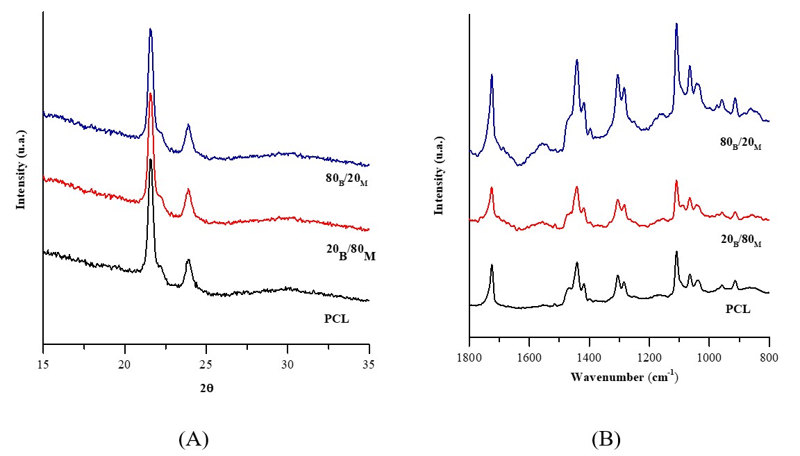
The XRD patterns and Raman spectra obtained for the scaffolds are shown in Figure 1. In XRD (Figure 1A), it can be observed two characteristic peaks of pure PCL, the first at 21.6º, representing the crystalline plane of reflection d (110), and the second at 23.9º, representing the d (200) plane[41]. It was observed that the characteristic peaks of PCL are also present in the scaffolds with the incorporation of Barbatimão and Melaleuca. The XRD patterns showed no new crystalline phases, which was expected due to amorphous nature of the added compounds.
X-ray diffraction diagram (A) and Raman spectrum (B) of scaffolds produced with PCL, Melaleuca auternifolia oil and Stryphnodendron adstringens extract.
Through Raman spectroscopy (Figure 1B), it was found planar and non-planar peaks of PCL. Bands at 1725 cm-1 are observed, representing the structure C=O, of 1466-1418 cm-1 relating to δCH2 and 1304-1284 cm-1, ωCH2, in 1110-1039 cm-1 characteristic of clusters and 958-914 cm-1 the structure C-COO[42]. It was also observed that the characteristic peaks of PCL were not altered when a higher concentration of Melaleuca was added[43]. However, it can be observed an intensity enhancement of the peaks when the concentration of Barbatimão was higher than that of Melaleuca. The increased intensity across the spectrum might be attributed to the complex polyphenolic structure of the Barbatimão extract[44].
Maximum stress values of the samples are presented in Table 2. It can be seen that the 20B/80M sample obtained the lowest result, while the 80B/20M sample has maximum stress values that are very similar and higher than the PCL.
This may suggest that the composition of Melaleuca oil makes PCL more fragile, while Barbatimão extract makes it more resistant. It is worth pointing out that, previous to statistical comparisons, the Shapiro-Wilk's normality test was performed and justified the suitability of a parametric hypothesis test for the mechanical data (W = 0.920; p = 0.463). Despite the differences in maximum tension, the values can be considered statistically identical, according to the one-way ANOVA test (F = 2.17; p = 0.179), for a 95% confidence interval. This demonstrates that the addition of Barbatimão extract and Melaleuca oil did not significantly modify the maximum tension of the PCL. The associated statistical analyzes results can be found in Supplementary Material.
Table 3 shows the results obtained in the contact angle test, which is important for evaluating the hydrophobicity of the scaffolds. PCL showed a hydrophobic characteristic, with an angle greater than 90°, a result also found in the literature[45,46]. This characteristic was also found in sample 20B/80M, however in sample 80B/20M it presented an angle of 84.8°, showing it to be slightly hydrophilic[47]. Hydrophilicity in a dressing is an important parameter, as it facilitates the absorption of exudate and the maintenance of moisture at the wound site[48]. However, it is known that a hydrophobic surface on a dressing reduces the adhesion of bacteria to the target region[49]. In this sense, samples with more hydrophobic characteristics (PCL and 20B/80M) have the potential to prevent the formation of biofilms and significantly reduce bacterial resistance at the site of application, while the slightly hydrophilic sample (80B/20M) can promote greater cell adhesion during wound healing[50].
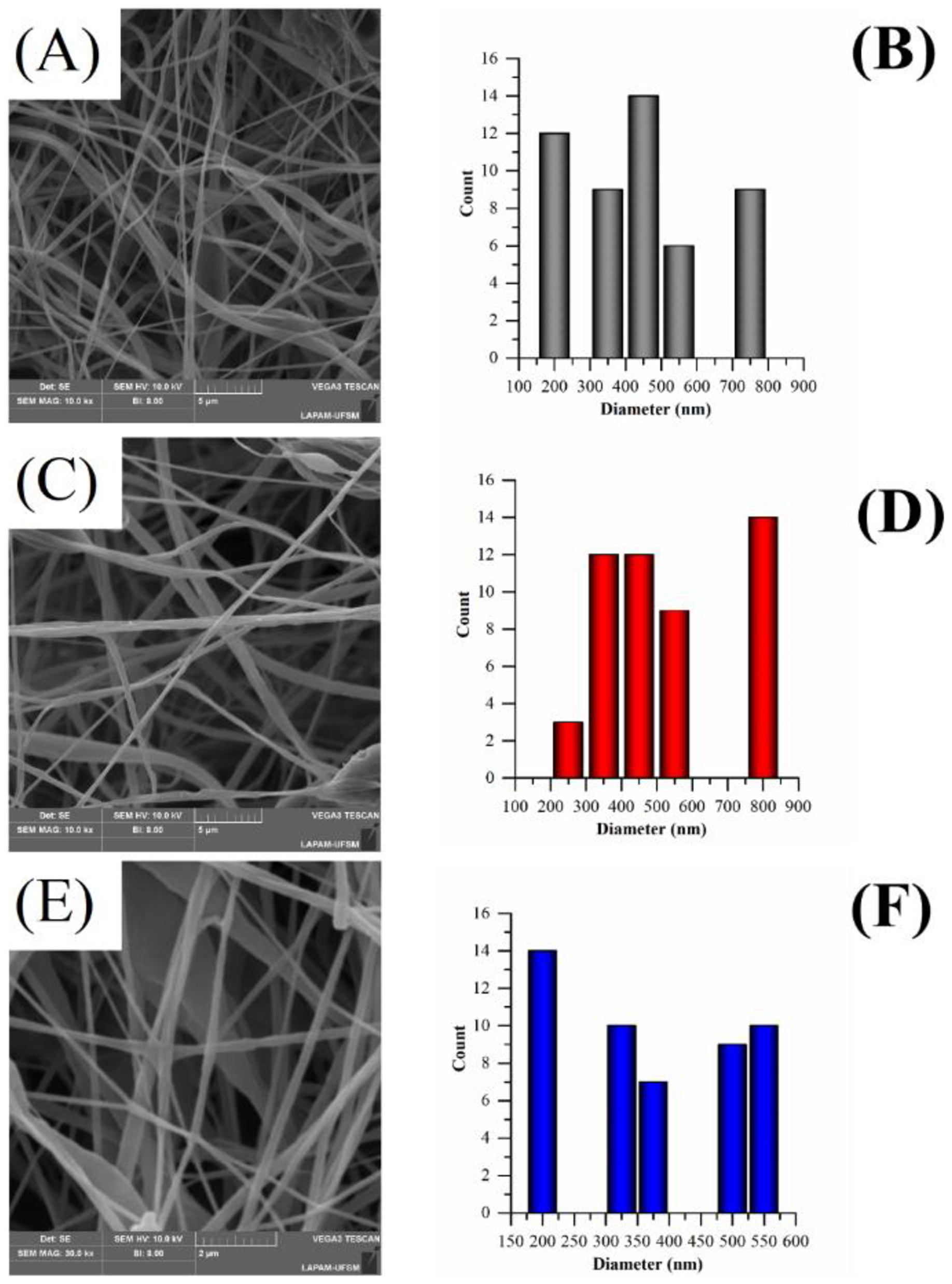
Scanning electron microscopy (Figure 2) showed that the resulting scaffolds had fibers in their structures, but none of the samples resulted in uniform fibers; all of them have very heterogeneous fibers, loaded with polymeric glomerates[51,52]. The scaffolds had average sizes of approximately 800 nm for the samples with PCL and 20B/80M, while the 80B/20M samples’ average size was around 200 nm.
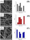
SEM of the PCL (A), 20B/80M (C) and 80B/20M (E) scaffolds, and the average distribution of the PCL (B), 20B/80M (D) and 80B/20M (F) polymeric scaffolds.
Figure 3 shows the results of the MTT cell viability test against PBMC cells. Basically, all samples did not show cytotoxicity to PBMC, as cell viability was greater than 70%[53]. It was noted that PCL (84.5%) resulted in a decrease in cell viability, when compared to NC. This also satisfies the biocompatibility requirements because, in the literature, decreases in cell viability of up to 30% in relation to NC are an indication that the material is not cytotoxic[54]. For the 20B/80M sample, it obtained a cellular observation 6% greater than the NC, providing non-cytotoxic characteristics to PMBC. The 80B/20M sample, however, presented a favorable environment for cell proliferation, with a viability percentage of 113.34%, when compared to the NC.
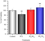
Results obtained in the cell viability test, via MTT, for PCL, 20B/80M and 80B/20M samples. Data were presented as mean *p < 0.05, **p < 0.01 and ***p < 0.001 versus NC.
The cell viability results were promising and are in line with the literature, where PCL-based scaffolds were non-cytotoxic to PBMC and other cell lines. In the case of sample 80B/20M, it is assumed that the increase in cell viability is due to the properties of Barbatimão and Melaleuca[55], where Barbatimão, in higher concentration in this sample, presents topical or oral use as a good route of administration. Furthermore, the incubation time of 24 h is sufficient to guarantee the safety of such biomaterials intended for topical applications[53].
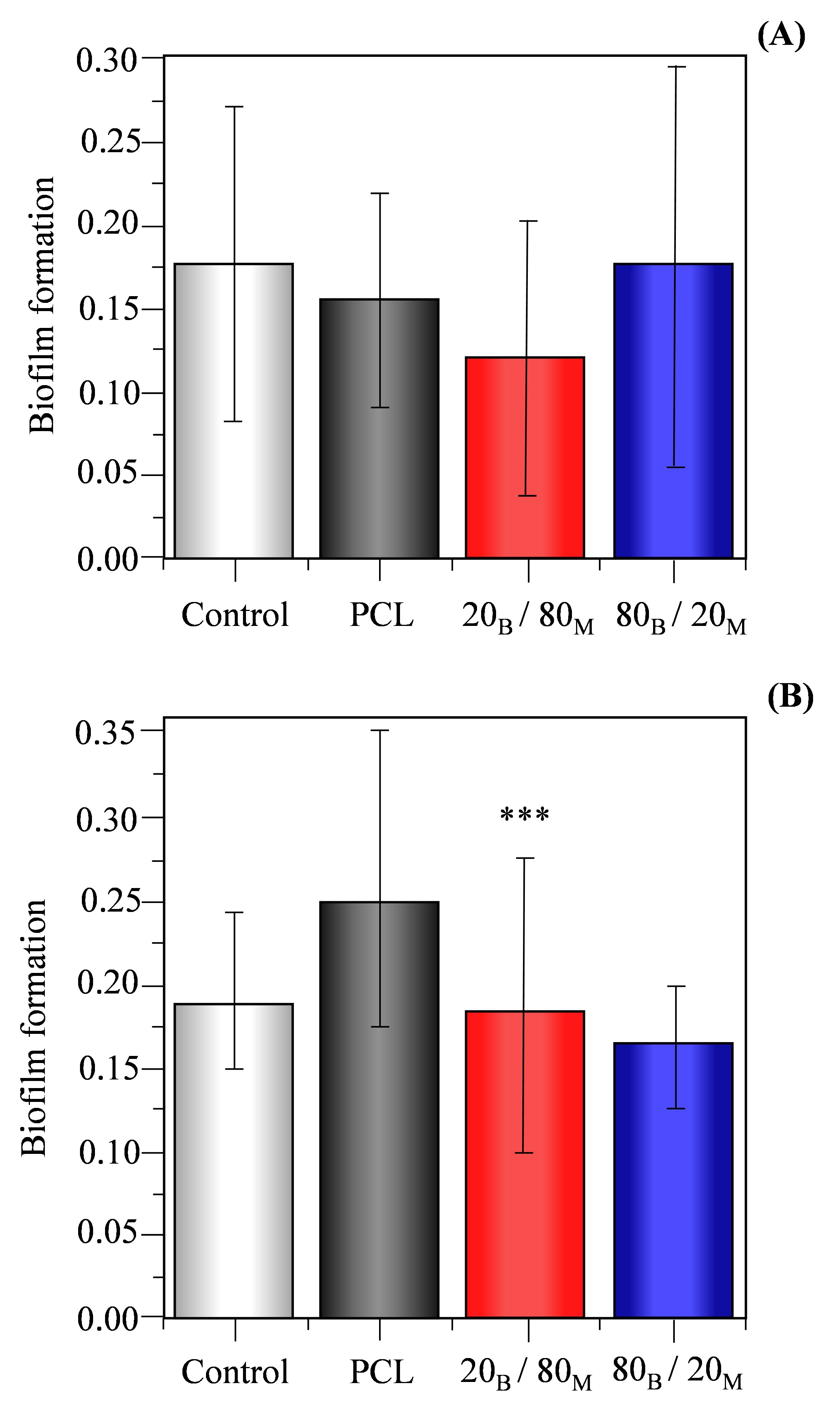
Figure 4 shows the results of the antibiofilm test against the bacteria gram-positive, Staphylococcus aureus and gran-negative Pseudomonas aeruginosa. Figure 4A shows the results obtained for the P. aeruginosa strain, in which the samples did not show a statistical difference compared to the control group (p = 0.965). The PCL sample showed a reduction of 5.0% compared to the control, while the 20B/80M sample showed a 3.6% reduction in the formation of bacterial biofilm. In the case of 80B/20M sample, it showed an increase of 5.5% compared to PC, not inhibiting biofilm formation. The biofilm-forming capacity at 20B/80M is due to the ability of Melaleuca alternifolia to combat pathogenic microorganisms during the treatment of infectious diseases, due to the presence of the polyphenol epigallocatechin-3-O-gallate (EGCG), whose antimicrobial properties may justify the antibiofilm efficiency[56]. In PCL, biofilm inhibition can be attributed to its hydrophobic character, which makes it difficult for bacteria to adhere to the surface and, consequently, reduces the possibility of biofilm formation[57].
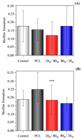
Results of the antibiofilm test against Pseudomonas aeruginosa (A) and Staphylococcus aureus (B). Data were presented as mean *p < 0.05, **p < 0.01 and ***p < 0.001 versus PC.
Regarding the bacteria S. aureus (Figure 4B), only the 80B/20M sample showed a significant difference (p<0.001) compared to the control group. PCL proved to form biofilms, with an increase of 11.4% compared to PC, while the 20B/80M sample showed results close to PC, with only 3.5% less than PC. However, when comparing PC and sample 80B/20M, it was observed a greater inhibition of biofilm, with a reduction of 33.3%. This greater biofilm inhibition capacity might be due to the bioactive properties of Barbatimão, such as antimicrobial, antifungal activity and recently, efficiency against the formation of biofilms[58]. It was possible to observe that only 80B/20M samples presented characteristics of non-biofilm formation against S. aureus, indicating that Barbatimão could induce an antimicrobial property against gram-positive strains. As mentioned before, the associated statistical analyzes results can be found in Supplementary Material.
4. Conclusions
It was possible to produce and evaluate scaffolds with different concentrations of Barbatimão and Melaleuca. Through the results obtained by X-ray diffraction and Raman spectroscopy, it can be concluded that there were no changes in the structure and crystallinity of PCL. Optical microscopy demonstrated that fibers were formed, but the scaffolds presented heterogeneous fibers, with some polymer agglomerates. The contact angle test demonstrated that the PCL 80B/20M mixture had a less hydrophobic characteristic compared to PCL and the other samples. The MTT assay showed that all the samples were not toxic to PBMC. The antibiofilm formation assay demonstrated that the proposed scaffolds did not inibited the biofilm formation against P. aeruginosa. On the other hand, samples with higher concentrations of Barbatimão displayed an enhanced reduction (33.3%) on biofilm formation against S. aureus. Therefore, the inclusion of Melaleuca alternifolia oil and Stryphnodendron adstringens extract in different quantities did not change the initial characteristics of PCL and, the obtained materials appear as a possibility in the manufacture of scaffolds for application in skin dressings.
Supplementary Material
Supplementary material accompanies this paper.
Table S1
Table S2
Table S3
Table S4
Table S5
Table S6
Table S7
Figure S1
This material is available as part of the online article from https://doi.org/10.1590/0104-1428.20250123
6. Acknowledgements
The authors acknowledge the Rio Grande do Sul Research Foundation (FAPERGS) for the financial support (Call 14/2022 - ARD/ARC, Grant No. 23/2551-0000825-4).
-
Data Availability:
All data supporting the findings of this study are available from the corresponding author upon request.
-
How to cite:
Pinheiro, L. D. S. M., Oviedo, V. R., Gomes, D. T., Carvalho, L. O., Fernandes, L. S., Sagrillo, M. R., Bertuol, D. A., & Rodrigues Jr., L. F. (2026). Electrospun scaffolds loaded with Stryphnodendron adstringens extract and Melaleuca alternifolia oil. Polímeros: Ciência e Tecnologia, 36(3), e20260031. https://doi.org/10.1590/0104-1428.20250123
7. References
-
1 Guo, B., & Ma, P. X. (2018). Conducting polymers for tissue engineering. Biomacromolecules, 19(6), 1764-1782. https://doi.org/10.1021/acs.biomac.8b00276 PMid:29684268.
» https://doi.org/10.1021/acs.biomac.8b00276 -
2 Lovett, M., Lee, K., Edwards, A., & Kaplan, D. L. (2009). Vascularization strategies for tissue engineering. Tissue Engineering. Part B, Reviews, 15(3), 353-370. https://doi.org/10.1089/ten.teb.2009.0085 PMid:19496677.
» https://doi.org/10.1089/ten.teb.2009.0085 -
3 Guo, B., & Ma, P. X. (2014). Synthetic biodegradable functional polymers for tissue engineering: a brief review. Science China. Chemistry, 57(4), 490-500. https://doi.org/10.1007/s11426-014-5086-y PMid:25729390.
» https://doi.org/10.1007/s11426-014-5086-y -
4 van Vlierberghe, S., Dubruel, P., & Schacht, E. (2011). Biopolymer-based hydrogels as scaffolds for tissue engineering applications: a review. Biomacromolecules, 12(5), 1387-1408. https://doi.org/10.1021/bm200083n PMid:21388145.
» https://doi.org/10.1021/bm200083n -
5 Hao, D., Swindell, H. S., Ramasubramanian, L., Liu, R., Lam, K. S., Farmer, D. L., & Wang, A. (2020). Extracellular matrix mimicking nanofibrous scaffolds modified with mesenchymal stem cell-derived extracellular vesicles for improved vascularization. Frontiers in Bioengineering and Biotechnology, 8, 633. https://doi.org/10.3389/fbioe.2020.00633 PMid:32671037.
» https://doi.org/10.3389/fbioe.2020.00633 -
6 Murray, R. Z., West, Z. E., Cowin, A. J., & Farrugia, B. L. (2019). Development and use of biomaterials as wound healing therapies. Burns & Trauma, 7, s41038-018-0139-7. https://doi.org/10.1186/s41038-018-0139-7
» https://doi.org/10.1186/s41038-018-0139-7 -
7 Okur, M. E., Karantas, I. D., Şenyiğit, Z., Üstündağ Okur, N., & Siafaka, P. I. (2020). Recent trends on wound management: new therapeutic choices based on polymeric carriers. Asian Journal of Pharmaceutical Sciences, 15(6), 661-684. https://doi.org/10.1016/j.ajps.2019.11.008 PMid:33363624.
» https://doi.org/10.1016/j.ajps.2019.11.008 -
8 Nicks, B. A., Ayello, E. A., Woo, K., Nitzki-George, D., & Sibbald, R. G. (2010). Acute wound management: revisiting the approach to assessment, irrigation, and closure considerations. International Journal of Emergency Medicine, 3(4), 399-407. https://doi.org/10.1007/s12245-010-0217-5 PMid:21373312.
» https://doi.org/10.1007/s12245-010-0217-5 -
9 Puca, V., Marulli, R. Z., Grande, R., Vitale, I., Niro, A., Molinaro, G., Prezioso, S., Muraro, R., & Di Giovanni, P. (2021). Microbial species isolated from infected wounds and antimicrobial resistance analysis: data emerging from a three-years retrospective study. Antibiotics, 10(10), 1162. https://doi.org/10.3390/antibiotics10101162 PMid:34680743.
» https://doi.org/10.3390/antibiotics10101162 -
10 Pallavali, R. R., Degati, V. L., Lomada, D., Reddy, M. C., & Durbaka, V. R. P. (2017). Isolation and in vitro evaluation of bacteriophages against MDR-bacterial isolates from septic wound infections. PLoS One, 12(7), e0179245. https://doi.org/10.1371/journal.pone.0179245 PMid:28719657.
» https://doi.org/10.1371/journal.pone.0179245 -
11 Krishnan, K. A., & Thomas, S. (2019). Recent advances on herb-derived constituents-incorporated wound-dressing materials: a review. Polymers for Advanced Technologies, 30(4), 823-838. https://doi.org/10.1002/pat.4540
» https://doi.org/10.1002/pat.4540 -
12 Dhilip Kumar, S. S., & Abrahamse, H. (2020). Advancement of nanobiomaterials to deliver natural compounds for tissue engineering applications. International Journal of Molecular Sciences, 21(18), 6752. https://doi.org/10.3390/ijms21186752 PMid:32942542.
» https://doi.org/10.3390/ijms21186752 -
13 Khan, A. U. R., Xiangyang, S., Ahmad, A., & Mo, X.-M. (2018). Electrospinning of crude plant extracts for antimicrobial and wound healing applications. SM Journal of Biomedical Engineering, 4(1), 1024. Retrieved in 2025, December 26, from https://www.researchgate.net/publication/352687194
» https://www.researchgate.net/publication/352687194 - 14 Husen, A. (Ed.) (2023). Herbs, shrubs, and trees of potential medicinal benefits. Boca Raton: CRC Press.
-
15 Himesh, S., Singhai, A. K., & Sharma, S. (2012). Quantification of ascorbic acid in leaves of Annona squamosa. International Journal of Pharmacy and Pharmaceutical Sciences, 4(3), 144-147. Retrieved in 2025, December 26, from https://web.archive.org/web/20180413062416id_/http://ijppsjournal.com/Vol4Issue3/3588.pdf
» https://web.archive.org/web/20180413062416id_/http://ijppsjournal.com/Vol4Issue3/3588.pdf -
16 Dwivedi, R., Kumar, S., Pandey, R., Mahajan, A., Nandana, D., Katti, D. S., & Mehrotra, D. (2020). Polycaprolactone as biomaterial for bone scaffolds: review of literature. Journal of Oral Biology and Craniofacial Research, 10(1), 381-388. https://doi.org/10.1016/j.jobcr.2019.10.003 PMid:31754598.
» https://doi.org/10.1016/j.jobcr.2019.10.003 -
17 Cipitria, A., Skelton, A., Dargaville, T. R., Dalton, P. D., & Hutmacher, D. W. (2011). Design, fabrication and characterization of PCL electrospun scaffolds: a review. Journal of Materials Chemistry, 21(26), 9419-9453. https://doi.org/10.1039/c0jm04502k
» https://doi.org/10.1039/c0jm04502k -
18 Lim, M. M., Sun, T., & Sultana, N. (2015). In vitro biological evaluation of electrospun polycaprolactone/gelatine nanofibrous scaffold for tissue engineering. Journal of Nanomaterials, 2015(1), 303426. https://doi.org/10.1155/2015/303426
» https://doi.org/10.1155/2015/303426 -
19 Feng, B., Ji, T., Wang, X., Fu, W., Ye, L., Zhang, H., & Li, F. (2020). Engineering cartilage tissue based on cartilage-derived extracellular matrix cECM/PCL hybrid nanofibrous scaffold. Materials & Design, 193, 108773. https://doi.org/10.1016/j.matdes.2020.108773
» https://doi.org/10.1016/j.matdes.2020.108773 -
20 Permyakova, E. S., Polčak, J., Slukin, P. V., Ignatov, S. G., Gloushankova, N. A., Zajíčková, L., Shtansky, D. V., & Manakhov, A. (2018). Antibacterial biocompatible PCL nanofibers modified by COOH-anhydride plasma polymers and gentamicin immobilization. Materials & Design, 153, 60-70. https://doi.org/10.1016/j.matdes.2018.05.002
» https://doi.org/10.1016/j.matdes.2018.05.002 -
21 Lima, T. C. D., Cardoso, M. V., Modesto, T., Oliveira, A. L. B., Silva, M. N., & Monteiro, M. C. (2016). Breve revisão etnobotânica, fitoquímica e farmacologia de Stryphnodendron adstringens utilizada na Amazônia. Revista Fitos, 10(3), 329-338. https://doi.org/10.5935/2446-4775.20160025
» https://doi.org/10.5935/2446-4775.20160025 -
22 Sper, F. L., Amêndola, I., Ramos, L. P., Santos, J. G., Freitas, E. T., Meccatti, V. M., Ferraz, L. F. F., Oliveira, J. R., Santamaria, M. P., Ribeiro, M. C. M., & Oliveira, L. D. (2021). Effect of Stryphnodendron adstringens (Mart.) Coville extract in aerobic and anaerobic microorganisms and mammalian cells. Research, Society and Development, 10(11), e364101119953. https://doi.org/10.33448/rsd-v10i11.19953
» https://doi.org/10.33448/rsd-v10i11.19953 -
23 Melo, R. N., Goes, V. F. F., Canelli, A. P., Aro, A. A., Moreira, B. O., Correia, J. R., Catirse, A. B. C. E. B., Reis, A. C., Costa, S. M., & Godoi, A. P. T. (2023). Antibacterial activity of barbatimão (Stryphnodendron adstringens) against periodontopathogens and cytotoxic effects on fibroblasts. Letters in Applied Microbiology, 76(6), ovad054. https://doi.org/10.1093/lambio/ovad054 PMid:37291703.
» https://doi.org/10.1093/lambio/ovad054 -
24 Gomes, P. W. P., Pamplona, T. C. D. L., Navegantes-Lima, K. C., Quadros, L. B. G., Oliveira, A. L. B., Santos, S. M., Silva, C. Y. Y., Silva, M. J. C., Souza, J. N. S., Quirós-Guerrero, L. M., Boutin, J. A., Monteiro, M. C., & Silva, M. N. (2021). Chemical composition and antibacterial action of Stryphnodendron pulcherrimum bark extract, “barbatimão” species: evaluation of its use as a topical agent. Arabian Journal of Chemistry, 14(6), 103183. https://doi.org/10.1016/j.arabjc.2021.103183
» https://doi.org/10.1016/j.arabjc.2021.103183 -
25 Xavier-Silva, K. R., Bailão, E. F. L. C., Camilo-Cotrim, C. F., Leonardo-Silva, L., Caldeira, A. J. R., Almeida, L. M. A., de Paula, J. A. M., Borges, L. L., & Ayres, F. M. (2024). Biological activities of Stryphnodendron adstringens bark decoction. Acta Scientiarum. Biological Sciences, 46(1), e68903. https://doi.org/10.4025/actascibiolsci.v46i1.68903
» https://doi.org/10.4025/actascibiolsci.v46i1.68903 -
26 Zhang, X., Guo, Y., Guo, L., Jiang, H., & Ji, Q. (2018). In vitro evaluation of antioxidant and antimicrobial activities of melaleuca alternifolia essential oil. BioMed Research International, 2018(1), 2396109. https://doi.org/10.1155/2018/2396109 PMid:29854733.
» https://doi.org/10.1155/2018/2396109 -
27 Assmann, C. E., Cadoná, F. C., Bonadiman, B. S. R., Dornelles, E. B., Trevisan, G., & Cruz, I. B. M. (2018). Tea tree oil presents in vitro antitumor activity on breast cancer cells without cytotoxic effects on fibroblasts and on peripheral blood mononuclear cells. Biomedicine & Pharmacotherapy = Biomédecine & Pharmacothérapie, 103, 1253-1261. https://doi.org/10.1016/j.biopha.2018.04.096 PMid:29864906.
» https://doi.org/10.1016/j.biopha.2018.04.096 -
28 Southwell, I. A., Hayes, A. J., Markham, J., & Leach, D. N. (1993). The search for optimally bioactive Australian tea tree oil. Acta Horticulturae, (344), 256-265. https://doi.org/10.17660/ActaHortic.1993.344.30
» https://doi.org/10.17660/ActaHortic.1993.344.30 -
29 Carson, C. F., & Riley, T. V. (1995). Antimicrobial activity of the major components of the essential oil of Melaleuca alternifolia. The Journal of Applied Bacteriology, 78(3), 264-269. https://doi.org/10.1111/j.1365-2672.1995.tb05025.x PMid:7730203.
» https://doi.org/10.1111/j.1365-2672.1995.tb05025.x -
30 Cox, S. D., Mann, C. M., Markham, J. L., Bell, H. C., Gustafson, J. E., Warmington, J. R., & Wyllie, S. G. (2000). The mode of antimicrobial action of the essential oil of Melaleuca alternifolia (tea tree oil). Journal of Applied Microbiology, 88(1), 170-175. https://doi.org/10.1046/j.1365-2672.2000.00943.x PMid:10735256.
» https://doi.org/10.1046/j.1365-2672.2000.00943.x -
31 Lima, L. L., Bierhalz, A. C. K., & Moraes, Â. M. (2020). Influence of the chemical composition and structure design of electrospun matrices on the release kinetics of Aloe vera extract rich in aloin. Polymer Degradation & Stability, 179, 109233. https://doi.org/10.1016/j.polymdegradstab.2020.109233
» https://doi.org/10.1016/j.polymdegradstab.2020.109233 -
32 Unalan, I., Endlein, S. J., Slavik, B., Buettner, A., Goldmann, W. H., Detsch, R., & Boccaccini, A. R. (2019). Evaluation of electrospun poly(ε-caprolactone)/gelatin nanofiber mats containing clove essential oil for antibacterial wound dressing. Pharmaceutics, 11(11), 570. https://doi.org/10.3390/pharmaceutics11110570 PMid:31683863.
» https://doi.org/10.3390/pharmaceutics11110570 -
33 Ardekani-Zadeh, A. H., & Hosseini, S. F. (2019). Electrospun essential oil-doped chitosan/poly(ε-caprolactone) hybrid nanofibrous mats for antimicrobial food biopackaging exploits. Carbohydrate Polymers, 223, 115108. https://doi.org/10.1016/j.carbpol.2019.115108 PMid:31426968.
» https://doi.org/10.1016/j.carbpol.2019.115108 -
34 Bressa, N. R., Oviedo, V. R., Machado, A. M. B., Almeida, W. L., Volkmer, T. M., Santos, L. A. L., Sagrillo, M. R., & Rodrigues, L. F., Jr. (2022). Incorporation of astrocaryum vulgare (tucuma) oil into PCL electrospun fibers. Polímeros: Ciência e Tecnologia, 31(3), e2021036. https://doi.org/10.1590/0104-1428.20210056
» https://doi.org/10.1590/0104-1428.20210056 -
35 Kumar, P., Lee, J.-H., Beyenal, H., & Lee, J. (2020). Fatty acids as antibiofilm and antivirulence agents. Trends in Microbiology, 28(9), 753-768. https://doi.org/10.1016/j.tim.2020.03.014 PMid:32359781.
» https://doi.org/10.1016/j.tim.2020.03.014 -
36 Reneker, D. H., Kataphinan, W., Theron, A., Zussman, E., & Yarin, A. L. (2002). Nanofiber garlands of polycaprolactone by electrospinning. Polymer, 43(25), 6785-6794. https://doi.org/10.1016/S0032-3861(02)00595-5
» https://doi.org/10.1016/S0032-3861(02)00595-5 -
37 Luo, C. J., Stride, E., & Edirisinghe, M. (2012). Mapping the influence of solubility and dielectric constant on electrospinning polycaprolactone solutions. Macromolecules, 45(11), 4669-4680. https://doi.org/10.1021/ma300656u
» https://doi.org/10.1021/ma300656u -
38 Pinheiro, L. D. S. M., Sangoi, G. G., Righi, N. C., Vizzotto, B. S., Moreno Ruiz, Y. P., Galembeck, A., Pavoski, G., Espinosa, D. C. R., Machado, A. K., & Silva, W. L. D. (2024). PLA/PCL polymer nanocomposite with silver and copper nanoparticles and lavender essential oil: Synthesis, characterization and application in tissue engineering. Surfaces and Interfaces, 55, 105391. https://doi.org/10.1016/j.surfin.2024.105391
» https://doi.org/10.1016/j.surfin.2024.105391 -
39 Wilms, L. C., Hollman, P. C. H., Boots, A. W., & Kleinjans, J. C. S. (2005). Protection by quercetin and quercetin-rich fruit juice against induction of oxidative DNA damage and formation of BPDE-DNA adducts in human lymphocytes. Mutation Research, 582(1–2), 155-162. https://doi.org/10.1016/j.mrgentox.2005.01.006 PMid:15781220.
» https://doi.org/10.1016/j.mrgentox.2005.01.006 -
40 Stepanović, S., Vuković, D., Hola, V., Di Bonaventura, G., Djukić, S., Ćirković, I., & Ruzicka, F. (2007). Quantification of biofilm in microtiter plates: overview of testing conditions and practical recommendations for assessment of biofilm production by staphylococci. APMIS : Acta Pathologica, Microbiologica, et Immunologica Scandinavica, 115(8), 891-899. https://doi.org/10.1111/j.1600-0463.2007.apm_630.x PMid:17696944.
» https://doi.org/10.1111/j.1600-0463.2007.apm_630.x -
41 Abdelrazek, E. M., Hezma, A. M., El-khodary, A., & Elzayat, A. M. (2019). Spectroscopic studies and thermal properties of PCL/PMMA biopolymer blend. Egyptian Journal of Basic and Applied Sciences, 3(1), 10-15. https://doi.org/10.1016/j.ejbas.2015.06.001
» https://doi.org/10.1016/j.ejbas.2015.06.001 -
42 Schmidt, P., & Hendra, P. J. (1994). The application of Fourier-transform Raman spectroscopy to the determination of conformation in poly(ε-caprolactam) chains. Spectrochimica Acta. Part A: Molecular Spectroscopy, 50(11), 1999-2004. https://doi.org/10.1016/0584-8539(94)80211-4
» https://doi.org/10.1016/0584-8539(94)80211-4 -
43 Baranowska-Korczyc, A., Warowicka, A., Jasiurkowska-Delaporte, M., Grześkowiak, B., Jarek, M., Maciejewska, B. M., Jurga-Stopa, J., & Jurga, S. (2016). Antimicrobial electrospun poly(ε-caprolactone) scaffolds for gingival fibroblast growth. RSC Advances, 6(24), 19647-19656. https://doi.org/10.1039/C6RA02486F
» https://doi.org/10.1039/C6RA02486F -
44 Yokosuka, A., Kawakami, S., Haraguchi, M., & Mimaki, Y. (2011). Seven new triterpene glycosides from the pericarps of Stryphnodendron fissuratum. Phytochemistry Letters, 4(3), 259-266. https://doi.org/10.1016/j.phytol.2011.04.010
» https://doi.org/10.1016/j.phytol.2011.04.010 -
45 Kumar, T. S. S., Chakrapani, V. Y., Natesan, P. V., Raj, D. K., & Kumary, T. V. (2017). Effect of molecular weight on electro spun PCL based composite fibrous mats. Biomedical Journal of Scientific & Technical Research, 1(4), 1127-1133. https://doi.org/10.26717/BJSTR.2017.01.000379
» https://doi.org/10.26717/BJSTR.2017.01.000379 -
46 Ku, S. H., & Park, C. B. (2010). Human endothelial cell growth on mussel-inspired nanofiber scaffold for vascular tissue engineering. Biomaterials, 31(36), 9431-9437. https://doi.org/10.1016/j.biomaterials.2010.08.071 PMid:20880578.
» https://doi.org/10.1016/j.biomaterials.2010.08.071 -
47 De Gennes, P. G. (1985). Wetting: statics and dynamics. Reviews of Modern Physics, 57(3), 827-863. https://doi.org/10.1103/RevModPhys.57.827
» https://doi.org/10.1103/RevModPhys.57.827 -
48 Krysiak, Z. J., Knapczyk-Korczak, J., Maniak, G., & Stachewicz, U. (2021). Moisturizing effect of skin patches with hydrophobic and hydrophilic electrospun fibers for atopic dermatitis. Colloids and Surfaces. B, Biointerfaces, 199, 111554. https://doi.org/10.1016/j.colsurfb.2020.111554 PMid:33421924.
» https://doi.org/10.1016/j.colsurfb.2020.111554 -
49 Page, K., Wilson, M., & Parkin, I. P. (2009). Antimicrobial surfaces and their potential in reducing the role of the inanimate environment in the incidence of hospital-acquired infections. Journal of Materials Chemistry, 19(23), 3819-3831. https://doi.org/10.1039/b818698g
» https://doi.org/10.1039/b818698g -
50 Sridhar, R., Lakshminarayanan, R., Madhaiyan, K., Barathi, V. A., Lim, K. H. C., & Ramakrishna, S. (2015). Electrosprayed nanoparticles and electrospun nanofibers based on natural materials: applications in tissue regeneration, drug delivery and pharmaceuticals. Chemical Society Reviews, 44(3), 790-814. https://doi.org/10.1039/C4CS00226A PMid:25408245.
» https://doi.org/10.1039/C4CS00226A -
51 Mohamadi, P. S., Hivechi, A., Bahrami, H., Hemmatinegad, N., & Milan, P. B. (2022). Antibacterial and biological properties of coconut oil loaded poly(ε-caprolactone)/gelatin electrospun membranes. Journal of Industrial Textiles, 51(1, Suppl), 906S-930S. https://doi.org/10.1177/1528083721991595
» https://doi.org/10.1177/1528083721991595 -
52 Feng, D., Li, Y., Zheng, Y., Chen, J., Zhang, X., Li, K., Shen, J., & Guo, X. (2025). Study on the electrospinning fabrication of PCL/CNTs fiber membranes and their oil–water separation performance. Polymers, 17(12), 1705. https://doi.org/10.3390/polym17121705 PMid:40574240.
» https://doi.org/10.3390/polym17121705 -
53 International Organization for Standardization – ISO. (2009). ISO 10993-5: biological evaluation of medical devices - part 5: tests for in vitro cytotoxicity. Geneva. Retrieved in 2025, December 26, from https://nhiso.com/wp-content/uploads/2018/05/ISO-10993-5-2009.pdf
» https://nhiso.com/wp-content/uploads/2018/05/ISO-10993-5-2009.pdf -
54 Cho, S. J., Jung, S. M., Kang, M., Shin, H. S., & Youk, J. H. (2015). Preparation of hydrophilic PCL nanofiber scaffolds via electrospinning of PCL/PVP-b-PCL block copolymers for enhanced cell biocompatibility. Polymer, 69, 95-102. https://doi.org/10.1016/j.polymer.2015.05.037
» https://doi.org/10.1016/j.polymer.2015.05.037 -
55 Pellenz, N. L., Barbisan, F., Azzolin, V. F., Duarte, T., Bolignon, A., Mastella, M. H., Teixeira, C. F., Ribeiro, E. E., Cruz, I. B. M., & Duarte, M. M. M. F. (2018). Analysis of in vitro cyto- and genotoxicity of barbatimão extract on human keratinocytes and fibroblasts. BioMed Research International, 2018(1), 1942451. https://doi.org/10.1155/2018/1942451 PMid:30402464.
» https://doi.org/10.1155/2018/1942451 -
56 Souza-Moreira, T. M., Queiroz-Fernandes, G. M., & Pietro, R. C. L. R. (2018). Stryphnodendron species known as “barbatimão”: a comprehensive report. Molecules, 23(4), 910. https://doi.org/10.3390/molecules23040910 PMid:29662029.
» https://doi.org/10.3390/molecules23040910 -
57 Hong, G., Hu, Z., Zhou, Y., Chen, M., Wu, H., Lu, W., Jin, W., Yao, K., Xie, Z., & Shi, J. (2024). An integrated dual-layer heterogeneous polycaprolactone scaffold promotes oral mucosal wound healing through inhibiting bacterial adhesion and mediating hgf-1 behavior. Research, 7, 0499. https://doi.org/10.34133/research.0499 PMid:39691765.
» https://doi.org/10.34133/research.0499 -
58 Ishida, K., Mello, J. C. P., Cortez, D. A. G., Dias, B. P., Fo., Ueda-Nakamura, T., & Nakamura, C. V. (2006). Influence of tannins from Stryphnodendron adstringens on growth and virulence factors of Candida albicans. The Journal of Antimicrobial Chemotherapy, 58(5), 942-949. https://doi.org/10.1093/jac/dkl377 PMid:16973655.
» https://doi.org/10.1093/jac/dkl377
Edited by
-
Associate Editor:
Sebastião V. Canevarolo
All data supporting the findings of this study are available from the corresponding author upon request.