 Functionalized silica gel with Ag and ZnO nanoparticles on antimicrobial properties and their application in absorbent pads
Abstract
This study investigates the functionalization of silica gel, derived from renewable sources of rice husks, functionalized with silver (Ag) and zinc oxide (ZnO) nanoparticles for use in antimicrobial absorbent pads intended for active meat packaging applications. Silica matrices were synthesized and subsequently decorated with Ag through chemical reduction and ZnO through precipitation methods. A comprehensive characterization was performed to evaluate metal loading efficiency, leaching behavior in aqueous media, and antimicrobial activity against Pseudomonas aeruginosa, a Gram-negative bacterium associated with spoilage. Agar diffusion and minimum inhibitory concentration (MIC) tests revealed that Ag-functionalized silica exhibited significant antimicrobial efficacy, with MIC values ranging from 15 to 21 µg mL-1. In contrast, ZnO-functionalized silica demonstrated negligible inhibition, attributed to the known zinc homeostasis mechanisms of P. aeruginosa. The application of functionalized silica in absorbent pads significantly reduced bacterial counts during refrigerated storage of beef samples for 96 h, particularly with Ag-SiO2. These findings highlight the potential of Ag-based nanostructures supported on silica gel as effective antimicrobial components for sustainable active food packaging.
Keywords:
silica gel; antimicrobials; active packaging; metallic nanoparticles.
INTRODUCTION
Extending the shelf life of food or its “expiration date” is a constant pursuit of industry and researchers. Among the measures being tested are packages with antimicrobial properties that aim to maintain sensory and safety characteristics. In this regard, the use of metal-based nanomaterials and metal oxides has grown in recent years as antimicrobial additives.1-4 A significant advantage of using nanomaterials as antimicrobial agents in active packaging is that they allow for a reduction in the content of food preservatives to meet the needs of a specific market niche that seeks minimally processed foods with minimal levels of additives in their composition.5 Typically, these antimicrobial nanoparticles are incorporated into the polymer matrix that makes up the packaging.6
Antimicrobial agents can be natural or synthetic, reproducing the structure of a natural or mineral compound through insoluble salts, oxides, and metal ions. In antimicrobial active packaging, these agents are usually coupled to a polymer matrix or to the food product itself, and their mechanism of action varies depending on the composition and type of incorporation. Antimicrobial additives incorporated into packaging or food act to reduce the growth rate of the microbial population during the reproduction phase (latency phase) of microorganisms or to inactivate them. These active substances may be in direct contact with the food or only in the atmosphere inside the packaging, being released gradually to control the multiplication of pathogenic microorganisms and/or those that cause deterioration.7 For the correct application of active packaging with antimicrobials, it is also necessary to consider the type of product to be stored (bread, fish, meat, fruit, vegetables, processed meats) and its characteristics, availability of water for reactions, predominant type of microorganism, storage temperature, type of packaging, mechanism of action, and toxicity of the product to food, among other factors.8
In packaging containing fresh meat, the presence of Gram-negative bacteria such as Pseudomonas, Acinetobacter, and Flavobacterium, which grow on the surface, or Gram-positive bacteria like Lactobacillus, which predominate inside the product, is notable. These are the main microorganisms that can cause deterioration of this type of food.8 The presence of Pseudomonas sp. in meat products originates from the environment and can cause sensory and physicochemical alterations, such as surface slime and rancidity of fresh meats. Their action is favored by high water activity and inappropriate temperatures for food preservation.8
Pseudomonas spp. are ubiquitous Gram-negative bacteria commonly found in soil, water, and food environments; however, beyond their relevance as spoilage microorganisms in fresh meat, several strains represent a major concern in clinical settings. If a hospital environment is also considered, Pseudomonas aeruginosa is a leading cause of opportunistic infections in severely immunocompromised patients and exhibits high intrinsic and acquired resistance to multiple classes of antibiotics, including β-lactams. Of particular clinical significance are metallo-β-lactamase (MBL), producing strains, which demonstrate extensive resistance profiles and are implicated in hospital outbreaks of hard-to-treat infections. According to Queenan and Bush,9 MBL-mediated carbapenem resistance in P. aeruginosa represents a growing epidemiological threat, highlighting the urgent need for new antimicrobial strategies. Given the pathogenic potential of P. aeruginosa and its capacity to develop resistance to multiple antimicrobial agents, technologies capable of suppressing its growth acquire added societal and clinical relevance. In particular, antimicrobial systems effective against Pseudomonas may be especially valuable for food packaging solutions intended for sensitive environments, such as hospitals and healthcare facilities, where minimizing microbial dissemination is essential for patient safety and infection control.
Metallic nanoparticles (platinum, gold, silver, copper, etc.), metal oxides (zinc oxide, magnesium oxide, titanium dioxide, etc.), and organically modified nano-clays are among the most commonly used or tested in foods. The antimicrobial action of silver is well-known, with many studies10,11 currently investigating silver nanoparticle incorporation into food packaging materials. Zinc oxide has also been used as an antimicrobial agent added to active plastic films for fresh poultry meat packaging, showing inhibitory activity against S. aureus and S. typhimurium.12
The different forms of antimicrobial action are based on physicochemical characteristics, such as chemical composition and structure in terms of shape and size, as well as processing and application methods. In addition, synthesis conditions, such as temperature, pH, metal ion concentration, and reaction time, play important and decisive roles in the mechanism of action against microorganisms.13-15
Among recent alternatives for preserving fresh foods in active packaging, the use of silica gel functionalized with supported nanoparticles and metal oxides, which are employed in absorbent pads for high-moisture content products such as meats, can combine antimicrobial activity with high moisture adsorption capacity.1 The antimicrobial effect of these metallic nanoparticles may be associated with the generation of hydrogen peroxide,16 result from damage to the microorganism’s cell membrane and deoxyribonucleic acid (DNA),17 or arise from interaction with enzymes through thiol groups.
According to studies proposed by Hagiwara and Hyogo18 and Lotfiman and Ghorbanpour,19 the three most commonly used synthetic routes for the production of silica functionalized with metals and metal oxides are: (i) the sol-gel process where the synthesis of silica and nanoparticles occurs simultaneously; (ii) pre-formed nanoparticles are added during the sol-gel process; (iii) involves the synthesis of nanoparticles or the deposition of a nano-layer of metal or metal oxide onto a pre-formed silica, resulting in decorated or coated silica matrices, respectively.
The main advantage of using the third method is that it allows better control over the silica gel synthesis conditions and, consequently, over its morphological characteristics. Coating with an aluminosilicate layer is the most chemically complex method among those mentioned. However, in addition to not requiring heat treatment, it has the advantage of allowing a more uniform distribution of the functionalizing metal agent on the silica surface and the addition of combinations of different metal agents at different levels, with the aim of increasing and amplifying the antimicrobial effect. In this sense, the objective of this study is to evaluate the effect of functionalizing silica gel with silver and zinc oxide and its application in absorbent pads with a view to future use in active packaging for beef products.
EXPERIMENTAL
The methodology followed is described in Guido et al.,20 which corresponds to the first part of this work. As well as the tests for morphological characterizations corresponding to transmission electron microscopy (TEM) images and surface area tests, BET (Brunauer-Emmett-Teller) and BJH (Barrett-Joyner-Halenda) performed on the samples.
Materials
The sodium silicate solution produced from rice husk ash (density = 1.183 g cm-3 at 20 °C), used for the synthesis of silica gel, was provided by Oryzasil Sílicas Naturais Ltda., Itaqui-RS, Brazil. According to the supplier, the sodium silicate is derived from vegetable sources (rice husk). The other reagents used, all analytical grade (A.R.), were 95-97% sulfuric acid (Merck), 65% nitric acid (Neon), ammonium hydroxide (Dinâmica), ascorbic acid (Neon), silver acetate (Plat-Lab), sodium hydroxide (Neon), and zinc nitrate hexahydrate (Neon).
Methods
Silica gel synthesis
The first step carried out was the synthesis of silica gel. In a 1000 mL beaker, 600 mL of a previously prepared sodium silicate solution with a modulus (Wr) of 2.0 was added. The modulus is determined by the ratio of silica dioxide to alkali content observed according to ISO standards 1690:197621 and 1692:1976,22 respectively, with a SiO2 content of 20 g L-1. After ensuring that the temperature of the silicate solution was in the range of 25 ± 2 °C, sulfuric acid solution (1.070 g cm-3) was added dropwise under vigorous stirring using a bench mixer with a shear blade at a speed of 3000 rpm to achieve a pH of 6.0 within a period of 60 ± 5 min. After the acid addition was completed, the reaction mixture was stirred for an additional 60 min and then placed to evaporate in an oven at 105 °C for 24 h. The obtained hydrogel was then fragmented, suspended in 100 mL of preheated distilled water at 40 °C, filtered through filter paper, and washed in successive steps with 2.5 L of deionized water at 40 °C to remove sodium sulfate. The washed silica was then dried again at 105 °C for 24 h and stored in a desiccator.23-25
Functionalization of silica gel with Ag and ZnO
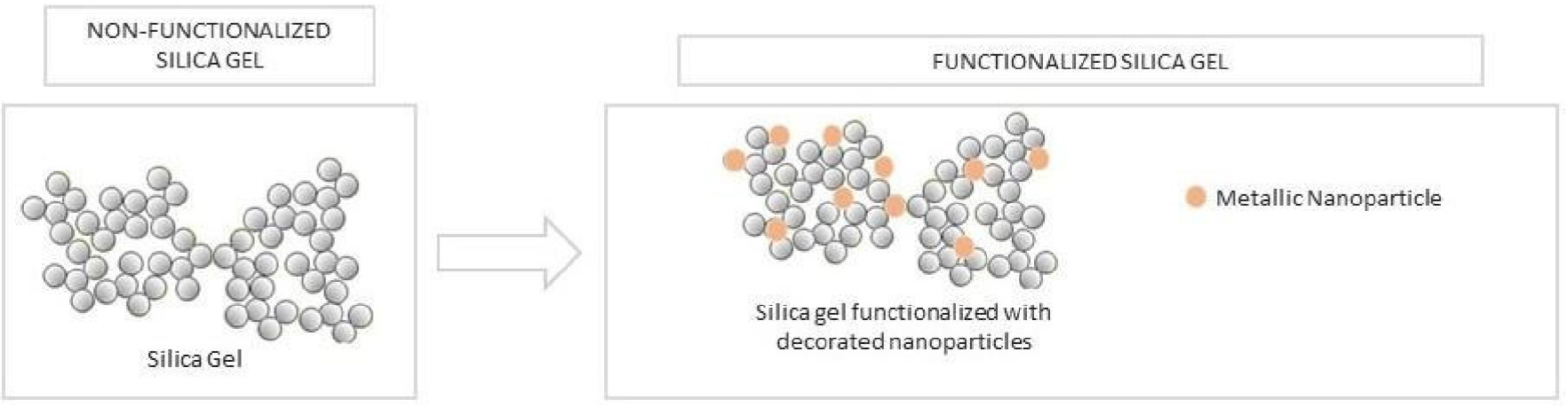
The second step involved decorating the previously prepared silica surface (with an approximately spherical geometry) with silver and zinc oxide using the chemical reduction method for silver26 and the precipitation method for ZnO27 as illustrated in Figure 1.
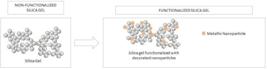
Representation of the functionalization process of silica gel with Ag and ZnO nanoparticles
The synthesis reaction of silica functionalized with silver (Ag SiO2) proceeds with the rapid conversion of silver salt into silver hydroxide and silver oxide through reaction with sodium hydroxide under light influence, and finally, the metallic silver is supported on the silica matrix26,28 functionalized with metallic nanoparticles decorating the silica surface. For this reaction, 5 g of silica were mixed in 50 mL of deionized water in a 250 mL round-bottom flask. Under constant stirring using a magnetic stirrer, 1.1 mL of 0.1 M NaOH solution was added, followed by 15 mL of 5% NH4OH. Immediately after, 1.0 mL of 0.005 M silver acetate solution was added dropwise and kept stirring for another 30 min, after which the mixture was neutralized with concentrated HNO3. After filtration and washing in successive stages with about 500 mL of distilled water at 40 °C, the precipitate was dried in an oven at 105 °C for 24 h and stored in a desiccator. According to the procedure described above, the nominal silver content in the functionalized silica was 108 ppm. Additionally, silica functionalized with 10,000 ppm of silver was synthesized by adding 10 mL of a 0.05 M silver acetate solution, without altering the dosages of the other reagents.
The synthesis of ZnO-SiO2 was carried out via the precipitation method according to the work proposed by Safavinia et al.,27 by adding 5 g of silica and 50 mL of deionized water into a 250 mL round-bottom flask. Then, 1.0 mL of 0.005 M zinc nitrate solution was added dropwise to the suspension under constant magnetic stirring, followed by 10 mL of 0.1 M NaOH solution. The reaction medium temperature was raised to 60 °C and maintained under stirring for 1 h. Afterward, the precipitate was filtered and washed in successive stages with about 500 mL of distilled water at 40 °C, dried in an oven at 105 °C for 24 h, and then subjected to calcination at 400 °C for 1 h. The obtained ZnO-SiO2 sample was stored in a desiccator. According to the procedure described above, the nominal Zn content in the functionalized silica was 654 ppm (or 814 ppm of ZnO) and another silica functionalized with 80,333 ppm of zinc (or 100,000 ppm of ZnO) was synthesized by mixing 10 g of SiO2 in 50 mL of deionized water under stirring with 20 mL of zinc acetate solution at 252.8 g L-1, followed by the addition of NaOH (0.1 M) until pH 9, with the procedure carried out as described in the step of producing SiO2 functionalized with 1.0 mL of zinc nitrate solution.
Development of active pads for fresh meat
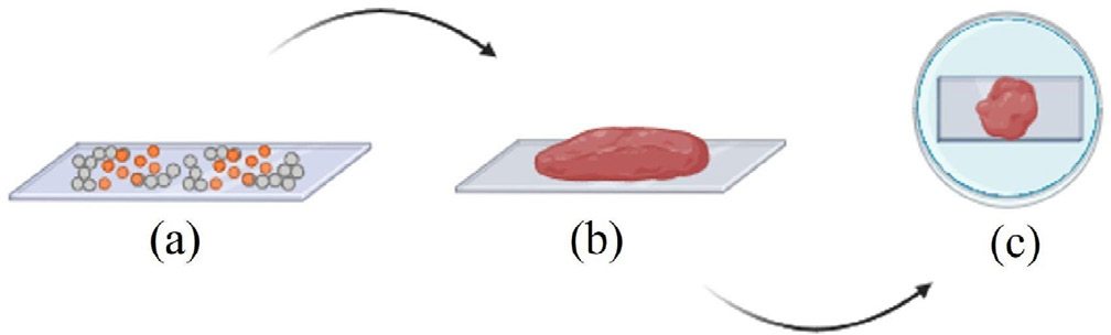
For the construction of each sachet, two pieces of polypropylene fabric measuring 8 cm × 6 cm were used, which were machine-sewn along almost the entire perimeter, leaving only a 1.5 cm opening on one side for filling with the absorbent material, and finally hand-sewn closed. The sachets were filled with 1 g of absorbent material in a (1:1) ratio of functionalized and non-functionalized silica or solely with silica gel, used as one of the control samples. The formed pads were arranged in a Petri dish, sterilized, and dried at 60 °C for 72 h. Figure 2 illustrates the stages of active pads production.
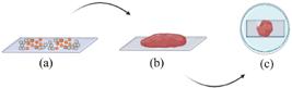
Stages of production and placement of the sachet in the Petri dish, where (a) represent the prototype of the active pads for meats with sachets filled with silica gel; (b) prototype developed for the evaluation of antimicrobial activities in meat samples; (c) testing the pads on a Petri dish
Characterizations
Determination of Ag and ZnO content in silica samples
The knowledge of the effective concentrations of Ag and ZnO nanoparticles in the silica gel samples is crucial for determining the minimum inhibitory concentration (MIC) for inhibiting the development of Pseudomonas aeruginosa. The samples were prepared according to the EPA 3051A29 method and analyzed by inductively coupled plasma optical emission spectrometry (ICP OES) following the SMEWW 3120 B30 and EPA 6010C31 methods. The nominal contents refer to the quantities of each element added during synthesis, based on the volumes and concentrations of the precursor solutions, while the effective contents refer to the quantities of each element present in the respective dried functionalized silica, determined by ICP OES. The EPA 3051A preparation method involves microwave-assisted acid digestion using nitric acid (HNO3) and hydrochloric acid (HCl). Since this method is not intended to achieve complete sample decomposition, the concentrations of the extracted analyte may not reflect the total sample content. The SMEWW 3120 B30 and EPA 6010C31 methods describe the determination of trace elements in aqueous solution. A standard aerosol is generated in a suitable nebulizer and spray chamber and injected into the plasma at temperatures of 6000 to 8000 K. The resulting ionization of a high percentage of atoms produces ionic emission spectra that are analyzed using a monochromator to examine emission wavelengths. The method also lists recommended analytical wavelengths and estimated instrumental detection limits.
Determination of specific migration of silver and zinc
The specific migration limits for contaminants present in pads materials in direct contact with food are regulated by Resolution of the Collegiate Board - RDC No. 88, of June 29, 2016, issued by the National Health Surveillance Agency (ANVISA, Brazil).32 For the extraction tests, approximately 2 g (with a precision of 0.1 mg) of silica gel sample and silica decorated with nanoparticles (previously dried for 24 h at 105 °C) were added to a Falcon tube along with 15 mL of distilled water. The suspension was allowed to stand for 8 days at a temperature of 20 ± 3 °C, and then filtered through a blue strip paper - slow filtration, into a 100 mL volumetric flask. The retained solid was further washed with distilled water until reaching a volume of 100 mL. The extracted metal content was then quantified by ICP OES using the SMEWW 3120 B30 method and the percentage was calculated based on the nominal quantity present in the functionalized silica sample.
Microbiological assessment: agar diffusion test
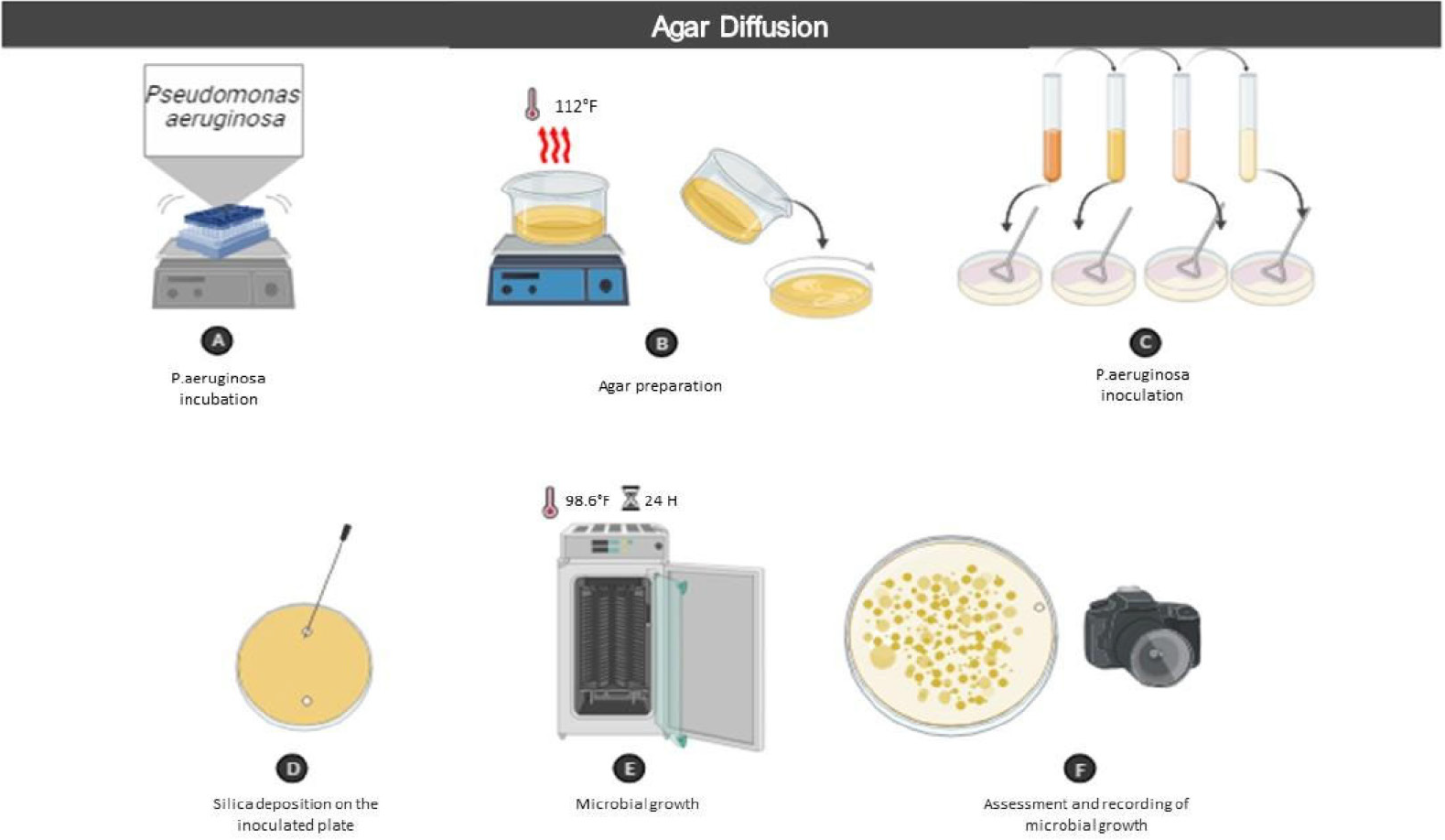
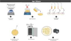
The agar diffusion assay, depicted in Figure 3, was conducted to qualitatively evaluate the antimicrobial effect of the functionalized materials against the microorganism under study. The test involved inoculating 100 μL of Pseudomonas aeruginosa suspension (American Type Culture Collection (ATCC) 27853) in serial dilutions of 10-2 and 10-3 onto Petri dishes prepared with selective agar base culture medium. Approximately 0.02 g of the functionalized silica was deposited in a well shape on two opposite sides of the plate, and it was then incubated at 37 °C for 24 h. The formation of inhibition zones around the sample was evaluated thereafter.
Microbiological assessment: determination of minimum inhibitory concentration
The tests for determining the minimum inhibitory concentration were performed only with the materials that showed inhibition potential against Pseudomonas aeruginosa in the agar diffusion assay to determine the concentration of functionalizing material required to inhibit bacterial growth. The broth macrodilution method in tryptic soy broth (TSB) was performed in duplicate at concentrations of 166.6; 96.6; 76.6; 58.3; 32.0; 26.3; 18.6; 10.6; 6.3; 3.6; 2; 1.3; and 0.6 mg mL-1, inoculated with 100 μL of bacteria and incubated at 37 °C for 24 h. The results were evaluated based on the turbidity of the liquid.
Evaluation of the effectiveness of functionalized silica for fresh meat
For the evaluation of the effectiveness of the active pads, beef from the bovine hindquarter was used, cut with a sterilized and flamed knife on a sterile surface into pieces measuring 1.5 × 1.0 × 1.0 cm. For clarity regarding the antimicrobial potential effects of the silica samples, plates were prepared with control samples without silica presence (B0), with silica presence without nanoparticles (B), and samples containing 1:1 ratios of functionalized/non-functionalized silica. In each Petri dish, with and without sachet, 2 pieces of meat weighing 5 ± 0.2 g each, previously washed with peptone water, were placed. The system was kept for 24, 72, and 96 h at a refrigeration temperature of 3 ± 2 °C. After each period, the samples were individually immersed in 30 mL of 0.9% saline solution and agitated for 10 min at 150 rpm. The obtained extract represented a 10-3 dilution and in 1:9 proportions. Then, 100 μL of the solutions were inoculated into plates containing agar specific for Pseudomonas aeruginosa and incubated at 37 °C for 48 h. The results were expressed in colony-forming units per unit volume (CFU mL-1).
RESULTS AND DISCUSSION
Determination of Ag and ZnO contents in functionalized silica samples
Some of the metal precursors may undergo homogeneous precipitation in solution, rather than binding exclusively to silica. This phenomenon is expected for both Ag and ZnO systems, especially in alkaline media, and contributes to the high metal losses observed during washing. However, the fraction of metal retained after thorough washing and filtration corresponds to species effectively associated with the silica matrix, rather than free precipitates. This is corroborated by the previous morphological characterization (TEM data and surface area reported in Guido et al.),20 which confirmed nanoparticles anchored to the silica surface, as well as by the low migration values measured, which are consistent with strongly bound metals. Thus, although some separate precipitation may occur, the reported effective metal content represents the portion stably incorporated into the silica structure.
Table 1 shows the effective contents of Ag and Zn elements present in the functionalized silica gel matrix. It can be observed that the metal contents in the samples produced indicate that there may have been a substantial loss of nanoparticles during the post-synthesis material washing stage. The silica gel used as the basis for the synthesis of the functionalized silica samples did not present detectable amounts of the metals in question.
The high metal losses during washing (Ag 87% and Zn 80%) are primarily attributed to (i) the weak interaction between the initially formed metal species and the silica surface before nucleation is complete, (ii) the solubility of precursor-derived species in aqueous media, and (iii) the absence of strong anchoring groups on the native silica surface. In silver functionalization carried out via chemical reduction, only a fraction of the Ag+ ions is reduced directly onto nucleation sites available on the silica surface. A substantial portion of the precursor remains in the liquid phase as soluble silver complexes which are readily removed during washing. Similar losses have been reported in the literature for Ag-silica systems; for example, Dulski et al.26 reported silver retention of only ~46%, indicating that extensive washing losses are expected when no additional surface modification or stabilizing ligands are used.
For ZnO deposition via precipitation, zinc hydroxide species initially form in suspension rather than exclusively on the silica surface. Because Zn(OH)2 and other intermediates remain partially soluble or colloidally dispersed in alkaline media, a large portion is removed during post-synthesis washing, especially without a prior surface functionalization step, silanization, to increase nucleation density and binding affinity. Susanna et al.33 similarly observed ZnO incorporation efficiencies ranging from 10 to 50%, depending on pH and precipitation kinetics. Therefore, the high apparent loss reflects the competition between homogeneous and heterogeneous nucleation, the chemical equilibrium of the precursor species, and the absence of surface binding functionalities that would otherwise promote stronger metal retention. This behavior is recognized in the literature for the single-step functionalization of bare silica particles without surface modification. It is important to note that, even with these losses, the effective metal loads obtained were sufficient to provide antimicrobial activity, meeting migration limits.
Determination of specific migration of Ag and Zn
The results of the specific migration tests for Ag and Zn are shown in Table 2. Considering that the material will be tested in beef, it is estimated that the product will exude about 5% liquid, and taking into account that the functionalized silica samples evaluated are capable of absorbing this fluid in a minimum amount equivalent to 200% of their mass, then the maximum amount of functionalized silica to be used in active pads would be 25 g for each kilogram of meat. This ensures adherence to the specific migration limits for silver (0.05 mg kg-1) and zinc (25 mg kg-1), as mandated by ANVISA RDC No. 88/2016.32
Results of specific migration tests of Ag and ZnO for silica samples subjected to 8 days of water extraction
Agar diffusion
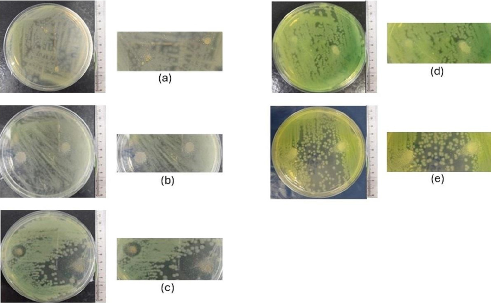
The agar diffusion test was performed qualitatively for both the functionalized silica samples and the silica gel used as a matrix (control sample). Figure 4 presents photographic images of the microbiological diffusion results on agar for the control sample (B) (Figure 4a), i.e., where no metallic nanoparticles were present. The results demonstrate that the silica gel without functionalizing material did not show halo formation for Pseudomonas aeruginosa. In the samples of silica decorated/anchored with nominal silver of 108 ppm (Figure 4b) and 10,000 ppm (Figure 4c), it is observed that the Ag SiO2 sample, with 10,000 ppm of Ag, showed promising results regarding the inhibition of Pseudomonas aeruginosa, as evidenced by the formation of an inhibition halo. However, with a nominal silver content of 108 ppm, bacteria grew on the functionalized silica, with no halo formation. The samples of silica decorated/anchored with zinc oxide nanoparticles with a concentration of 800 ppm (Figure 4d) and 100,000 ppm (Figure 4e) in nominal values, proved to be ineffective in inhibiting Pseudomonas aeruginosa, as no halo formation was observed at any of the tested concentrations.
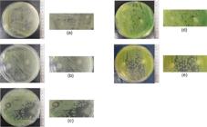
Photographic images of: (a) control sample SiO2; (b) Ag-SiO2 108 ppm; (c) Ag-SiO2 10,000 ppm; (d) ZnO-SiO2 800 ppm; (e) ZnO-SiO2 10,000 ppm
The results obtained for ZnO were divergent from those presented by Černík and Padil;34 however, the authors consider the action of complex metallic nanoparticles due to their morphology and have difficulty explaining why zinc exhibits antimicrobial activity against some Gram-negative bacteria and not others. The prevailing hypothesis is that reactive oxygen species (ROS), particularly superoxide ions and hydroxyl or hydroperoxyl radicals, are spontaneously produced by ZnO nanoparticles, leading bacteria to death by increasing intracellular oxidative stress. Another theory is that ZnO nanoparticles may cause damage to the cell membrane and DNA of microorganisms through direct or electrostatic interaction with cell surfaces.27,35
The evidence found in this study is consistent with recent publications,36 which justify that Pseudomonas aeruginosa possesses a cellular homeostatic mechanism for intracellular zinc control. This is because this opportunistic pathogen has resistance attributed to efficient components of active expulsion, metal resistance genes, and biofilm formation.37 In summary, this system of transporter proteins works by expelling excess metals from within the cells to the extracellular environment, also known as the homeostasis mechanism of some minerals like Zn in bacteria organisms, confirmed in genetic research. This may be an evolutionary dynamic of bacteria to survive the action of antibiotics.38,39
Some strategies that allow for the homeostasis of these metals have been described40 and characterized in Pseudomonas aeruginosa, supporting the hypothesis that all these systems serve to ensure the survival of the bacterium in environments limited or contaminated with Zn that have a direct relationship between virulence and antibiotic resistance, indicating that the different systems involved in resistance to these metals are not redundant, but follow a precise strategic plan, ensuring rapid adaptation of the cell to variations in the concentrations of these metals.
Silver nanoparticles, in turn, showed an inhibition halo at a concentration of 10,000 ppm, justified by the numerous antimicrobial action mechanisms of silver nanoparticles involving the release of silver ions and their interaction with thiol (sulfhydryl) groups in enzymes and proteins. Silver ions damage the bacterial cell envelope and its contents, increasing cell size and causing structural abnormalities in the cytoplasmic membrane, cytoplasmic contents, and outer cell layers. Additionally, silver ions interact with nucleic acids, inhibiting cell division.41,42
Determination of minimum inhibitory concentration
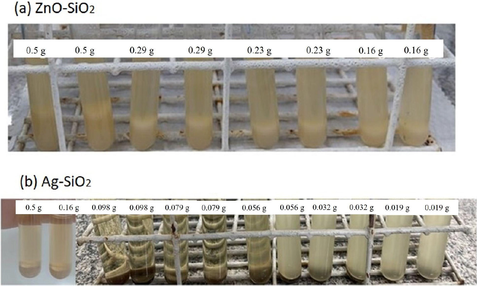
Based on the results obtained in the agar diffusion tests, MIC assays were performed for the silver-functionalized sample with a nominal concentration of 10,000 ppm. On the other hand, although the samples containing ZnO nanoparticles did not show promising results in the diffusion test, even for the highest nominal concentration of ZnO tested (100,000 ppm), it was decided to also evaluate the minimum inhibitory concentration of this material, the results of which are shown in Figure 5.
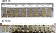
Photographic images of the broth 10 microdilution test in TSB for evaluating the minimum inhibitory concentration of functionalized silica: (a) ZnO-SiO2 and (b) Ag-SiO2 against Pseudomonas aeruginosa bacteria. The nominal concentration of Ag and ZnO in the functionalized silica was 100,000 ppm. The amounts of functionalized silica added to 3 mL of TSB broth for the sample with ZnO and 5 mL for the sample with Ag are indicated in the tubes
The sample functionalized with a nominal content of 100,000 ppm of ZnO did not show inhibition potential for Pseudomonas aeruginosa at the highest content applied in this study, of 166.6 μg mL-1. Considering the effective content of Zn present in the sample, determined by ICP OES (16.461 ppm), the maximum corrected zinc concentration was 2.744 μg mL-1. In a study by Zeelie,43 which investigated the effects of zinc ions on the inhibition of this bacterium, a minimum inhibitory concentration of 1917 μg mL-1 was identified, which does not correspond to the values found in this study but can be explained by the use of zinc in ionic form. On the other hand, more recent studies44 have already established the lower sensitivity of Gram-negative bacteria to zinc.
Table 3 shows the concentrations of Ag-SiO2 and silver nanoparticles corresponding to the amount of functionalized silica added to the tubes. As observed in Figure 5 and indicated, the range of minimum inhibitory concentration found for Pseudomonas aeruginosa is from 15 to 21 μg mL-1 (for 0.056 to 0.079 g of functionalized silica added to 5 mL of TSB broth), considering the effective silver content in the Ag-SiO2 functionalized silica, determined by ICP OES. These values are consistent with other data reported in the literature40,45-49 (1 to 15 μg mL-1) for silver particles, although obtained by different chemical routes.
Tested concentrations for the determination of the minimum inhibitory concentration of Ag-SiO2 functionalized silica in Pseudomonas aeruginosa bacteria. The gray-highlighted rows correspond to the range of silver concentrations in TSB broth, in which the supernatant solution changes its appearance from turbid (lower concentrations) to clear (higher concentrations)
These factors demonstrate that different forms of antimicrobial action are based on physicochemical characteristics, such as chemical composition and structure in terms of shape and size, as well as on the synthesis methods used. In addition, parameters such as temperature, pH, metal ion concentration, and reaction time play important and decisive roles in the mechanism of action against microorganisms.13-15
Evaluation of functionalized silicas in active pads for fresh meat
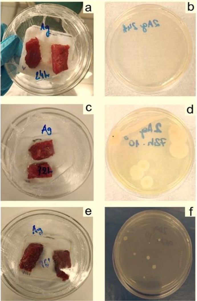
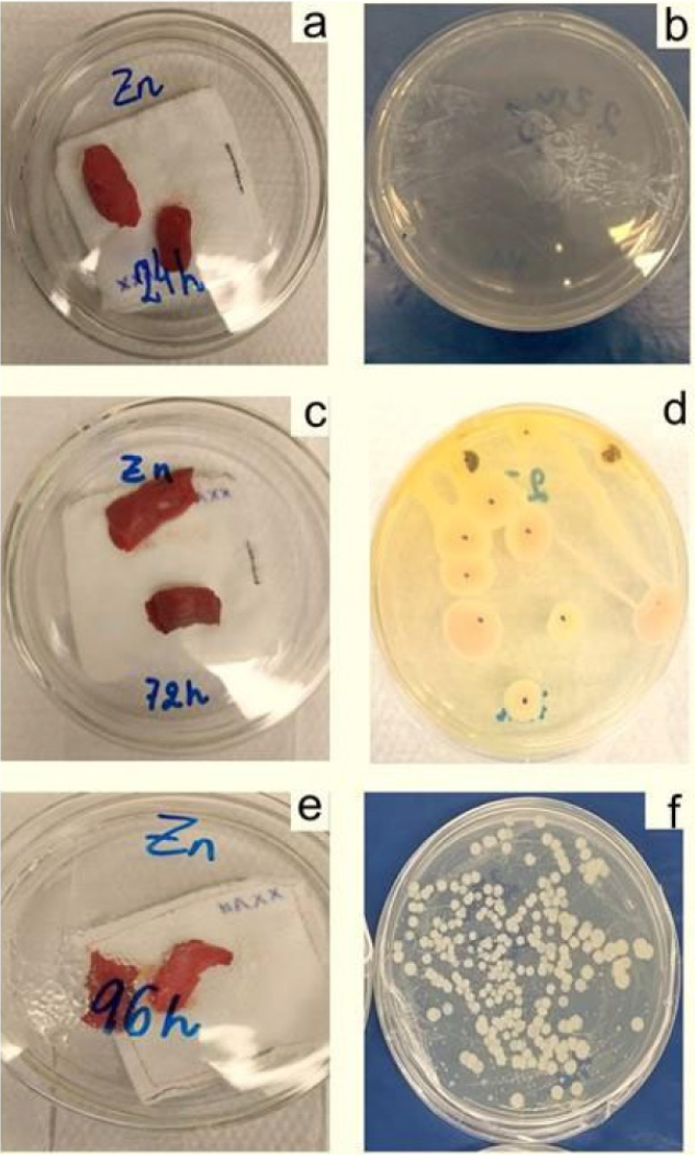
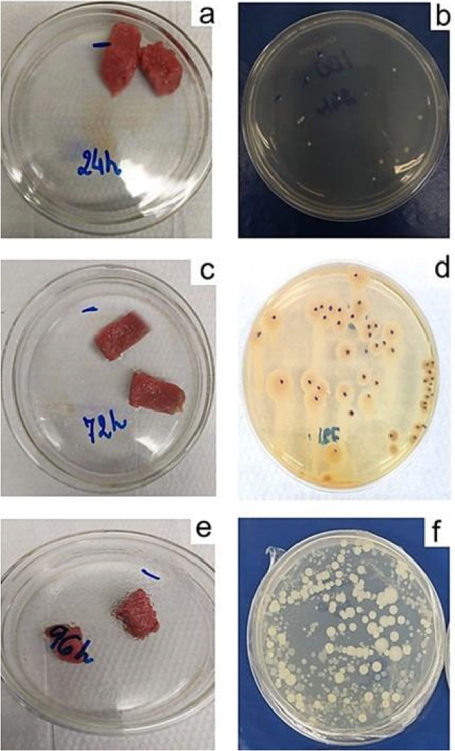
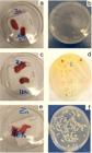
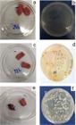
To evaluate the effectiveness of silica functionalized with agents to control microbial growth in fresh beef pads, the materials were used as filling in absorbent sachets, arranged in Petri dishes with lids simulating tray-type packaging. In each dish, two pieces of beef weighing approximately 5 g each were placed on the pad containing about 1 g of absorbent material. Figures 6, 7, and 8 show the Petri dishes with beef closed, wrapped in PVC film, and kept refrigerated for 24 to 96 h (a, c, and e), as well as the colony count in 0.1 mL of the samples diluted in 30 mL of 0.9% saline solution that were spread on agar plates and incubated for 24 h at 37 °C (b, d, f).
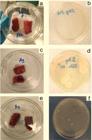
Photographic images showing the preservation time of 24 (a), 72 (c), and 96 (e) h of fresh meat, stored at 3 ± 2 °C in active pads containing silica functionalized with Ag-SiO2, and the colony count of Pseudomonas aeruginosa in 0.1 mL in 24 (b), 72 (d) and 96 h (f)
Photographic images showing the preservation time of 24 (a), 72 (c), and 96 (e) h of fresh meat, stored at 3 ± 2 °C in active pads containing silica functionalized with ZnO-SiO2, and the colony count of Pseudomonas aeruginosa in 0.1 mL in 24 (b), 72 (d) and 96 h (f)
Photographic images reporting the preservation time of 24 (a), 72 (c), and 96 (e) h of fresh meat, stored at 3 ± 2 °C in Petri dish without pads (B0), and the colony count of Pseudomonas aeruginosa in 0.1 mL in 24 (b), 72 (d) and 96 h (f)
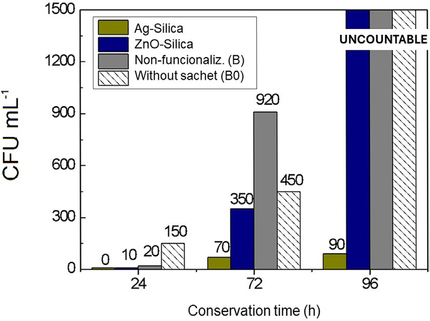
The results of the Pseudomonas aeruginosa colony count in CFU mL-1 present in fresh meat during storage are also presented in Figure 9.
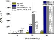
Pseudomonas aeruginosa colony count (CFU mL-1) as a function of the conservation time (h) of fresh meat
From Figures 7 to 9, it is possible to observe that the samples of silica functionalized with Ag have a greater capacity to inhibit the growth of Pseudomonas aeruginosa compared to the other samples evaluated at all time points in this study. These results are consistent with the studies conducted by Pormohammad and Turner50 and Muddassir et al.51 The ZnO-SiO2 sample, although not showing activity in diffusion tests, was more effective (up to 72 h of storage) than the samples of unmodified silica (B) and without sachet (B0), for which there was no significant difference in microbiological count, suggesting that for controlling the growth of this pathogen in fresh meat, only adsorption of exudate is not sufficient.
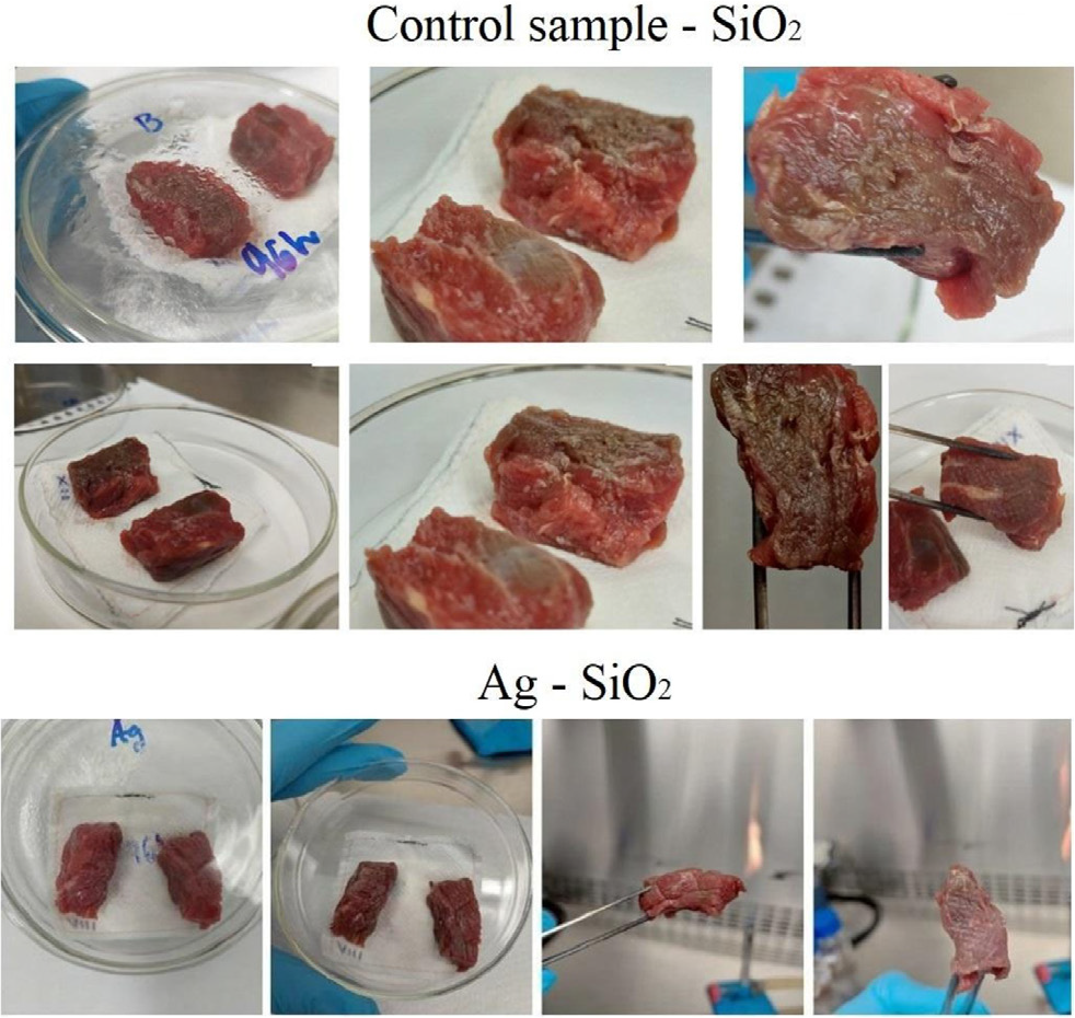
By comparing the microbiological count results in Figure 9 with the visual results observed in the photographic records of the functionalized silica and the non-functionalized silica (B), it confirms the evidence that the action of silver nanoparticles in controlling the microbial development of Pseudomonas aeruginosa can extend the shelf life of fresh meats.
The use of silver nanoparticles in controlling Pseudomonas aeruginosa is described as efficient because it addresses a bacterium that is highly resistant to antibiotics and is considered a public health problem in some countries.44 Furthermore, silver nanoparticles are considered effective for inhibiting both Gram-positive and Gram-negative bacteria,52 with potential applications in medicine53 and the food industry.54
Figure 10 presents a photographic comparison visually illustrating the test with beef using the control silica (B) and the Ag-SiO2 sample. It is observed that the part of the beef exposed to the non-functionalized silica showed surface slime formation, a situation attributed to microbiological deterioration and confirmation of microorganism proliferation. On the other hand, the beef sample exposed to the Ag-SiO2 sample showed no change in coloration and no surface slime formation.
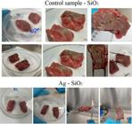
Photographic records of meat samples stored for 96 h in the presence of unmodified silica - control (B) and in the presence of Ag-SiO2
CONCLUSIONS
This study demonstrates the successful development of antimicrobial absorbent pads based on silica gel functionalized with Ag and ZnO nanoparticles, using rice husk-derived silicate as a sustainable precursor. The silver-functionalized silica exhibited marked antimicrobial activity against Pseudomonas aeruginosa, both in vitro and when applied to fresh meat storage, validating its applicability in active packaging systems. Despite the significant metal loss observed after synthesis (87% for Ag and 80% for Zn), the effective residual silver content was sufficient to meet regulatory migration limits while providing antimicrobial protection. On the other hand, ZnO functionalized silica showed limited efficacy, probably due to bacterial resistance mechanisms and low effective Zn content. These results support the potential implementation of Ag-SiO2 in commercial absorbent pads, contributing to extended shelf life and microbial safety in meat products.
ACKNOWLEDGMENTS
The authors would like to thank the company Oryzasil Sílicas Naturais for kindly providing the RHA and silicate samples, to Prof. Dr. Sabrina Arcaro for providing the simulated body fluid, to CAPES for granting the research grant, and to FAPESC (TO 2021TR001860).
DATA AVAILABILITY STATEMENT
All data are available in the text;
REFERENCES
-
1 Yildirim, S.; Röcker, B.; Pettersen, M. K.; Nilsen-Nygaard, J.; Ayhan, Z.; Rutkaite, R.; Radusin, T.; Suminska, P.; Marcos, B.; Coma, V.; Compr. Rev. Food Sci. Food Saf. 2018, 17, 165. [Crossref]
» Crossref -
2 Capelezzo, A. P.; Mohr, L. C.; Dalcanton, F.; Barreta, C. R. D. M.; Martins, M. A. P. M.; Fiori, M. A.; de Mello, J. M. M.; Quim. Nova 2018, 41, 367. [Crossref]
» Crossref -
3 Razura-Carmona, F. F.; Altamirano-Medina, T. P.; Bueno-Durán, A. Y.; Sáyago-Ayerdi, S. G.; Girón-Pérez, M. I.; P.U., B.-R.; Pérez-Larios, A.; Sánchez-Burgos, J. A.; Sánchez Burgos, J. A.; Revista Bio Ciencias 2023, 10, e1473. [Crossref]
» Crossref -
4 Kashiwaqui, N. Y.; Sumini, M.; Kitagawa, H. Y.; Thomé, M. L. F. L.; Montini, V. H.; Kobayashi, R. K. T.; Nakazato, G.; Brazilian Journal of Development 2023, 9, 22782. [Crossref]
» Crossref -
5 Braga, L. R.; Peres, L.; Boletim do Centro de Pesquisa de Processamento de Alimentos 2010, 28, 69. [Link] acessado em dezembro 2025
» Link -
6 Radusin, T. I.; Ristić, I. S.; Pilić, B. M.; Novaković, A. R.; Food Feed Res. 2016, 43, 119. [Crossref]
» Crossref -
7 Fadiji, T.; Rashvand, M.; Daramola, M. O.; Iwarere, S. A.; Processes 2023, 11, 590. [Crossref]
» Crossref -
8 de Alcantara, M.; de Morais, I. C. L.; de Matos, C.; de Souza, O. C. C.; Rev. Bras. Hig. Sanid. Anim. 2012, 6, 1. [Link] acessado em dezembro 2025
» Link -
9 Queenan, A. M.; Bush, K.; Clin. Microbiol. Rev. 2007, 20, 440. [Crossref]
» Crossref -
10 Panea, B.; Ripoll, G.; González, J.; Fernández-Cuello, Á.; Albertí, P.; J. Food Eng. 2014, 123, 104. [Crossref]
» Crossref -
11 Azlin-Hasim, S.; Cruz-Romero, M. C.; Cummins, E.; Kerry, J. P.; Morris, M. A.; J. Colloid Interface Sci. 2016, 461, 239. [Crossref]
» Crossref -
12 Akbar, A.; Anal, A. K.; Food Control 2014, 38, 88. [Crossref]
» Crossref -
13 Shobha, G.; Moses, V.; Ananda, S.; Int. J. Pharm. Sci. Invent. 2014, 3, 28. [Link] acessado em dezembro 2025
» Link -
14 Jaworski, S.; Wierzbicki, M.; Sawosz, E.; Jung, A.; Gielerak, G.; Biernat, J.; Jaremek, H.; Łojkowski, W.; Woźniak, B.; Wojnarowicz, J.; Stobiński, L.; Małolepszy, A.; Mazurkiewicz-Pawlicka, M.; Łojkowski, M.; Kurantowicz, N.; Chwalibog, A.; Nanoscale Res. Lett. 2018, 13, 116. [Crossref]
» Crossref -
15 Mikhailov, O. V.; Mikhailova, E. O.; Materials 2019, 12, 3177. [Crossref]
» Crossref - 16 Gogotsi, Y. G.; Uvarova, I. V.; Nanostructured Materials and Coatings for Biomedical and Sensor Applications, 1st ed.; Springer Science Business Media: Netherlands, 2003.
-
17 Li, F.; Lei, C.; Shen, Q.; Li, L.; Wang, M.; Guo, M.; Huang, Y.; Nie, Z.; Yao, S.; Nanoscale 2013, 5, 653. [Crossref]
» Crossref - 18 Hagiwara, S. Z.; Hyogo, O. M.; US Pat 5,244,667 1993
-
19 Lotfiman, S.; Ghorbanpour, M.; Surf. Coat. Technol. 2017, 310, 129. [Crossref]
» Crossref -
20 Guido, Z. N. S.; Braum, M. V.; Possolli, N. M.; De Bonna, R.; Angioletto, E.; Zimmermann, M. V. G.; Mater. Res. 2025, 28, e20240444. [Crossref]
» Crossref - 21 ISO 1690:1976: Sodium and Potassium Silicates for Industrial Use: Determination of Silica Content - Gravimetric Method by Insolubilization; ISO: Genebra, 1976.
- 22 ISO 1692:1976: Sodium and Potassium Silicates for Industrial Use: Determination of Total Alkalinity - Titrimetric Method; ISO: Genebra, 1976.
-
23 Besbes, M.; Fakhfakh, N.; Benzina, M.; Phys. Procedia 2009, 2, 1087. [Crossref]
» Crossref -
24 Sdiri, A.; Higashi, T.; Bouaziz, S.; Benzina, M.; Arabian J. Chem. 2014, 7, 486. [Crossref]
» Crossref -
25 Aimjaijit, W.; Chaiwiwatworakul, P.; Chuangchote, S.; Appl. Mech. Mater. 2016, 839, 70. [Crossref]
» Crossref -
26 Dulski, M.; Peszke, J.; Włodarczyk, J.; Sułowicz, S.; Piotrowska-Seget, Z.; Dudek, K.; Podwórny, J.; Malarz, K.; Mrozek-Wilczkiewicz, A.; Zubko, M.; Nowak, A.; Mater. Sci. Eng., C 2019, 103, 109790. [Crossref]
» Crossref -
27 Safavinia, L.; Akhgar, M. R.; Tahamipour, B.; Ahmadi, S. A.; Iran. J. Biotechnol. 2021, 19, 86. [Crossref]
» Crossref -
28 Dulski, M.; Malarz, K.; Kuczak, M.; Dudek, K.; Matus, K.; Sułowicz, S.; Mrozek-Wilczkiewicz, A.; Nowak, A.; Nanomaterials 2020, 10, 2551. [Crossref]
» Crossref - 29 EPA SW-846 Test Method 3051A: Microwave Assisted Acid Digestion of Sediments, Sludges, Soils, and Oils, Washington, 2007.
- 30 APHA Method 3120 B: Standard Methods for the Examination of Water and Wastewater, Washington, DC, 2020.
- 31 EPA SW-846 Method 6010C: Inductively Coupled Plasma-Atomic Emission Spectrometry, Washington, DC, 2013.
-
32 Agência Nacional de Vigilância Sanitária (ANVISA); Resolução da Diretoria Colegiada (RDC) No. 88, de 29 de junho de 2016, Aprova o Regulamento Técnico sobre Materiais, Embalagens e Equipamentos Celulósicos Destinados a Entrar em Contato com Alimentos e Dá outras Providências; ANVISA, Brasil, 2016. [Link] acessado em dezembro 2025
» Link -
33 Susanna, A.; Armelao, L.; Callone, E.; Dirè, S.; D’Arienzo, M.; Di Credico, B.; Giannini, L.; Hanel, T.; Morazzoni, F.; Scotti, R.; Chem. Eng. J. 2015, 275, 245. [Crossref]
» Crossref -
34 Černík, M.; Padil, V. V. T.; Int. J. Nanomed. 2013, 8, 889. [Crossref]
» Crossref -
35 Tiwari, V.; Mishra, N.; Gadani, K.; Solanki, P. S.; Shah, N. A.; Tiwari, M.; Front. Microbiol. 2018, 9, 1218. [Crossref]
» Crossref -
36 Hofmann, L.; Hirsch, M.; Ruthstein, S.; Int. J. Mol. Sci. 2021, 22, 2050. [Crossref]
» Crossref -
37 Pang, Z.; Raudonis, R.; Glick, B. R.; Lin, T.-J.; Cheng, Z.; Biotechnol. Adv. 2019, 37, 177. [Crossref]
» Crossref -
38 Irie, Y.; Roberts, A. E. L.; Kragh, K. N.; Gordon, V. D.; Hutchison, J.; Allen, R. J.; Melaugh, G.; Bjarnsholt, T.; West, S. A.; Diggle, S. P.; mBio 2017, 8, 10.1128/mbio.00374-17. [Crossref]
» Crossref -
39 Thi, M. T. T.; Wibowo, D.; Rehm, B. H. A.; Int. J. Mol. Sci. 2020, 21, 8671. [Crossref]
» Crossref -
40 Coriolano, D. L.; de Souza, J. B.; Bueno, E. V.; Medeiros, S. M. F. R. S.; Cavalcanti, I. D. L.; Cavalcanti, I. M. F.; Braz. J. Microbiol. 2021, 52, 267. [Crossref]
» Crossref -
41 Jung, W. K.; Koo, H. C.; Kim, K. W.; Shin, S.; Kim, S. H.; Park, Y. H.; Appl. Environ. Microbiol. 2008, 74, 2171. [Crossref]
» Crossref -
42 Yin, I. X.; Zhang, J.; Zhao, I. S.; Mei, M. L.; Li, Q.; Chu, C. H.; Int. J. Nanomed. 2020, 15, 2555. [Crossref]
» Crossref -
43 Zeelie, J. J.; Analyst 1998, 123, 503. [Crossref]
» Crossref -
44 Webster, T. J.; Seil, I.; Int. J. Nanomed. 2012, 7, 2767. [Crossref]
» Crossref -
45 Kora, A. J.; Arunachalam, J.; World J. Microbiol. Biotechnol. 2011, 27, 1209. [Crossref]
» Crossref -
46 Park, H.-J.; Park, S.; Roh, J.; Kim, S.; Choi, K.; Yi, J.; Kim, Y.; Yoon, J.; J. Ind. Eng. Chem. 2013, 19, 614. [Crossref]
» Crossref -
47 Arokiyaraj, S.; Vincent, S.; Saravanan, M.; Lee, Y.; Oh, Y. K.; Kim, K. H.; Artif. Cells, Nanomed., Biotechnol. 2017, 45, 372. [Crossref]
» Crossref -
48 Liao, S.; Zhang, Y.; Pan, X.; Zhu, F.; Jiang, C.; Liu, Q.; Cheng, Z.; Dai, G.; Wu, G.; Wang, L.; Chen, L.; Int. J. Nanomed. 2019, 14, 1469. [Crossref]
» Crossref -
49 da Silva, R. T. P.; Petri, M. V.; Valencia, E. Y.; Camargo, P. H. C.; de Torresi, S. I. C.; Spira, B.; Photodiagn. Photodyn. Ther. 2020, 31, 101908. [Crossref]
» Crossref -
50 Pormohammad, A.; Turner, R. J.; Antibiotics 2020, 9, 853. [Crossref]
» Crossref -
51 Muddassir, M.; Raza, A.; Munir, S.; Basirat, A.; Ahmed, M.; Butt, M. S.; Dar, O. A.; Ahmed, S. S.; Shamim, S.; Naqvi, S. Z. H.; Sci. Rep. 2022, 12, 20685. [Crossref]
» Crossref -
52 Morones, J. R.; Elechiguerra, J. L.; Camacho, A.; Holt, K.; Kouri, J. B.; Ramírez, J. T.; Yacaman, M. J.; Nanotechnology 2005, 16, 2346. [Crossref]
» Crossref -
53 Mohammad, Z. H.; Ahmad, F.; Ibrahim, S. A.; Zaidi, S.; Discover Food 2022, 2, 12. [Crossref]
» Crossref -
54 Rashidi, L.; Khosravi-Darani, K.; Crit. Rev. Food. Sci. Nutr. 2011, 51, 723. [Crossref]
» Crossref
Edited by
-
Associate Editor handled this article:
Eduardo H. S. Sousa