| Levy et al.(18)
|
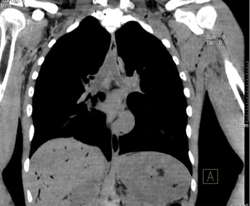
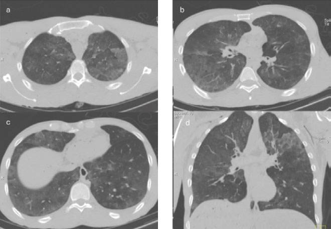
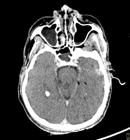
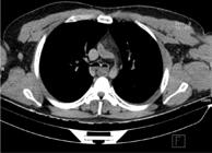
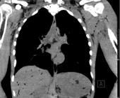
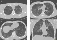
PMCT: fluid in the mastoid air cells and paranasal sinuses, with hyperdense material in 25% of the bodies; fluid in the subglottic trachea, main bronchi, and secondary bronchi, with frothy fluid in 21% and dense material in 50%; crazy-paving pattern in the lungs; bilateral pleural effusion; gastric distention with dense sediment in 21%; and pulmonary edema and congestion in 100%. Autopsy: hyperdense material in the paranasal sinuses (in 25%), airways (in 50%), and stomach (in 21%) in the same proportion as on PMCT; frothy fluid in the airways, confirmed in only 19% of the cases; and features not identified on PMCT, including abrasions on the face and extremities, together with minor contusions, in 54%, as well as occult fractures in the skull base and in a rib. |
| Christe et al.(2)
|
PMCT: fluid in the paranasal sinuses, especially in the maxillary and sphenoid sinuses, in all cases; airway content in all cases (dense in 30% and liquid in 70%), mostly in the main bronchi; 40% of the tracheal volume filled with fluid; fluid in the pharynx in 80%; crazy-paving pattern in 60%; pulmonary edema in 50%; pleural effusion in 70%; hemodilution in the right atrium (density, 50 HU); and distention of the stomach and duodenum. Autopsy: Some airway content in all cases; 33% of the tracheal volume filled with fluid; fluid in the pharynx in 50%; crazy-paving pattern in 60%; pleural effusion in 80%; stomach distention similar to that seen on PMCT; and the classic finding of Paltauf spots in 70% of the cases. |
| Usui et al.(7)
|
PMCT: pulmonary findings classified as type 1 (in 34%), characterized by diffuse ground-glass opacities with interstitial thickening (crazy-paving pattern); type 2 (in 41%), characterized by poorly defined centrilobular nodules and diffuse ground-glass opacities; type 1 + type 2 (in 11%); type 3, characterized by consolidation (in 5%); type 4, characterized by emphysema, with or without fibrosis (in 4%); or type 5, defined as that which is unclassifiable (in 4%). Autopsy: pulmonary findings classified as type 1, characterized by marked edema (in 97% of the cases), the cut lung surface having a brick-red, protruding appearance, with a large amount of clear red fluid and white or bloody foam coming from each cut surface and bronchi; type 2, characterized by a protruding and reddish cut surface similar to that seen in type 1, but with less edema and no white foam; or type 4, characterized by pulmonary fibrosis. |
| Plaetsen et al.(12)
|
PMCT: fluid in the maxillary/ethmoid sinuses (in 98%), sphenoid sinuses (in 88%), and frontal sinuses (in 83%); fluid density of 19 HU and 20 HU in the right and left maxillary sinuses, respectively; fluid in the mastoid air cells in 12%, in the nasal cavities in 78% (frothy in 54%), in the nasopharynx in 98% (frothy in 40%), in the oropharynx in 95% (frothy in 40%), and in the trachea in 83%; ground-glass opacities in the lungs in 89% and consolidation in 11%, being diffuse in 54%, uneven (crazy-paving pattern) in 27%, and predominantly in the bases and posterior portions in 15%; pleural effusion in 71%, more commonly found in those drowned in salt water; depression of the right hemidiaphragm; blood density of 60 HU in the inferior vena cava and 59 HU in the right atrium (drowned in fresh water); pericardial effusion; fluid in one or two portions of the esophagus (in 91% and 49%, respectively); fluid distention of the stomach, with contents in multiple layers in 27%; and distention of the duodenum and jejunum (in 34% and 32%, respectively). Autopsy: Frothy fluid was less commonly observed, confirming the transient nature of this finding. Measurement of hemodilution in the right atrium and inferior vena cava was less reproducible and more time-consuming using the absorbent paper test. |
| Kawasumi et al.(8)
|
PMCT: fluid in the maxillary and sphenoid sinuses in victims of drowning in salt water and fresh water, with significantly greater density in those drowned in salt water than in those drowned in fresh water (47.28 HU vs. 32.56 HU). Autopsy: All cases were determined to be cases of wet drowning. None of the bodies showed fractures in the skull or facial bones. |
| Leth and Madsen(15)
|
PMCT: lung volume: 3,136 cm3; lung density: -1,073 HU; blood density in the right ventricle: 35 HU (drowning in fresh water) and 40 HU (drowning in salt water); and blood density in the pulmonary artery trunk: 52 HU (drowning in fresh water) and 54 HU (drowning in salt water). Autopsy: lung weight: 1,337 g; and lung density: 448 g/liter. Lung density was found to be was significantly lower in the bodies of drowning victims than in those of individuals who died from other causes. |
| Gotsmy et al.(19)
|
PMCT: gastric content layers in the following proportions: one layer in 41.8%, two in 40%, and three in 18.2%; and upper gastric content layers commonly more hypodense than the lower ones. When three layers were present, the upper layer always consisted of frothy material, whereas the lower layers contained denser components. Autopsy: gastric content layers in the following proportions: one layer in 50.9%, two in 34.5%, and three in 12.7%. In 28 of the 55 cases, a significant discrepancy was identified between the number of gastric content layers observed on PMCT and the number observed at autopsy. |
| Kakimoto et al.(5)
|
PMCT: The fluid volume in the maxillary sinus was significantly higher in drowning cases than in non-drowning cases. A total maxillary sinus fluid volume > 1.04 ml was more useful in indicating drowning than was a pleural effusion volume > 175 ml and a lung weight > 829 g. The combination of maxillary sinus fluid volume and pleural effusion volume was more effective in predicting drowning than was either index in isolation. Maxillary sinus fluid volume was less influenced by the postmortem interval, remaining valid for up to 1 week after death. Autopsy: The pleural effusion volume was significantly higher in drowning cases than in non-drowning cases. The mean lung weight among the cases of drowning was 1,093 g. |
| Heo et al.(9)
|
PMCT: sphenoid sinus fluid volume: mean, 2.7 ± 2.8 ml; and median, 1.65 ml. Autopsy: sphenoid sinus fluid volume: mean, 1.9 ± 1.7 ml; and median, 1.55 ml. |

 Contribution of computed tomography in the postmortem diagnosis of drowning: a systematic review
 Thumbnail
 Thumbnail
 Thumbnail
 Thumbnail
 Thumbnail
 Thumbnail