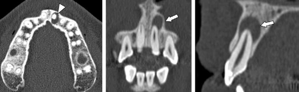
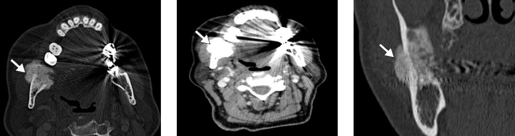
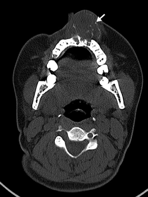
| Radicular cyst |
Radiolucent, unilocular, well-defined, and radicular (< 1 cm), with thin cortical margins and slight bone expansion |
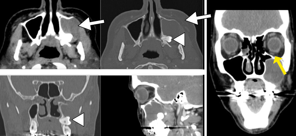
| Dentigerous cyst |
Radiolucent, unilocular, well-defined, surrounding the crown of an impacted tooth, with continuous margins and possible expansion |
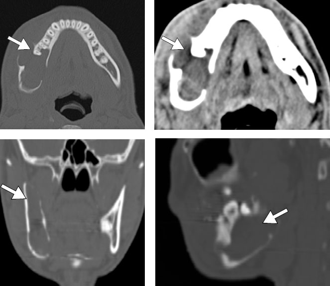
| Odontogenic keratocyst |
Radiolucent, unilocular or multilocular, with thin, sclerotic margins, discreet expansion, and root resorption; anteroposterior growth and potential cortical erosion |
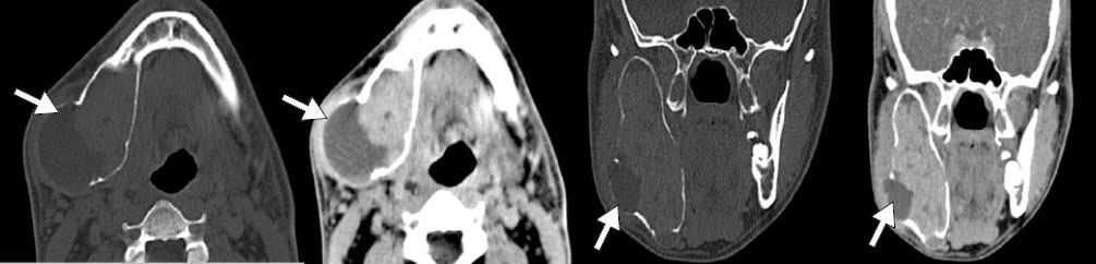
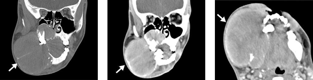
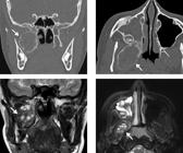
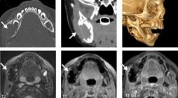
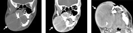
| Ameloblastoma |
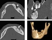
Expansile, unilocular or multilocular, soap-bubble pattern, root resorption, and cortical thinning; on MRI, solid areas with enhancement and restriction |
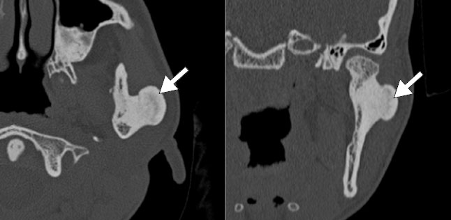
| Ossifying fibroma |
Well-defined with sclerotic margins, variable density (lytic, mixed, or ground-glass), and bone remodeling; juvenile variant more aggressive |
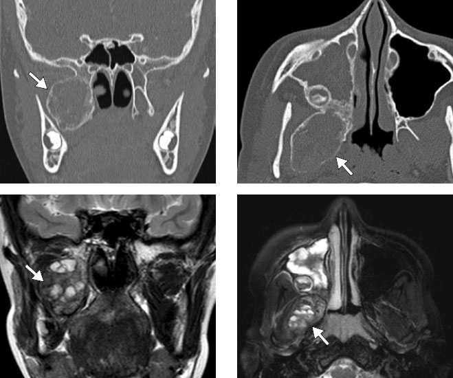
| Aneurysmal bone cyst |
Expansile, unilocular or multilocular, with enhancing septa and fluid-fluid levels on CT/MRI; may be associated with preexisting lesions |
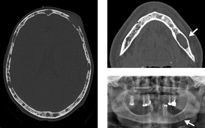
| Multiple myeloma |
Radiolucent, ovoid, well-defined, without periosteal expansion or reaction; may exhibit a “punched-out” pattern |
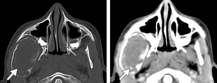
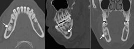
| Giant cell granuloma |
Radiolucent, unilocular or multilocular, expansile tumor with cortical erosion and remodeling; differential diagnosis includes a brown tumor |
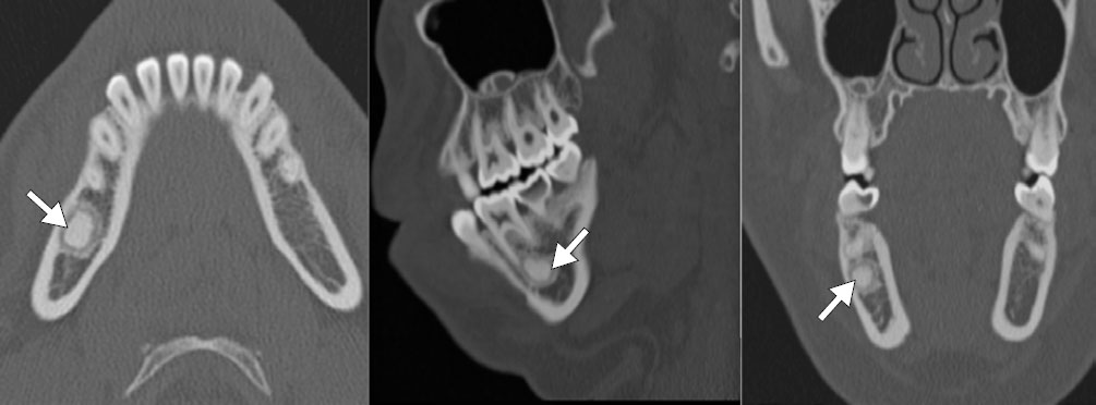
| Odontoma |
Radiopaque or mixed, well-defined, with a radiolucent halo; can be compound (tooth-like) or complex (amorphous mass) |
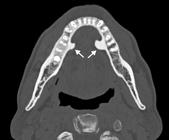
| Osteoma |
Radiopaque, sclerotic, and well-circumscribed, usually pedunculated and unrelated to the teeth; multiple cases suggest Gardner’s syndrome |
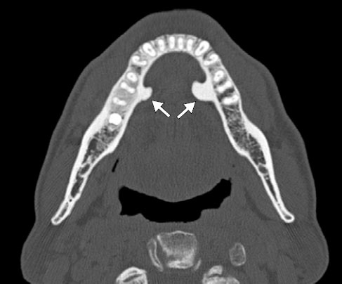
| Torus |
Dense, homogeneous, and well-defined cortical exostosis, without halo or expansile effect |
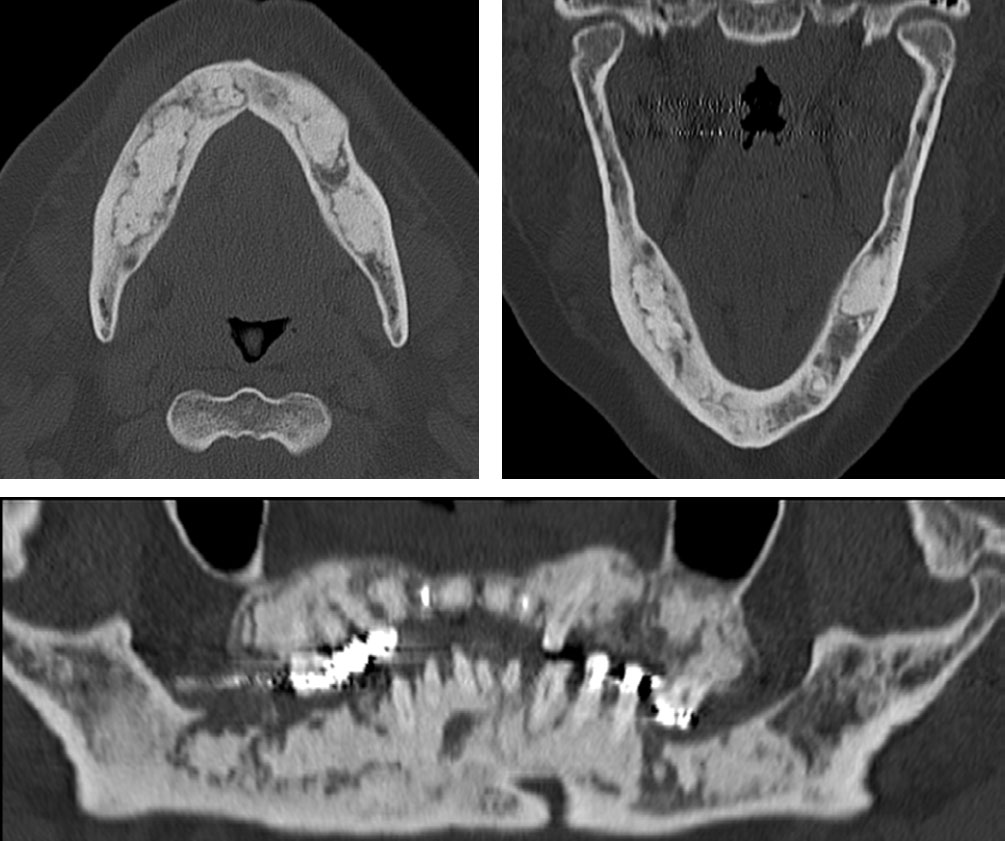
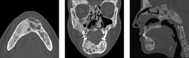
| Cementum-osseous dysplasia |
Initially radiolucent, evolving to ground-glass or radiopacity, with well-defined margins and a multifocal pattern |
| Brown tumor |
Radiolucent, expansile, unilocular or multilocular lesion with cortical thinning and associated diffuse demineralization. Related to hyperparathyroidism |
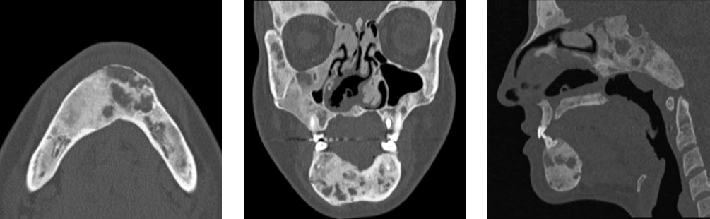
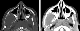
| Fibrous dysplasia |
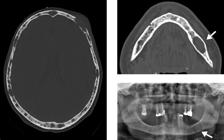
Ground-glass, poorly defined margins, infiltrative pattern, and longitudinal growth; can be monostotic or polyostotic |
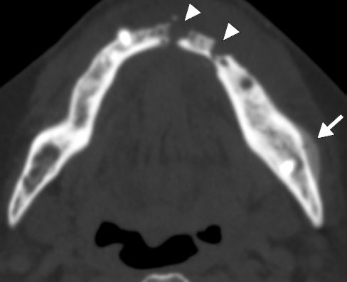
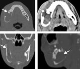
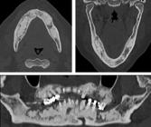
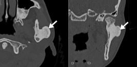
| Osteoradionecrosis |
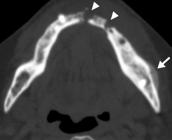
Irregular lytic and sclerotic areas, with bone sequestra and cortical interruption |
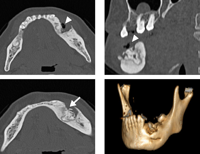
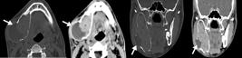
| Osteomyelitis |
Lytic, sclerotic, or mixed pattern, with chronic periosteal reaction, sequestra, and obliteration of the fat planes; may result from untreated dental infection |
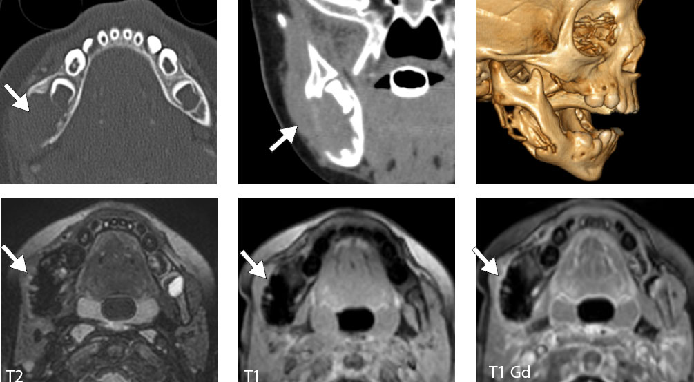
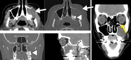
| Malignant tumors |
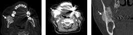
Extensive, ill-defined margins, infiltrative pattern, necrosis, heterogeneous enhancement, and soft-tissue invasion |

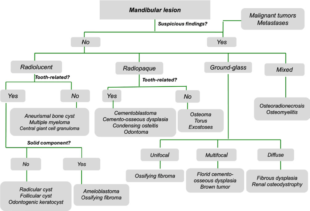
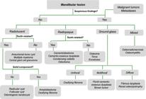
 Benign and malignant lesions of the mandible and maxilla: imaging findings and systematic approach
 Thumbnail
 Thumbnail
 Thumbnail
 Thumbnail
 Thumbnail
 Thumbnail
 Thumbnail
 Thumbnail
 Thumbnail
 Thumbnail
 Thumbnail
 Thumbnail
 Thumbnail
 Thumbnail
 Thumbnail
 Thumbnail
 Thumbnail
 Thumbnail
 Thumbnail