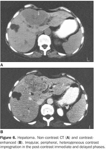
OBJECTIVE: To describe the different tomographic findings in hypodense hepatic lesions in children and its differential diagnosis. MATERIALS AND METHODS: Computed tomographic studies were obtained from 50 patients (age range: 0-16 years) with low-density liver lesions previously diagnosed by ultrasound. Images were made before and after administration of intravenous contrast medium. Image findings were analyzed and afterwards correlated with anatomopathological diagnosis. RESULTS: Forty-seven of 50 cases were confirmed, 30 by anatomopathological diagnosis. Most of then were benign lesions, hemangioma in 20%. Such lesions presented a homogeneous contrast absorption, mainly at the delayed phase, differing from malignant lesions. Metastasis was the most frequently found malignant lesion (18%). CONCLUSION: Computed tomographic study is of great value in complementing the diagnosis of hypodense hepatic lesions in children, and must follow ultrasound diagnosis as a routine procedure.
Hypodense hepatic injuries; Computed tomography; Childhood