 Abdominoperineal Resection of the Rectum in a Patient with Human Immunodeficiency Virus and Squamous Cell Carcinoma of the Anal Canal: Case Report
ABSTRACT
Introduction: Squamous cell carcinoma of the anal canal is an uncommon neoplasm, frequently associated with human papillomavirus (HPV), and presents therapeutic challenges, especially in immunosuppressed patients, such as those living with human immunodeficiency virus (HIV). Early identification based on risk factors is essential to increase the chances of therapeutic success.
Case report: A 40-year-old HIV-positive male patient was diagnosed with residual squamous cell carcinoma of the anal canal following initial treatment for an anal fissure. Due to the persistence of the disease after chemoradiotherapy, he underwent abdominoperineal resection of the rectum. The clinical course was dismal. As a result of early bilateral progression of inguinal lymph node disease, the patient died, demonstrating the high tumor aggressiveness in the context of immunosuppression.
Conclusion: This case demonstrates the complexity of managing squamous cell carcinoma of the anal canal in an HIV-positive patient and underscores the aggressiveness of the disease in refractory scenarios. The outcome reinforces the importance of surveillance and early diagnosis in high-risk populations to improve therapeutic response and clinical outcomes.
Key words:
Carcinoma, Squamous Cell/complications; Anal Canal/surgery; HIV; Papillomavirus Infections; Proctectomy
RESUMO
Introdução: O carcinoma espinocelular de canal anal é uma neoplasia incomum, frequentemente associada ao papilomavírus humano (HPV), e apresenta desafios terapêuticos, principalmente em pacientes imunossuprimidos, como os que vivem com vírus da imunodeficiência humana (HIV). A identificação precoce baseada nos fatores de risco é fundamental para ampliar as chances de sucesso terapêutico.
Relato do caso: Paciente masculino, 40 anos, soropositivo, com diagnóstico de carcinoma espinocelular de canal anal residual após tratamento inicial para fissura anal. Diante da persistência da doença após quimiorradiação, foi submetido à ressecção abdominoperineal do reto. A evolução foi desfavorável. Em decorrência da progressão precoce da doença linfonodal na região inguinal, de forma bilateral, o paciente evoluiu a óbito, evidenciando a elevada agressividade tumoral no contexto de imunossupressão.
Conclusão: Este caso evidencia a complexidade do manejo do carcinoma espinocelular de canal anal em paciente soropositivo e ressalta a agressividade da doença em cenários refratários. O desfecho reforça a importância da vigilância e do diagnóstico precoce em populações de alto risco, visando melhorar a resposta terapêutica e os resultados clínicos.
Palavras-chave:
Carcinoma de Células Escamosas/complicações; Canal Anal/cirurgia; HIV; Infecções por Papilomavírus; Proctectomia
RESUMEN
Introducción: El carcinoma espinocelular de canal anal es una neoplasia infrecuente, a menudo asociada al virus del papiloma humano (VPH), y presenta desafíos terapéuticos, principalmente en pacientes inmunosuprimidos, como aquellos que viven con virus de inmunodeficiencia humana (VIH). La identificación temprana basada en factores de riesgo es fundamental para ampliar las posibilidades de éxito terapéutico.
Informe del caso: Paciente masculino de 40 años, seropositivo, con diagnóstico de carcinoma epidermoide residual del canal anal tras tratamiento inicial por fisura anal. Ante la persistencia de la enfermedad después de la quimiorradioterapia, se sometió a una resección abdominoperineal del recto. La evolución fue desfavorable. Como consecuencia de la progresión temprana y bilateral de la enfermedad ganglionar en la región inguinal, el paciente falleció, lo que pone de manifiesto la elevada agresividad tumoral en el contexto de inmunosupresión.
Conclusión: Este caso evidencia la complejidad del manejo del carcinoma espinocelular de canal anal en un paciente seropositivo y resalta la agresividad de la enfermedad en escenarios refractarios. El desenlace refuerza la importancia de la vigilancia y el diagnóstico temprano en poblaciones de alto riesgo, con el fin de mejorar la respuesta terapéutica y los resultados clínicos.
Palabras clave:
Carcinoma de Células Escamosas/complicaciones; Canal Anal/cirugía; VIH; Infecciones por virus del Papiloma; Proctectomía
INTRODUCTION
Anal neoplasms account for approximately 2.7% of all digestive system cancers, being anal canal squamous cell carcinoma (ASCC) the most prevalent type, although rare. This condition is a human papillomavirus (HPV) associated malignancy with nearly 90% association, particularly subtype 161,2. Immunosuppression associated with the human immunodeficiency virus (HIV), most of all in individuals with low count of the cluster differentiation 4 (CD4), increases the risk and influences the initial phases of the development of the neoplasm2.
For most of the patients with localized disease, the standard therapeutic approach is chemoradiotherapy3. However, in cases of persistence of locoregional relapse of the disease post initial management, an abdominoperineal resection (APR) is the main surgical salvage strategy for ASCC4. This technique consists in the resection of sigmoid colon, rectum, anus, sphincter apparatus and levator ani, in addition to permanent terminal colostomy3.
Immunocompetent patients with ASCC have better overall survival compared with immunosuppressed in regard to prognosis. For the general population, the therapeutic focus lies in sphincter sparing through Nigro protocol. Immunosuppressed patients subvert the immune system compromising the immune surveillance capacity5,6. The distinct molecular characterization results in lower response rate to chemoradiation and higher incidence of local relapse, reducing the survival rates, anticipating the necessity of APR as surgical salvage strategy6. Cases described in the literature reinforce the late diagnosis and atypical clinical presentations in refractory settings consolidate the reserved prognosis for this subgroup7.
Due to the complexity of the management, this report is justified by the rarity of the neoplasm and the challenges in managing immunosuppressed patients. The aim of this study is to report a clinical case of an inpatient of "Hospital da Fundação Oswaldo Aranha (H.FOA)" in the city of Volta Redonda, Rio de Janeiro, with main diagnosis of ASCC associated with HIV-positive serology.
It is a descriptive case report based on the analysis of the patient's medical chart. The Ethics Committee approved the study, report number 6,881,611 (CAAE (submission for ethical review): 79764824.5.0000.5237) in compliance with Directive number 466/20128 of the National Health Council (CNS). The patient signed the Informed Consent Form.
CASE REPORT
Male, 40-year old homosexual patient, with a 5-year HIV infection history in daily antiretroviral therapy (ART), keeping CD4 above 900 cells/mm³ during the whole process. Sought oncologic care at "Hospital da Fundação Oswaldo Aranha (H.FOA)" on December 21, 2023, complaining of pain and bleeding at evacuation, symptoms initially attributed to an anal fissure. Reported that on October 5, 2023 was submitted to surgical procedure to correct anal fistula at a public hospital in the city of Volta Redonda, whose anatomopathological test revealed ASCC, being referred to a high complexity oncologic unit (Unacon).
The first anatomopathological macroscopy exam performed on November 7, 2023 revealed irregular heterogeneous firm fragments. At microscopy, findings compatible with well-differentiated and invasive ASCC have been found, without angiolymphatic or perineural invasions. The therapeutic protocol adopted was chemoradiotherapy with capecitabine 500 mg in three consecutive cycles between February 1 and 29, 2024, in addition to radiotherapy on the anal canal and glutes, totaling a 6,000 cGy dose in 30 fractions (200 cGy/day), divided in initial phase of 4,600 cGy and reinforcement of 1,400 cGy applied between January 25 and April 2, 2024. The treatment was suspended for seven days in March 2024 due to radiodermatitis grade 2.
Computed tomography of the thorax, upper abdomen and pelvis was performed on May 14, 2024 to evaluate the therapeutic response. Parietal thickening of the anal canal was observed, suggesting persistence of the disease (figure 1), with no evidence of lung metastasis or mediastinal lymph nodes. On May 15, 2024, rectosigmoidoscopy with biopsy revealed a 7-cm extensive ulcerated, circumferential lesion affecting the anus and perianal region. The biopsy report was conclusive for ASCC of the perianal and anal regions with residual disease.

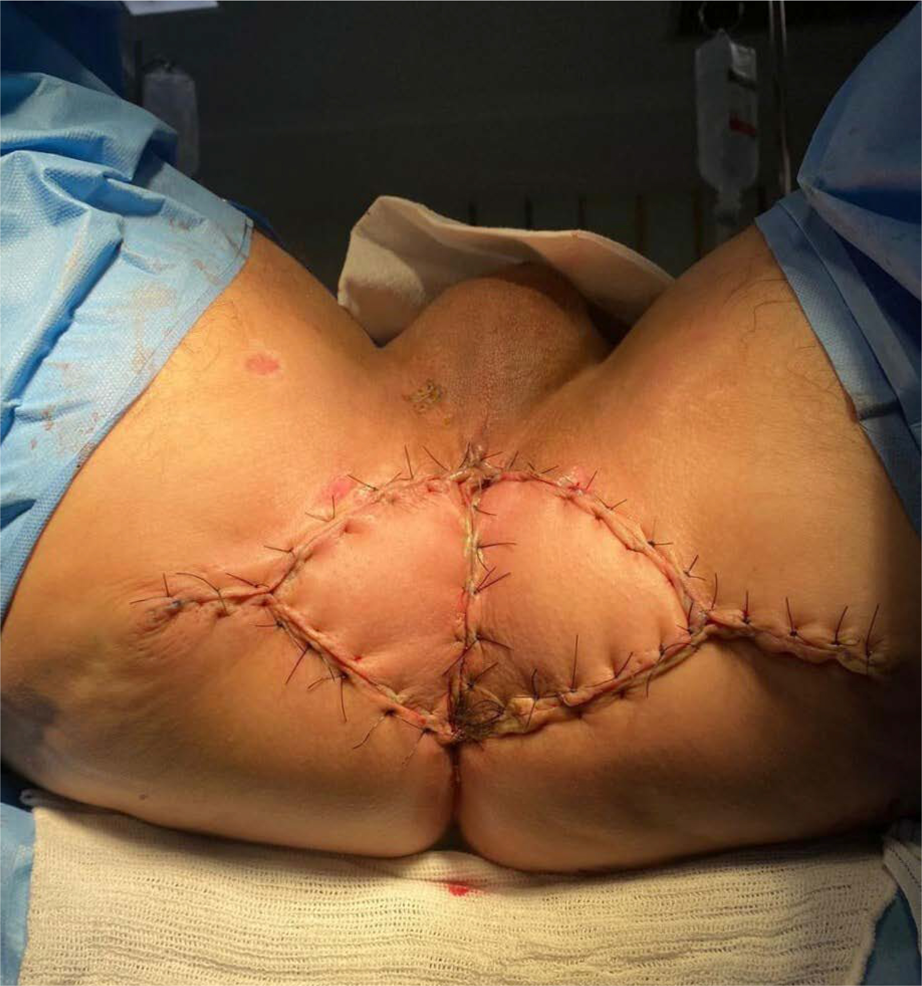
The patient was submitted to APR of the rectum on August 8, 2024. In lithotomy position on the surgical bed, the perineal lesion with severe actinic radiodermatitis was observed with presence of fibrinoid necrosis and fibrous induration of adjacent tissues compatible with actinic chronic damage secondary to localized radiotherapy (figure 2 A). Through full midline incision, the intervention initiated in the abdomen, without evidence of metastases. The sigmoid colon was moved, preserving the ureter and left gonadal vessels. The inferior mesenteric artery was ligated in its origin followed by D2 systematic lymphadenectomy. The upper rectal artery was ligated and the rectosigmoid segment was severed at the sacral promontory.

A: Perineal lesion at immediate pre-operation; B: Double Y myocutaneous flaps transposition to close perineal defect
A total mesorectal excision up to levator ani was performed, respecting the anatomic planes and keeping the integrity of the mesorectal envelope. Levator ani ligaments were severed with proper homeostasis. At perineal stage, the anal canal was excised and pelvic floor was closed with double-Y myocutaneous flap (figure 2 B), followed by subcutaneous closure with Donatti stitches (figure 3). A terminal colostomy was performed in the left lower quadrant of the abdomen.

Post-operation was favorable, the patient was admitted to the Intensive Care Unit (ICU) remaining homeostatically stable. Received antibiotic therapy with ciprofloxacin 200 mg for six days and metronidazole 0.5% single dose. The colostomy functioning was satisfactory in the first 24 hours. The psychological evaluation was uneventful. The patient was discharged on August 15, 2024 without complications.
Three months after the surgical procedure at outpatient follow-up, an inguinal lymphadenomegaly appeared. On November 22, 2024, the patient was submitted to incisional biopsies of bilateral inguinal lymph nodes, whose anatomopathological result revealed SCC affecting fibroadipose tissues with neoplastic infiltration in the skin, characterizing cutaneous metastasis. On December 13, 2024, magnetic resonance imaging of the pelvis revealed total anorectal amputation with exuberant post-surgical fibrosis in addition to complex organized collection in perineal soft parts with no signs of local neoplastic relapse. Partial infiltration of the muscles of levator ani was observed more pronounced at right and right, irregular and heterogeneous superficial inguinal lymphadenomegaly suggestive of secondary compromise.
These findings confirmed metastasis compatible with locally residual advanced disease, being indicated palliative treatment. Between January 11 and 23, 2025, the patient was submitted to pelvic radiotherapy, total dose of 2,500 cGy in five daily fractions of 500 cGy for locoregional control. Concomitantly, a second chemotherapy line with IV capecitabine 500 mg and oxaliplatin 250mg diluted in 500 ml of saline solution 0.9% was applied, completing three cycles of this regimen between January 21 and March 21, 2025.
After the oncological evaluation conducted on February 21, 2025, the persistence of the nodal recurrence was confirmed. In May 2025, due to refractory incoercible hiccups, an adverse event attributed to the toxicity of platin, the conduct was to change the chemotherapy to IV-infused paclitaxel 165 mg diluted in saline solution 500 ml 0.9% every 21 days. The new protocol initiated on May 16 and ended on June 24, 2025.
On July 8, 2025 in a routine outpatient visit of Unacon's oncologic clinic, the patient presented hemoglobin 4 g/dL, being admitted to H.FOA, progressing to sudden clinical worsening and death within few hours from admission. Despite the interventions, the poor response to chemoradiation classified the case as reserved prognosis, exposing the aggressiveness of the disease.
DISCUSSION
Anal carcinoma corresponds to 4% of the malignant tumors of the anal region7. Furthermore, the most common subtype is SCC. Although more common in women, its occurrence is increasing in men who have sex with men and in persons living with HIV6. The physiopathology is strongly associated with well-established risk factors such as HPV and HIV infection, number of sexual partners, receptive anal sexual intercourse and smoking9.
HIV and HPV infection are the main factors involved in the development of SCC of the anal canal. HIV provokes a subversion of the immune system, leading to a continuous loss of T CD4+ cells which compromises the ability of the organism to fight infections and neoplasms10. HPV, on its turn, is a sexually transmitted DNA virus, type 16 is the most common found in anal cancer9.
Staging is critical for prognostic and therapeutic definition, and the majority of the cases is diagnosed in localized or localized advanced stages1,11. The treatment remains based on the Nigro protocol that associates concomitant chemo and radiotherapy, while surgery is indicated for cases of therapeutic failure5,6. In these settings, APR remains as the main salvage strategy although associated with high post-operative morbidity and limited benefit of adjuvant therapies4,12.
The patient of the present case was classified as a high-risk profile. His therapeutic journey followed the standard protocol: was submitted to chemoradiation but due to the persistence of the disease, was prescribed abdominoperineal resection as a salvage measure. In this setting, recurring inflammatory stimuli and alterations of scar tissue of anal fistula contributed to the carcinogenesis, especially in immunosuppressive contexts13. Furthermore, the diagnosis is frequently postponed because local fibrosis and edema can lead the examiner to confound tumor infiltration with benign inflammatory adhesions13.
CONCLUSION
The early identification of anal carcinoma is critical in risk groups like HIV-infected individuals, immunosuppressed patients and individuals with persistent HPV infection. The presentation in the case mimicked an anal fistula and justified the histopathological investigation. Frequently, a late diagnosis associated with atypical clinical presentations directly correlates with dismal therapeutic outcomes and reduced survival rates.
DATA AVAILABILITY STATEMENT
All the content underlying the text is contained in the manuscript.
References
-
1 Rogers JE, Sirisaengtaksin A, Leung M, et al. Hepatic metastasectomy in squamous cell carcinoma of the anal canal: a case series of a curative approach. Cancers. 2023;15(15):3890. doi: https://doi.org/10.3390/cancers15153890
» https://doi.org/10.3390/cancers15153890 -
2 Roy J, Faraon B, Polcino M. Wide local excision of perianal squamous cell carcinoma associated with low-risk human papillomavirus (HPV) subtype. Cureus. 2025;17(4):e82335. doi: https://doi.org/10.7759/cureus.82335
» https://doi.org/10.7759/cureus.82335 -
3 Jethwa KR, Hallemeier CL. Research on anal squamous cell carcinoma. Cancers. 2022;14(1):42. doi: https://doi.org/10.3390/cancers14010042
» https://doi.org/10.3390/cancers14010042 -
4 Rosen R, Quezada-Diaz FF, Gönen M, et al. Oncologic outcomes of salvage abdominoperineal resection for anal squamous cell carcinoma initially managed with chemoradiation. J Clin Med. 2024;13(8):2156. doi: https://doi.org/10.3390/jcm13082156
» https://doi.org/10.3390/jcm13082156 -
5 Nigro ND, Vaitkevicius VK, Considine Júnior B. Combined therapy for cancer of the anal canal: a preliminary report. Dis Colon Rectum. 1974;17(3):354-6. doi: https://doi.org/10.1007/BF02586980
» https://doi.org/10.1007/BF02586980 -
6 Armstrong SA, Malley R, Wang H, et al. Molecular characterization of squamous cell carcinoma of the anal canal. J Gastrointest Oncol. 2021;12(5):2423-37. doi: https://doi.org/10.21037/jgo-20-610
» https://doi.org/10.21037/jgo-20-610 -
7 Costa MLF, Ferreira ACC, Machado TR, et al. Neoplasia de canal anal com metástase pulmonar: relato de caso. J Coloproctol. 2023;43(S 01):S1-S270. doi: https://doi.org/10.1055/s-0044-1780983
» https://doi.org/10.1055/s-0044-1780983 -
8 Conselho Nacional de Saúde (BR). Resolução n° 466, de 12 de dezembro de 2012. Aprova as diretrizes e normas regulamentadoras de pesquisas envolvendo seres humanos [Internet]. Diário Oficial da União, Brasília, DF. 2012 dez 12 [acesso 2026 mar 12]; Edição 112; Seção 1:59. Disponível em: https://www.gov.br/conselho-nacional-de-saude/pt-br/atos-normativos/resolucoes/2012/resolucao-no-466.pdf/view
» https://www.gov.br/conselho-nacional-de-saude/pt-br/atos-normativos/resolucoes/2012/resolucao-no-466.pdf/view -
9 Ebrahimi F, Rasizadeh R, Jafari S, et al. Prevalence of HPV in anal cancer: exploring the role of infection and inflammation. Infect Agent Cancer. 2024;19(63):1-10. doi: https://doi.org/10.1186/s13027-024-00624-0
» https://doi.org/10.1186/s13027-024-00624-0 -
10 van Heuvel Y, Schatz S, Rosengarten JF, et al. Infectious RNA: human immunodeficiency virus (HIV) biology, therapeutic intervention, and the quest for a vaccine. Toxins (Basel). 2022;14(2):138. doi: https://doi.org/10.3390/toxins14020138
» https://doi.org/10.3390/toxins14020138 -
11 Astaras A, Bornand T, Koessler S. Squamous rectal carcinoma: a rare malignancy, literature review and management recommendations. ESMO Open. 2021;6(4):100180. doi: https://doi.org/10.1016/j.esmoop.2021.100180
» https://doi.org/10.1016/j.esmoop.2021.100180 -
12 Naessens C, Evin C, Le-Malicot K, et al. Salvage abdominoperineal resection for anal squamous cell carcinoma after radiotherapy or chemoradiotherapy: analysis of the french prospective cohort FFCD-ANABASE. Eur J Surg Oncol. 2025;51(10):110346. doi: https://doi.org/10.1016/j.ejso.2025.110346
» https://doi.org/10.1016/j.ejso.2025.110346 -
13 Zhu QB, Zhang J, Liu CY, et al. Case report of squamous cell carcinoma secondary to recurrent anal fistula. Front Oncol. 2025;15:1673829. doi: https://doi.org/10.3389/fonc.2025.1673829
» https://doi.org/10.3389/fonc.2025.1673829
Edited by
-
Scientific-editor:
Anke Bergmann. Orcid iD: https://orcid.org/0000-0002-1972-8777