Impact of estrogen on morphology and programmed cell death in the meibomian glands of rats
ABSTRACT
Objective: To examine the relationship between hormonal changes, particularly estrogen, and lacrimal gland function in dry eye syndrome using a surgically induced menopausal rat model.
Methods: Twenty-four female rats were divided into four groups: control, ovariectomized, ovariectomized with estrogen replacement, and ovariectomized with phytoestrogen replacement. Rats with regular estrous cycles were selected via vaginal cytology. Ovariectomy was performed on ovariectomized groups, and ovariectomized with estrogen replacement and ovariectomized with phytoestrogen replacement groups received hormonal interventions. Histological analysis assessed collagen content, cellular volume, and apoptosis in lacrimal gland tissues.
Results: Collagen content significantly decreased in the ovariectomized group compared to control, emphasizing the importance of tissue integrity. Cellular volume increased in ovariectomized with estrogen replacement and ovariectomized with phytoestrogen replacement groups, indicating hormonal influence. Apoptosis levels varied, with higher levels in ovariectomized and ovariectomized with estrogen replacement groups, while ovariectomized with phytoestrogen replacement resembled Control Group.
Conclusion: Estrogen likely modulates lacrimal gland function and tear film stability. Collagen content underscores tissue's integrity role in gland function.
Keywords:
Meibomian gland dysfunction; Dry eye syndromes; Lacrimal apparatus; Estrogens
RESUMO
Objetivo: Examinar a relação entre mudanças hormonais, especialmente o estrogênio, e a função das glândulas lacrimais na síndrome do olho seco, usando um modelo de menopausa induzida cirurgicamente em ratos.
Métodos: Vinte e quatro ratas fêmeas foram divididas em quatro grupos: controle, ovariectomizadas, ovariectomizadas com reposição de estrogênio e ovariectomizadas com reposição de fitoestrógeno. Ratos com ciclos estrosos regulares foram selecionados por citologia vaginal. A ovariectomia foi realizada nos grupos ovariectomizados, com os grupos com reposição de estrogênio e com reposição de fitoestrógeno recebendo intervenções hormonais. A análise histológica avaliou o conteúdo de colágeno, o volume celular e a apoptose nos tecidos das glândulas lacrimais.
Resultados: O conteúdo de colágeno diminuiu significativamente no grupo de ovariectomizadas em comparação com o controle, destacando a importância da integridade tecidual. O volume celular aumentou nos grupos de ovariectomizadas com reposição de estrogênio e com reposição de fitoestrógeno, indicando influência hormonal. Os níveis de apoptose variaram, com níveis mais altos nos grupos de ovariectomizadas e de ovariectomizadas com reposição de estrogênio, enquanto o de ovariectomizadas com reposição de fitoestrógeno se assemelhou ao grupo controle.
Conclusão: O estrogênio provavelmente modula a função das glândulas lacrimais e a estabilidade do filme lacrimal. O conteúdo de colágeno destaca o papel da integridade tecidual na função glandular.
Descritores:
Disfunção da glândula tarsal; Síndromes do olho seco; Aparelho lacrimal; Estrogênios
INTRODUCTION
Dry eye disease (DED), also known as keratoconjunctivitis sicca, stands as one of the most prevailing afflictions encountered within the realm of ophthalmology. Its hallmark resides in the perturbation of the ocular tear film, stemming from either an inadequate production of tears or an excessive evaporation thereof. This disruption culminates in the impairment of the interpalpebral ocular surface, often conjoined with disconcerting ocular symptoms.(1)
The pathological presentations of dry eye manifest a notable diversity in the severity of clinical indications and symptoms, along with their intricate interplay with a spectrum of underlying conditions. The instability of the tear film engenders vulnerability to ocular surface detriment, increasing susceptibility to infections, and, in severe instances, precipitating substantial vision impairment.(2)
Extending beyond the realm of ocular discomfort, these symptoms exact a profound toll on daily activities, encompassing occupational endeavors, reading, computer utilization, and even the fundamental task of driving. Furthermore, the psychosocial ramifications of dry eye symptoms reverberate across an individual's overall health and well-being, eliciting discernible perturbations in the tapestry of life quality.(3)
The etiology of dry eye is intricately woven, encapsulating a multifactorial tapestry with congenital anomalies, nutritional imbalances, autoimmune pathologies, neurologic disorders, and endocrine dysregulations. External milieu perturbations, such as environmental conditions engendering decreased room humidity, coupled with certain infectious agents, may exacerbate this condition. Notably, specific bacterial strains, including Staphylococcus aureus, Staphylococcus epidermidis, Streptococcus, Corynebacterium, and Propionibacterium acnes, provide lipases capable of unsettling the equilibrium of the tear film stability.(4)
Alterations in tear composition serve as a sign of tear film instability, precipitating perturbations in the cellular metabolism of the ocular surface, concomitant decrease of local defense mechanisms, and leading to the inception of locally perpetuated inflammatory cascades. Consequently, a prevailing hallmark among sundry iterations of dry eye manifests as the presence of inflammatory markers and symptoms, whose clinical dynamics undergo modulation across the continuum of the ailment.(5)
The lack of tear production not only impairs the structural integrity of ocular surface tissues but also culminates in attenuated anabolic stimuli, concomitant elevation of inflammatory mediators, and the emergence of a hyperosmolar microenvironment. This heightened osmolarity engenders a cascade of repercussions, inducing direct epithelial impairment, dwindling cytoplasmic density, and the accumulation of mucous secreted by osmotically disturbed mucosecretory cells.(6)
Epidemiological inquiries underscore an augmented prevalence of dry eye, particularly amongst women, with a proclivity toward those beyond the age of 50.(3) In 1997, scholars noted that over 14% of the elderly population showed symptomatic dry eye, of whom 10% relied on artificial tear supplementation for alleviation. Thus, women and the elderly inhabit a heightened vulnerability sphere concerning the development of dry eye.(7)
An arresting nexus emerges between disturbances within the tear film and physiological fluctuations, chiefly observed during hormonal vicissitudes, such as menopause, pregnancy, and the utilization of hormonal contraceptives. This insinuates a plausible regulatory role of hormones in the orchestration of tear production.(8) In the year 2000, the proposition gained empirical support, with studies corroborating the presence of estrogen receptors within the basal cells of Meibomian glands.(9) Subsequently, in 2007, a study substantiated the putative impact of hormones on the incidence and progression of dry eye syndrome.(10)
Hormone replacement therapy, an arena fraught with global contention as to its risk- benefit paradigm in the context of dry eye, encompasses the administration of estrogen and progesterone to menopausal women.(2,11) As the global population enjoys increased longevity, the prevalence of dry eye syndrome is ready for amplification.(12)
The aim of this study is to examine the relationship between hormonal changes, particularly estrogen, and lacrimal gland function in dry eye syndrome using a surgically induced menopausal rat model.
METHODS
This study has obtained approval from the Ethics Committee on Animal Use (CEUA) in Experimentation of the Health Sciences Center at the Universidade Federal do Rio de Janeiro, under reference number DAHEICB076.
In the experimental study, 24 female adult Wistar rats with an average weight of 250 g were employed. These animals were housed in a vivarium under controlled conditions of temperature (24 ± 1°C) and light (12 hours/day) with a maximum of six animals per cage, having access to water and standard rodent chow ad libitum. The study received ethical approval from the animal research ethics committee.
Vaginal cytology was performed on the animals to select those with a regular estrous cycle lasting 4 to 5 days. To monitor the cyclical variations that occur during the estrous cycle, vaginal cytology was conducted using 0.9% NaCl smears daily over a period of 2 weeks for all animals. Only rats displaying a regular estrous cycle of 4 to 5 days were included in the study.
All animals in the Control Group (CTROL) underwent a surgical stress procedure, including anesthesia, surgical incision, and suturing only.
Bilateral oophorectomy was performed on the female rats under intraperitoneal anesthesia with xylazine (5 mg/kg) and ketamine (50 mg/kg). They were sacrificed 23 days post-oophorectomy. The animals were divided into four groups: control (CTROL); ovariectomized (OVX); OVX with 17-β estradiol (E2) replacement at a dose of 5.0 μg/100g body weight; and OVX with Trifolium pratense (TP) administration at a dose of 500 mg/kg body weight.
Ovariectomized rats received daily subcutaneous injections of 17β-estradiol (Sigma) for 16 days, starting 1 week after oophorectomy, at a dose of 5.0 μg/100 g body weight. This dose of estradiol corresponds to the one used in postmenopausal hormone replacement therapy in women. 17β-estradiol was dissolved in a small amount of ethanol and suspended in propylene glycol to achieve a concentration of 5.0 μg/100g body weight. Both the CTROL and untreated OVX animals received injections of the vehicle (propylene glycol).
Ovariectomized animals received TP phytoestrogen at a daily dose of 500 mg/kg, containing 8% isoflavones, administered through gavage for 16 days. This dose was chosen for its clinical relevance in treating menopausal symptoms. TP phytoestrogen was acquired from Galena Quimica e Farmaceutica Ltda, which conducted high- performance liquid chromatography to confirm the isoflavone content in the extract.
Since these animals were part of an ongoing research project in the endocrine physiology laboratory where some blood measurements were essential for other studies, decapitation was performed to avoid alterations in blood levels at the end of the 16-day treatment period. Fragments of the middle third of the upper eyelids, encompassing the entire thickness (from eyelid skin to tarsal conjunctiva), with approximately 4 mm in width, were excised.
Tissues from 24 animals (four groups) were subjected to analysis. For all assessed criteria, histological sections containing fragments of the upper eyelids (right and left) were obtained for each animal. The samples were paired (left and right upper eyelids) from the same animal and fixed in buffered 10% formalin for 24 hours. Subsequently, they were embedded in paraffin for later histological examination using light microscopy (LM).
For collagen analysis, cellular density, and cellular volume assessment by LM, three slides with 20 fields (images) per slide were required for each animal, with a magnification of 40x.
Histological analysis was performed using Image-Pro Plus software. Morphometric analysis (assessment of cellular density and volume) involved the acquisition of 3 μm thick cross-sectional slices from the paraffin blocks, stained with hematoxylin and eosin (HE). The histological slides were used for histomorphometric analysis via LM of the meibomian gland acini.
Collagen was identified using Picrosirius Red staining in selected histological sections. Apoptosis evaluation of programmed cell death in the Meibomian gland acini was conducted using the Tunel technique on 3 μm thick cross-sectional slices from the paraffin blocks. For apoptosis assessment, the ApopTag® Peroxidase in situ apoptosis detection kit (cat S7100, Chemicon, United States) was employed.
GraphPad Prism software was utilized for data storage and statistical analysis. T- tests and analysis of variance (Anova) were employed, with significance set at p < 0.05. The obtained data were presented using graphs and tables.
RESULTS
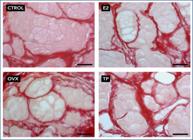
In the assessment of collagen, the mean values and standard deviations were as follows: 2.392 ± 0.1239 %/μm² in the CTROL Group, 0.3285 ± 0.3775 %/μm² in the OVX Group, 0.2399 ± 0.0945 %/μm² in the E2 Group, and 0.3400 ± 0.1417 %/μm² in the TP Group. These results are presented in table 1.
Upon comparing these results between the groups, a highly significant difference (p < 0.0001) was observed between CTROL versus TP and OVX versus TP. A significant difference (p < 0.05) was found for comparisons between CTROL versus OVX, OVX versus E2, and E2 versus TP. These findings are illustrated in figure 1.

(A) Collagen area across the groups; (B) collagen area among the groups. (C) cellular length across the groups; (D) cellular density across the groups; (E) Cellular positivity across the groups.
In the assessment of cell length, the mean values and standard deviations were found to be 3.829 ± 1.502 μm in the CTROL Group, 3.598 ± 1.579 μm in the OVX Group, 4.159 ± 1.798 μm in the E2 Group, and 4.158 ± 1.697 μm in the TP Group. These results are presented in table 1B.
No statistically significant differences were observed when comparing the results of cell length assessment between the different groups. However, there was a slight increase in cell length in the E2 and TP Groups. These findings are illustrated in Figure 1C and further detailed in figure 2.
In the assessment of cell density, the mean values and standard deviations were as follows: 1,064.0 ± 368.9 cells in the CTROL Group, 929.0 ± 302.6 cells in the OVX Group, 1,125.0 ± 155.7 cells in the E2 Group, and 1,099 ± 185.1 cells in the TP Group. These results are presented in table 1C.
There were no statistically significant differences between the groups. However, the OVX Group exhibited a decrease in cell density compared to the other groups, without statistical significance. The E2 Group showed a slight increase in density compared to the other groups, but without statistical significance. These findings are illustrated in figure 1D and further detailed in figure 3.

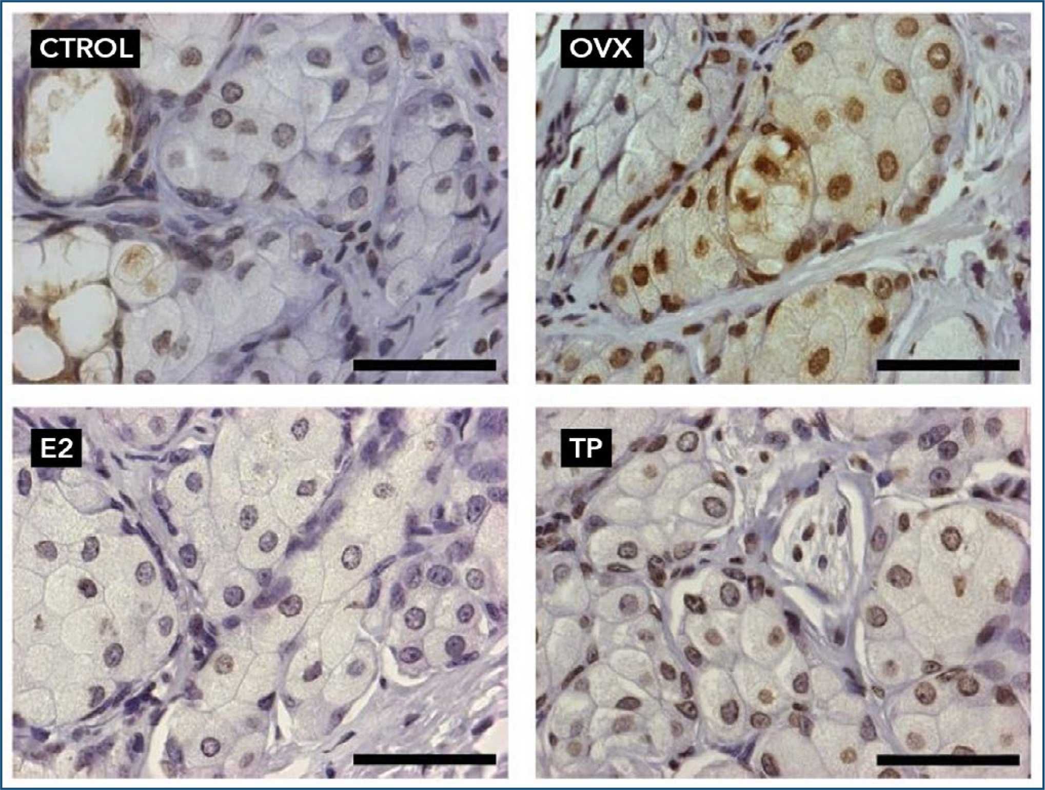
Values of mean and standard deviation for cell apoptosis are presented in table 1D.
In figure 1E, it can be observed that the data for the CTROL and TP Groups do not differ significantly from each other, showing no statistical significance. On the other hand, the remaining groups differ significantly from each other with p < 0.001. These findings are illustrated in figure 1E and further detailed in figure 4.

DISCUSSION
Dry eye disease, also known as keratoconjunctivitis sicca, is a prevalent ocular condition characterized by an insufficient tear film, resulting in ocular discomfort and visual disturbances.(13) This study aimed to assess the impact of hormonal interventions on various parameters related to the lacrimal gland and dry eye syndrome.
The lacrimal gland is a pivotal component of tear production and regulation, and emerging evidence suggests that hormonal fluctuations may significantly influence its function.(14) Our study focused on evaluating the effects of different hormonal interventions, including estrogen replacement therapy and phytoestrogen supplementation, in a surgically induced menopausal rat model.
An important aspect of lacrimal gland health is the regulation of collagen content within the glandular tissue. Collagen plays a crucial role in maintaining tissue integrity, and alterations in its composition can affect glandular function.(15) Our results demonstrated a substantial decrease in collagen content in the OVX Group compared to the Control Group. However, estrogen replacement (E2) and phytoestrogen supplementation (TP) appeared to attenuate this decrease, although statistical significance was not reached.
Tear film stability is a complex process involving interactions within the lacrimal gland, and hormonal influences are well-documented.(16) In our study, the evaluation of cellular volume showed a trend towards an increase in the E2 and TP Groups compared to the OVX and Control Groups. Although this difference did not reach statistical significance, it suggests a potential role of hormones in modulating lacrimal gland cellular function and tear film stability.(17)
The assessment of lacrimal gland cellular density revealed no statistically significant differences between the groups. However, the OVX Group displayed a trend towards reduced cellular density compared to the other groups, although this trend lacked statistical significance. These findings are consistent with previous research highlighting hormonal influences on lacrimal gland morphology and cellular density. (18)
Apoptosis, or programmed cell death, is a crucial process for maintaining tissue homeostasis.(19) Our study evaluated apoptotic activity in lacrimal gland tissue, revealing differences between groups. The control and TP Groups exhibited similar apoptotic levels, suggesting that phytoestrogen supplementation may help preserve lacrimal gland tissue integrity. In contrast, the OVX and E2 Groups showed significantly higher levels of apoptosis. This observation underscores the complex interplay between hormones and lacrimal gland health, as excessive apoptosis can lead to glandular dysfunction.
The influence of hormonal interventions on lacrimal gland health and dry eye syndrome is consistent with recent literature emphasizing the role of hormones, particularly estrogen, in ocular health.(20) Estrogen receptors have been identified in various ocular tissues, including the lacrimal gland, supporting their involvement in tear film regulation and glandular function.(21,22)
Phytoestrogens, such as genistein and equol, have also called attention for their potential benefits in mitigating dry eye symptoms and improving meibomian gland function.(23,24) These plant-derived compounds have estrogenic properties and have been investigated for their role in modulating lacrimal gland health.(25)
Furthermore, recent studies have contributed to our understanding of the impact of phytoestrogens on lacrimal gland health, revealing a significant association between phytoestrogen consumption and dry eye relief through their estrogen-like effects.(26)
An in vivo study was conducted in mice to explore the impact of phytoestrogens on meibomian gland dysfunction. The study demonstrated that supplementation with phytoestrogens improved the function of the meibomian glands, highlighting the potential therapeutic benefits of these compounds in the management of dry eye syndrome.(17)
In a rat model, the effects of genistein on meibomian gland dysfunction were explored, uncovering a significant improvement in meibomian gland health following genistein treatment.(27) This research underscores the potential of phytoestrogens as a targeted therapy for meibomian gland dysfunction, a common contributor to dry eye syndrome.
CONCLUSION
Our study contributes to the growing body of research on the influence of hormonal interventions on lacrimal gland health and dry eye syndrome. These findings hold promise for the development of effective treatments for individuals suffering from dry eye disease, a condition that can significantly impact their quality of life.
REFERENCES
- 1 Lemp MA. Report of the National Eye Institute/Industry workshop on Clinical Trials in Dry Eyes. CLAO J. 1995;21(4):221-32
- 2 Schaumberg DA, Buring JE, Sullivan DA, Reza DM. Hormone replacement therapy and dry eye syndrome. JAMA. 2001;286;2114-9.
- 3 Schaumberg DA, Sullivan DA, Buring JE, Dana MR. Prevalence of dry eye syndrome among US women. Am J Ophthalmol. 2003;136(2):318-26.
- 4 McCulley JP, Shine WE. The lipid layer of tears: dependent on meibomian gland function. Exp Eye Res. 2004 Mar;78(3):361-5.
- 5 Aguilar AJ, Júnior NK, Rocha FJ, Rocha EM. Olho seco. In: Gomes JA, Alves MR. Superfície ocular. Rio de Janeiro: Cultura Médica; 2006. p. 57-68.
- 6 Aguilar AJ. La hiperosmolaridad del film lagrimal em el ojo seco. Arq Bras Oftal. 2008;71:69-71.
- 7 Schein OD, Muñoz B, Tielsch JM, Bandeen-Roche K, West S. Prevalence of dry eye among the elderly. Am J Ophthalmol. 1997;124(6):723-8.
- 8 Auw-Haedrich C, Feltgen N. Estrogen receptor expression in meibomian glands and its correlation with age and dry-eye parameters. Graefes Arch Clin Exp Ophthalmol. 2003;241(9):705-9.
- 9 Esmaeli B, Harvey JT, Hewlett B. Immunohistochemical evidence for estrogen receptors in meibomian glands. Ophthalmology. 2000;107(1):180-4.
- 10 Erdem U, Ozdegirmenci O, Sobaci E, Sobaci G, Göktolga U, Dagli S. Dry eye in post-menopausal women using hormone replacement therapy. Maturitas. 2007;56(3):257-62.
- 11 Evans V, Millar TJ, Eden JA, Willcox MD. Menopause, hormone replacement therapy and tear function. Adv Exp Med Biol. 2002;506(Pt B):1029-33.
- 12 Brewitt H, Sistani F. Dry eye disease: the scale of the problem. Surv Ophthalmol. 2001;45 Suppl 2:S199-202.
- 13 Murphy C, Alvarado J. Just what the doctor ordered: estrogen therapy for dry eye. Invest Ophthalmol Vis Sci. 2004;45(2):365–6.
- 14 Liu S, Hatton MP, Khandelwal P, Sullivan DA, Sullivan BD. Secretory vesicles in glandular epithelia: insights from lacrimal and salivary gland acinar cells. Int Rev Cell Mol Biol. 2014;308:53–84.
- 15 Craig JP, Nichols KK, Akpek EK, Caffery B, Dua HS, Joo CK, et al. TFOS DEWS II Definition and Classification Report. Ocul Surf. 2017;15(3):276-83.
- 16 Sullivan DA, Jensen RV, Suzuki T, Richards SM, Do CV, Schwartz DA, et al. Estrogen and progesterone influence the production of secretory proteins in the lacrimal gland. Adv Exp Med Biol. 2009;664:127–36.
- 17 Paranjpe V, Galor A, Grambergs R, Mandal N. The role of sphingolipids in meibomian gland dysfunction and ocular surface inflammation. Ocul Surf. 2022;26:100-110.
- 18 Franco PC, Simões RS, Carbonel AA, Sasso GR, Florencio-Silva R, Baracat EC, Girão MB, Soares Júnior JM, Simões MJ. The influence of phytoestrogens or estrogens on the proliferation of the rat endocervical mucosa. Rev Assoc Med Bras (1992). 2020;66(2):174-9.
- 19 Sullivan DA, Rocha EM, Aragona P, Clayton JA, Ding J, Golebiowski B, et al. TFOS DEWS II Sex, Gender, and Hormones Report. Ocul Surf. 2017;15(3):284-333.
- 20 Prelevic GM, Kocjan T, Markou A. Hormone replacement therapy in postmenopausal women. Min Endocrinol. 2005;30(1):27-36.
- 21 Gustafsson JA. Estrogen receptor beta – a new dimension in estrogen mechanism of action. J Endocrinol. 1999;163:379-83.
- 22 Razandi M, Pedram A, Greene GL, Levin ER. Cell membrane and nuclear estrogen receptors (ERs) originate from a single transcript: studies of ERalpha and ERbeta expressed in Chinese hamster ovary cells. Mol Endocrinol. 1999;13(2):307-19.
- 23 Razandi M, Pedram A, Greene GL, Levin ER. Cell membrane and nuclear estrogen receptors (ERs) originate from a single transcript: studies of ERalpha and ERbeta expressed in Chinese hamster ovary cells. Mol Endocrinol. 1999;13(2):307-19.
- 24 Chhadva P, Goldhardt R, Galor A. Meibomian gland disease: the role of gland dysfunction in dry eye disease. Ophthalmology. 2017;124(11S):S20-S26.
- 25 Knight DC, Eden JA. A review of the clinical effects of phytoestrogens. Obstet Gynecol. 1996;87(5 Pt 2):897-904.
- 26 Scuderi G, Contestabile MT, Gagliano C, Iacovello D, Scuderi L, Avitabile T. Effects of phytoestrogen supplementation in postmenopausal women with dry eye syndrome: a randomized clinical trial. Can J Ophthalmol. 2012;47(6):489-92.
- 27 Dong ZY, Ying M, Zheng J, Hu LJ, Xie JY, Ma Y. Evaluation of a rat meibomian gland dysfunction model induced by closure of meibomian gland orifices. Int J Ophthalmol. 2018;11(7):1077-83.