
 Two new species of Protomicrocotyle Johnston & Tiegs, 1922 (Monogenea: Protomicrocotylidae) parasites of Caranx hippos (Linnaeus, 1776) (Carangiformes: Carangidae), from Tecolutla, Veracruz, Mexico
Abstract
Two new species of Protomicrocotyle are described as parasites of the gill filaments of Caranx hippos from the Gulf of Mexico coast, characterized using morphological, morphometric, and molecular methods. Multivariate statistics (principal component and discriminant analyses) were used for the morphometric analyses. Pairwise genetic distances (p-distances) and cluster analyses using the Neighbor-joining method were performed using partial sequences of CO1 mtDNA and 28S rDNA of the new species and partial sequences of other species of Protomicrocotylidae from GenBank. Protomicrocotyle eamsae sp. nov. differs morphologically from other congeners by the presence of a retractile glandulomuscular organ in the prohaptor, and Protomicrocotyle pritchardae sp. nov. differs from other congeners by the body length and by the arrangement, shape, and number of testes. The new species was recovered as a distinct group, separate from other protomicrocotylids, based on CO1 mtDNA and 28S rDNA analyses. The results of the morphometric analysis of 91 morphological variables and the molecular analysis support the assignment of the new species to Protomicrocotyle in Caranx hippos from Tecolutla, Veracruz, Mexico.
Keywords:
Monogenea; morphometry; molecular; genetic distance; Neighbor-Joining; Protomicrocotyle
Resumo
Duas novas espécies de Protomicrocotyle são descritas como parasitos dos filamentos branquiais de Caranx hippos da costa do Golfo do Mexico, caracterizadas usando métodos morfológicos, morfométricos e moleculares. Estatísticas multivariadas (análises de componentes principais e discriminantes) foram usadas para as análises morfométricas. Distâncias genéticas pareadas (p-distances) e análises de agrupamento usando o método Neighbor-joining foram realizadas usando sequências parciais de CO1 mtDNA e 28S rDNA da nova espécie e sequências parciais de outras espécies de Protomicrocotylidae do GenBank. Protomicrocotyle eamsae sp. nov. difere morfologicamente de outros congêneres pela presença de um órgão glandulomuscular retrátil no prohaptor, e Protomicrocotyle pritchardae sp. nov. difere de outros congêneres pelo comprimento do corpo e pelo arranjo, forma e número de testículos. A nova espécie foi recuperada como um grupo distinto, separado de outros protomicrocotilídeos, com base nas análises de CO1 mtDNA e 28S rDNA. Os resultados da análise morfométrica de 91 variáveis morfológicas e da análise molecular corroboram a atribuição da nova espécie ao Protomicrocotyle em Caranx hippos de Tecolutla, Veracruz, Mexico.
Palavras-chave:
Monogenea; morfometria; molecular; distância genética; Neighbor-Joining; Protomicrocotyle
Introduction
Monogenea are flatworms that are distinguished by the presence of a specialized attachment structure, the haptor, located at the posterior end of the body. They have a monoxenous life cycle that is limited exclusively to the aquatic environment (Buchmann & Bresciani, 2006; Pulido-Flores, 2024). Typically, they are found on the skin, gills, or fins of fish, both freshwater and marine, although some monogeneans can also infect amphibians and reptiles (Buchmann & Bresciani, 2006; Kearn, 2014; Mendoza-Garfias et al., 2017; Pulido-Flores, 2024). Some hypotheses based on analyses of molecular data suggest that Monogenea is paraphyletic (does not form a monophyletic group) (Brabec et al., 2023; Justine, 1991a, b; Mollaret et al., 1997); however, pending studies that combine morphological and molecular data in a Total Evidence analysis (Kluge, 1998, 2004), we continue to consider Monogenea to be monophyletic (Pulido-Flores, 2024).
Monogeneans encompass an estimated global diversity of 5,000 known species, comprising 716 genera and 63 families (Caira & Littlewood, 2013). In Mexico, this class is represented by the second-highest number of recorded species of helminth, with approximately 313 nominal species and 54 undetermined taxa. Collectively, these records correspond to 162 genera included in 29 families (García-Prieto et al., 2014; Mendoza-Garfias et al., 2017).
Monogeneans assigned to Protomicrocotylidae are parasites on the gill arches of marine fish, particularly carangid fish (Caballero y Caballero & Bravo-Hollis, 1965, 1967; Kritsky et al., 2011; Ramalingam, 1960; Yamaguti, 1963). Currently, the family is represented by nine genera: Abortipedia Unnithan, 1962, Bilaterocotyle Chauhan, 1945, Bilaterocotyloides Ramalingam, 1961, Chauhanocotyle Khoche & Dad, 1975, Lethacotyle Manter & Price, 1953, Neomicrocotyle Ramalingam, 1960, Protomicrocotyle Johnston & Tiegs, 1922, Vallisiopsis Subhapradha, 1951, and Youngiopsis Lebedev, 1972, with approximately 43 species assigned to the family (Lebedev, 1986; Ramírez-Cruz et al., 2023; WoRMS, 2026).
All known species of Protomicrocotyle are parasites of fish belonging to the family Carangidae Nelson, 1984, with the majority having been reported as parasites of the genera Caranx Lacepède, 1801 (Boada et al., 2012; Bravo-Hollis, 1966, 1979; Caballero y Caballero & Bravo-Hollis, 1965; Hargis, 1957; Iruegas-Buentello, 1999; Kritsky et al., 2011; Luque & Alves, 2001; Mendoza-Garfias et al., 2017; Montoya-Mendoza et al., 2017; Ramalingam, 1960; Ramírez-Cruz et al., 2023; Vianna et al., 2020; Violante-González et al., 2019; Wahl, 1972; Yamaguti, 1953); a single species was described from Trachinotus Lacepède. To date, 10 species have been described: P. mirabilis (MacCallum, 1918) Johnston & Tiegs, 1922 (type species) from the Atlantic Ocean in C. hippos (Linnaeus) from the New York Aquarium (MacCallum, 1918); P.manteri Bravo-Hollis, 1966 from La Paz, Baja California Sur, Mexico, in Trachinotus paitensis Cuvier (= T. paloma Jordan & Starks) (Bravo-Hollis, 1966); P.nayaritensis Bravo-Hollis, 1979 from Isla Isabel, Nayarit, Mexico, in C. caninus Günther, (originally identified as C.hippos caninus) (Bravo-Hollis, 1979); P.ivoriensis Wahl, 1972 from Ebrié Lagoon, Ivory Coast, Western Africa, in C. hippos (Wahl, 1972); P.celebesensis Yamaguti, 1953 from Celebes Island, Indonesia, from Caranx sp. (Yamaguti, 1953); P.madrasensis Ramalingam, 1960 in C. affinis Rüppell; P.mannarensis Ramalingam, 1960; P.minutum Ramalingam, 1960 in C.sexfasciatus Quoy & Gaimard from India (Ramalingam, 1960); P.carangis Pillai & Pillai, 1978 from Kerala coast, India, in C. ignobilis (Forsskål) [= C.sansun (Fabricius)] (Pillai & Pillai, 1978); and the recently described species, P. veracruzensis Ramírez-Cruz, Monks, Manríquez-Morán, Violante-González & Pulido-Flores, 2023 from Casitas and Puerto de Veracruz, Veracruz, Mexico, in C.latus Agassiz (Ramírez-Cruz et al., 2023).
As part of an ongoing studies of the biodiversity of monogenean parasites of marine fish from the Gulf of Mexico, two new species of Protomicrocotyle were found on the gill filaments of C. hippos in the locality of Tecolutla, Veracruz. The new species are illustrated and described herein using morphological, morphometrics and molecular data.
Material and Methods
Specimen collection
Fourteen specimens of Caranx hippos were collected (November 2021) from the coastal waters off Tecolutla, Veracruz (20° 28′ 39″ N; 97° 00′ 30″ W) (Figure 1). Fish were obtained through commercial purchases from local fisherman. Each fish was sexed, measured, and photographed in order to facilitate taxonomic identification, in accordance with the recommendations set forth in the specialized literature (Nelson, 2006). Fish were maintained on ice until necropsied.

Necropsies were carried out in the field. Gill arches were extracted from the gill cavity of each fish, placed in a labeled bag with sea water, and kept on ice pending examination. Each branchial arch was examined using a Leica Zoom 2000 stereoscopic microscope. Monogeneans were collected from the gill filaments and water in each bag was examined for detached worms. Specimens for morphological study were relaxed with hot water and stored in A.F.A. (Lamothe-Argumedo, 1997; Pritchard & Kruse, 1982), and specimens for molecular analyses were stored at 100% ethanol.
Type material and vouchers of the new species were deposited in the Colección Nacional de Helmintos (CNHE), the Harold W. Manter Laboratory of Parasitology Collection (HWML), University of Nebraska-Lincoln, and the Colección de Helmintos (CHE) of the Centro de Investigaciones Biológicas of the Universidad Autónoma del Estado de Hidalgo (UAEH).
Morphological study
Specimens of Protomicrocotyle were stained using Gomori’s trichrome, Mayer’s carmalum, or Delafield’s hematoxylin, dehydrated in an ethanol series, cleared in methyl salicylate, and mounted individually as whole mounts on slides in Canada balsam. Specimens were identified based on morphological keys and specialized literature (Bravo-Hollis, 1979; Kritsky et al., 2011; Ramalingam, 1960; Ramírez-Cruz et al., 2023; Yamaguti, 1963). The protocol for morphological measurements followed Ramírez-Cruz et al. (2023).
Specimens were examined using a Leica DM LB2 compound optical binocular microscope equipped with differential interference contrast optics; measurements were taken using a calibrated ocular micrometer and presented in micrometers (µm) in the following format: minimum, maximum, and in parentheses, mean, standard deviation, and the number of individuals or anatomical structures that were measured (n). Description of the shape of anatomical structures was based on Clopton (2004). Line drawings were made using a drawing tube and processed in Adobe® Illustrator® CS6 2012 and Adobe Photoshop® CS6 2012 software (Adobe® Systems Inc., San Jose, California). Photographs were taken using a Digital Microscope VHX-7000 series (Unidad Central de Laboratorios, UAEH).
Morphometric analyses
For comparative purposes, some type, paratypes and vouchers specimens from the CNHE, HWML and CHE were examined: Protomicrocotyle mirabilis (CNHE-12822–12825, HWML-216985–216994, CHE-P00147); P. manteri (CNHE-21; 134; 344; 345; 346; 347; 348; 3114; 3115), P. nayaritensis (CNHE-158; 159), P. veracruzensis (holotype CNHE-12819; paratypes CNHE-12820–12821, HWML-216978–216984 and 2166998, CHE-P00148), Neomicrocotyle pacifica (Meserve, 1938) Yamaguti, 1968 (CNHE: 371; 372; 3116).
A total of 91 morphological variables from 117 specimens of Protomicrocotyle were analyzed (88 continuous and three meristic). The data matrix was integrated with the data of the aforementioned species and that of the newly identified species. For analysis, the quantitative variables (continuous and meristic) were transformed into the Natural logarithm (Ln). Multivariate analyses included Principal Component Analyses (PCA) and Linear Discriminant Analyses (LDA). The PCA is a standard method for reducing the dimensionality of morphometric and ecological data (Hammer et al., 2001). In this study, the eigenvalues and eigenvectors of the variance-covariance matrix were used for reduction of dimensionality. The variance-covariance matrix was used because all variables were measured with the same metric unit (µm transformed to Ln). The weighting coefficients (loadings table = component loadings) were used to determine the relationship and influence that each original variable had on the principal components. The scree plot was used to visualize the "breaking point" where the components stop providing meaningful information and the convex hulls tool is activated in the scatter plot to automatically enclose points from the same group within a polygon, which facilitates the observation of the groups.
The new variables are linear combinations of the original variables and were used for the discriminant analyses. The variables that did not contribute information were eliminated, and the Linear Discriminant Analysis (LDA) was performed with the new matrix. A total of 55 morphological variables, identified from 117 specimens of Protomicrocotyle, were analyzed. The matrix with the transformed data was used. The variance-covariance matrix was used for standard procedure of the LDA (Hammer et al., 2001). The distribution of groups was visualize using the scatter plot of the convex hulls. The original resubstituting matrix and the Jackknife Matrix (Cross-Validation) were used to determine the original accuracy percentage (% Correctly Classified [% CC]) and to verify the robustness of the statistical model (Hammer et al., 2001). The PCA and LDA analyzes were performed in PAST v.4.17 (Hammer et al., 2001). Plots were edited for publication in Adobe® Photoshop®.
DNA extraction, amplification and sequencing
DNA extraction and amplification were carried out on specimens collected in the field during 2021 and identified as species of Protomicrocotyle. Whole genomic DNA was extracted using a DNeasy Blood and Tissue Kit (Qiagen, Hilden, Germany) following the manufacturer’s protocol. Fragments of CO1 mtDNA were amplified using the primers JB3-F (ASmit1) (5’-TTTTTTGGGCATCCTGAGGTTTAT-3’) and JB4-R (ASmit2) (5’-TAAAGAAAGAACATAATGAAAATG-3’) (Bowles et al., 1993; Ramírez-Cruz et al., 2023; Tambireddy et al., 2016). The primers LSU5-F (5’-TAGGTCGACCCGCTGAAYTTAAGC-3’) and EC-D2-R (5’-CCTTGGTCCGTGTTTCAAGACGGG-3’) were used for 28S rDNA amplification (Littlewood et al., 1997; Ramírez-Cruz et al., 2023; Tkach et al., 2003).
Polymerase Chain Reactions (PCR) were performed in a total of 25 µL solution consisting of 4 µL of template DNA, 4.85 µL of master solution [0.15 µL of Taq DNA polymerase (5 µ/mL; BioTecMol), 1 µL dNTPs (2.5 mM; Promega), 0.2 µL of each primer (10 nM), 1.8 µL of 5x PCR buffer (BioTecMol), 1.5 µL of MgCl2 (25 mM; BioTecMol)], and 16.15 µL of distilled water. The cycling conditions for CO1 mtDNA and 28S rDNA included initial denaturation at 94°C for 5 min, followed by 38 cycles of 94°C for 30 s, 45°C for 30 s, and 72°C for 1:10 min, and a final extension of 7:00 min at 72°C.
The PCR products were visualized using an electrophoresis agarose gel and were purified using a polyethylene glycol (PEG) protocol for purification of the DNA and removal of residual primers, dNTPs, and other superfluous products (Monzalvo et al., 2024; Ramírez-Cruz et al., 2023; Schmitz & Riesner, 2006). The PCR products were transferred to a 1.5 mL microtube containing 20 µl of 20% PEG solution (PEG 2.5 M NaCl) and incubate at 37°C for 10 minutes. The PCR products were then centrifuged at 14,000 revolutions per minute (r.p.m.) for 10 minutes, the supernatant solution was removed with a micropipette (avoid contact with the DNA button), 150 µl of cold (-20°C) 95% ethanol was added, and the tube was centrifuged again at 14,000 r.p.m. for 10 minutes. The supernatant solution was removed with a micropipette, avoiding contact with the DNA button. The microtubes was placed in a DNA concentrator (Savant SC110A Speedvac) at 50–60 °C for approximately 10 minutes, until all the ethanol had evaporated. The DNA button was resuspended in 100 µl of distilled water. Finally, the quality of the sample was evaluated using electrophoresis on an agarose gel (Alejos-Velázquez et al., 2014; Lis, 1980).
Samples were sequenced using a BigDye Terminator v3.1 Cycle Sequencing Kit in 10 µL reactions, and a 3730xl DNA Analyzer (Thermo Fisher Scientific), using the JB3-F, and LSU5-F primers for CO1 mtDNA and 28S rDNA, respectively. Sequencing was performed at the Instituto de Biología, UNAM, Mexico. Sequence data and electropherograms were inspected and edited using Pregap4 and Gap4 modules by Staden software V.1.6 (Staden, 1996).
Alignment, genetic distances and the Neighbor-Joining tree
Partial sequences obtained for the CO1 mtDNA and the 28S rDNA regions were aligned with sequences from other species of Protomicrocotyle and of Allodiscocotyla diacanthi Unnithan, 1962 (Allodiscocotylidae; outgroup) retrieved from GenBank (Table 1). Pairwise genetic distances (p-distance) between all sequences were calculated in MEGA 11 (Tamura et al., 2021) (Table 1). A distance matrix was used for clustering analysis and the presentation of tree topology. The Neighbor-Joining (NJ) method was used to builds a tree from a matrix of pairwise evolutionary distances relating to the set of taxa being studied, therefore, the algorithm of the method finds the pairs of sequences that minimize the total length of the topology of the tree in each iteration (Gascuel & Steel, 2006; Saitou & Nei, 1987). The NJ analyses from CO1 mtDNA and 28S rDNA was performed in MEGA 11 with bootstrap analysis based on 1,000 resampling of each data set (Tamura et al., 2021). Trees were edited in Adobe® Photoshop®.
List of monogeneans included in the genetic distances analyses and Neighbor Joining analysis. For each sequence, the GenBank accession number of partial sequences of cytochrome b and the conserved ribosomal regions of the 28S rDNA is given. New sequences obtained in the present study are in bold.
Results
Morphology study
Class Monogenea van Beneden, 1858
Subclass Polyopisthocotylea Odhner, 1912
Order Mazocraeidea Bychowsky, 1937
Family Protomicrocotylidae Johnston & Tiegs, 1922
Genus Protomicrocotyle Johnston & Tiegs, 1922
Protomicrocotyle eamsae sp. nov.
Description (Figures 2, 3, 4, 5, 6; Tables 2, 3). Based on eight specimens, stained and mounted on slides to be viewed from ventral side of the body. Measurements and data of other species are presented in Tables 2 and 3.

Line drawings of the holotype (CNHE 12575) of Protomicrocotyle eamsae sp. nov. from the gills of Caranx hippos. (A) Complete body, ventral view. The arrows indicate the slightly lobed lateral ends of the haptoral lappet. Bar 200 μm; (B) Male copulatory organ. Bar 50 μm; (C) Spine of MCO. Bar 50 μm; (D) Vaginal vestibule. Bar 50 μm; (E) Spine of vaginal vestibule. Bar 25 μm.
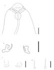
Line drawings of the paratype (CNHE 12576) of Protomicrocotyle eamsae sp. nov. (A) Details of the anterior region, ventral view. Bar 50 μm; (B) Lateral anchors. Bar 16 μm; (C) Median anchors. Bar 16 μm; (D) Hooks. Bar 16 μm.

(A) Reproductive organs of Protomicrocotyle eamsae sp. nov. (CNHE 12575) Bar 50 μm; (B) Egg with two long polar filaments (Paratype CNHE 12576). Bar 50 μm.

Light micrographs of the paratype (HWML 218464) of Protomicrocotyle eamsae sp. nov. (A) Complete body, ventral view. Bar 250 μm; (B) Details of the prohaptor, ventral view. Bar 100 μm; (C) Body trunk, ventral view. Bar 100 μm; (D) Details of the haptoral lappet. Bar 25 μm. The arrows indicate the slightly lobed lateral ends of the haptoral lappet. Mayer’s Carmalum stain.

Light micrographs of the paratype (HWML 218465) of Protomicrocotyle eamsae sp. nov. (A) Anterior region, ventral view. Bar 100 μm; (B) Details of the masculine copulatory organ and vaginal vestibule. Bar 50 μm; (C) Aperture oral and pseudo-suckers. Bar 50 μm; (D) Clamp. Bar 50 μm; (E) Lateral anchor. Bar 50 μm. Mayer's Carmalum stain.
Measurements (in µm) of haptoral armature (anchors, and hooks) of species of Protomicrocotyle from the Gulf of Mexico.
Body (Figure 2A, 5A). Body fusiform; anterior end conical. Body length (including the haptor) 1135–1879 (1682 ± 213, n = 8); width 390–622 (474 ± 72, n = 8) (Figure 2A, 5A, 5C). Tegument with striations along body surface, imparting a textured appearance, more obvious in the anterior region (Figures 2A, 5A, 6A). Paired prohaptoral suckers anterolateral, septate, muscular, oblique, and very shallowly dolliform; right sucker 36–50 (42 ± 6, n = 8) long, 23–30 (27 ± 2, n = 8) wide; left sucker 37–53 (43 ± 5, n = 8) long, 24–31 (28 ± 3, n = 8) wide (Figures 2A, 3A, 5A, 5B, 6A, 6C). Aperture oral ventral, subterminal, finely ovoid, 25–29 (26 ± 2, n = 6) long, 20–28 (24 ± 3, n = 6) wide. Glandomuscular organ (sensuBravo-Hollis, 1966) present (Figures 3B, 5A, 5B, 6A, 6C). Pre-pharynx absent; pharynx broadly elliptoid, muscular, 30–51 (41 ± 5, n = 8) long, 38–48 (41 ± 3, n = 8) wide (Figures 2A, 3A, 6A, 6C). Esophagus 320 (n = 1) long and 20 (n = 1) wide, with lateral diverticula. Cecal bifurcation posterior to male copulatory organ (MCO), 470 (n = 1) from anterior end of worm. Ceca lateral to midline, extend posteriorly to haptoral region but failing to reach haptoral lappet; cecal diverticula present that extend to margins of body (Figures 2A, 5A).
Haptor (Figures 2A, 5A, 5D). Haptor asymmetrical, armed with a row of four gastrocotylid-type clamps, each having a short muscular peduncle; haptoral groove between first and second clamps (Figures 5A, 2A, 6D). Haptor 245–430 (334 ± 55, n = 8) long, 275–350 (307 ± 30, n = 8) wide at the level of second clamp (Figure 5A). Clamps similar in size: first clamp 29–50 (39 ± 9, n = 6) long by 36–48 (42 ± 5, n = 6) wide; second clamp 28–44 (33 ± 4, n = 5) long by 41–48 (45 ± 3, n = 5) wide; third clamp 28–49 (40 ± 9, n = 6) long by 37–53 (46 ± 6, n = 6) wide; and fourth clamp 28–47 (39 ± 7, n = 7) long by 37–55 (45 ± 8, n = 7) wide (Figure 2A, 6D). Haptoral lappet transversely elongate with slightly lobed lateral ends (Figures 2A, 5D); right lateral length 72–113 (100 ± 6, n = 7), left lateral length 90–126 (105 ± 12, n = 7), and central length 82–131 (114 ± 16, n = 7). Haptoral lappet 295–375 (337 ± 24, n = 8) wide, armed with two pairs of anchors and one pair of hooks (Figures 3B, 3C, 3D, 5D, 6E; Table 2).
Male reproductive structures (Figures 2B, 5B, 6A, 6B). Testes intercecal, 27–34 (31± 3, n = 6) in number, very shallowly doliform in shape (horizontally elongated), irregular in size, preovarian, arranged in two fields, each with a single row of testes. Testes 23–43 (34 ± 9, n = 8) long and 66–109 (89 ± 14, n = 8) wide (Figures 2A, 5A, 5C); length-width ratio L:W = 1: 1.8. Vas deferens dorsal to testes, goes in the direction of the anterior region; in the part anterior to the testicular region, widest in region between testicular region and cecal bifurcation, coiled, anterior to cecal bifurcation, narrow and straight to connection with MCO (Figures 2A, 5A). Male copulatory organ muscular, subspherical, 37–52 (45 ± 6, n = 8) long, 40–50 (44 ± 4, n = 8) wide, armed with 18–26 (24 ± 2, n = 8) spines (Figures 2B, 5A, 5B, 6A, 6B). Spines of MCO hooklike, 25–31 (28 ± 2, n = 8) long, 2–4 (3 ± 1, n = 8) wide, arranged in a circle with tips directed outward (Figures 2B, 2C, 6A, 6B). Genital atrium ventral, near midline, anterior to cecal bifurcation, opening 190–350 (257 ± 48, n = 8) from anterior end of body (Figures 2A, 5A, 5B, 6A), subspherical, with muscular edges and without any sclerotized structures.
Female reproductive structures (Figures 2A, 2D, 4, 6A, 6B). Germarium intercecal, post-testicular, comprised of oval germarial bulb with irregular edges, containing immature oocytes, 48–104 (74 ± 20, n = 6) long, 60–103 (80 ± 15, n = 6) wide (Figure 4A). Ascending duct reaches anteriorly past vitelline duct, bent slightly to the right (Figure 5A) or left (Figures 2A, 4A) side, forming an inverted U that crosses the ascending duct to descend towards the oötype (Figures 2A, 4A, 5A). Vaginal duct on left side, ventral to common vitelline duct, seminal receptacle dorsal to common vitelline duct; ducts connect to oötype. Genito-intestinal canal observed (Figure 4A). Uterus ascends from oötype to the genital atrium (Figure 4A). Vaginal pore ventral, anterior to vaginal vestibule. Vaginal vestibule 54–88 (75 ± 10, n = 8) long, 44–58 (51 ± 5, n = 8) wide (Figures 2A, 2B, 5A, 5B, 6A, 6B), located 270–445 (346 ± 56, n = 8) from anterior end of body and 70–180 (120 ± 24, n = 8) from MCO. Vaginal vestibule lateral to midline, on the side opposite to haptoral clamps (Figures 2A, 2D, 5A, 5B, 6A, 6B). Vaginal vestibule armed with 43–58 (52 ± 4, n = 6) spines. Spines 23–26 (25 ± 1, n = 8) long, 2–4 (2, n = 14) wide (Figure 2E). Spines flattened, variable in length with basal spines smallest, uppermost spines largest. Each spine with small needle-like spikes at the distal end (Figures 2A, 2D, 2E, 5A, 5B, 6A, 6B). Vaginal duct descends from vaginal vestibule to germarium; small elongate seminal receptacle dorsal to common vitelline duct, observed in some specimens, not measured (Figure 4A). Vitelline glands in two lateral fields, starting just posterior to cecal bifurcation, overlapping ceca anteriorly and posteriorly, reaching lateral margin of testes, uniting posterior to germarium, extending to posterior region of body but not into haptoral lappet (Figure 2A). Vitellogenic ducts unite dorsal to anterior part of germarium, to form common vitelline duct that is connected to oötype (Figures 4A, 5A).
Eggs (Figure 4B). Some specimens with single egg in uterus. Eggs eliptoid, with two polar filaments; length, not including polar filaments, 136–166 (155 ± 9, n = 4) long, 42–53 (48 ± 5, n = 4) wide. Anterior filament (as positioned in uterus) 209 (n = 1) long by 5 (n = 1) wide; posterior filament 260 (n = 1) long by 6 (n = 1) wide.
Taxonomic summary
Type Host:Caranx hippos (Linnaeus, 1766) (Carangidae).
Common name: Crevalle jack.
Site of infection: Gills filaments.
Type locality: Littoral waters of the Gulf of Mexico, off Tecolutla, Veracruz (20°28′39″N; 97°00′30″W) (Figure 1).
Specimens deposited: Holotype CNHE 12575; Paratypes CNHE 12576; HWML 218464–218466; CHE-P00155.
ZooBank registration: BB2B1819-B0E0-4FBD-BBE3-D59BB85FEBFF.
Genbank accession numbers: mtDNA: PZ380651 to PZ380654. 28S rDNA: PZ377455–PZ377458.
Etymology: The species is named in honor of Dra. Elizabeth A. Martínez-Salazar (Unidad Académica de Ciencias Biológicas, Universidad Autónoma de Zacatecas, Zacatecas, Mexico) for her contributions to the academic training of the first author, her contributions to the fields of Helminthology and Molecular Systematics, and her friendship.
Remarks
Morphological characteristics, including the number of testes, number and shape of the spines on the male copulatory organ, and the number of vaginal spines (Bravo-Hollis, 1966; Kritsky et al., 2011; Pillai & Pillai, 1978; Ramalingam, 1960; Wahl, 1972; Yamaguti, 1953), as well as the body size and shape, and the arrangement and shape of the testes (Kritsky et al., 2011; Ramírez-Cruz et al., 2023), have been employed as diagnostic characters for the differentiation of species of Protomicrocotyle. These and other characters indicate that the new species should be assigned to Protomicrocotyle and are useful for distinguishing the new species from other known members of the genus.
The testes of P. eamsae sp. nov. are arranged in two fields, as in P.mirabilis (Kritsky et al., 2011; Ramírez-Cruz et al., 2023), P. carangis (Pillai & Pillai, 1978) and P.mannarensis (Ramalingam, 1960); The testes of P. veracruzensis, P. minutum, and P.nayaritensis are arranged in rows with 1–2 adjacent testes on each side (Bravo-Hollis, 1966; Ramalingam, 1960), in P. madrasensis they appear to be in row with 2 adjacent testes on each side (Ramalingam, 1960), and those of P. ivoriensis have rows with 1–3 adjacent testes (Wahl, 1972). In the new species, the testes are shallowly doliform (horizontally elongated) (Figures 2A, 5A, 5C) and in P.mirabilis, the testes are very broadly eliptoid (Kritsky et al., 2011; Ramírez-Cruz et al., 2023). Furthermore, the new species has fewer testes (31, in two fields) than P.nayaritensis (46–48, in a single field) (Bravo-Hollis, 1979).
The body of P. eamsae sp. nov. is shorter in length (1682) than most other species of Protomicrocotyle reported from the coasts the Eastern Pacific Ocean and Gulf of Mexico (Table 3); the only species that is similar in size is P.minutum (1500), from India (Ramalingam, 1960).
Protomicrocotyle eamsae sp. nov. has fewer spines on the MCO and more spines on the vaginal vestibule than P. manteri (24 vs. 33–38 and 52 vs. 15, respectively) (Bravo-Hollis, 1966) (Table 3). The new species and P.nayaritensis are similar to P. manteri in having an average of 24 spines in the MCO and is different from P.nayaritensis, which has 48–54 spines in the MCO. Additionally, the new species, with 52 spines in the vaginal vestibule, can be distinguished from P.nayaritensis, which has 22–46 spines in the vaginal vestibule.
The testes of each field of P. eamsae sp. nov. are arranged in a single row, while in P.veracruzensis, the testes of each field occasionally have one or two smaller-sized testes interspersed between the larger testes to form an irregular column, rather than having all in a single row (Ramírez-Cruz et al., 2023). As well, the new species is shorter in body length than P.veracruzensis (1682 vs. 3858, respectively) and the new species has 31 testes and P.veracruzensis has 47 (Table 3).
Finally, this new species can be distinguished from P. mirabilis, P.veracruzensis, P.nayaritensis and the second new species described below by the presence of the retractile glandulomuscular organ in the prohaptor, which has only been documented in P.manteri from the Pacific Ocean (Bravo-Hollis, 1966). Additionally, it can be distinguished by the slightly lobed lateral ends of the haptoral lappet (Figures 2A, 5A, 5D).
Protomicrocotylepritchardae sp. nov.
Description (Figures 7 –10; Tables 2, 3). Based on 11 specimens, stained, and mounted to be viewed from ventral side of the body. Some comparative measurements and data of other species are presented in Tables 2 and 3.

Line drawings of the holotype (CNHE 12577) of Protomicrocotyle pritchardae sp. nov. from the gills of Caranx hippos. (A) Complete body, ventral view. Bar 100 μm; (B) Anterior region. Bar 50 μm; (C) Masculine copulatory organ. Bar 100 μm; (D) Spine of MCO. Bar 100 μm; (E) Vaginal vestibule. Bar 100 μm; (F) Spine of vaginal vestibule. Bar 25 μm; (G) Clamp with muscular peduncle. Bar 32 μm.

(A) Reproductive organs of Protomicrocotyle pritchardae sp. nov. (CNHE 12577). Bar 100 μm; (B) Egg with two long polar filaments (paratype CNHE 12578). Bar 50 μm; (C) Lateral anchor (CNHE 12578). Bar 16 μm; (D) Median anchor (CNHE 12578). Bar 16 μm; (E) Hook (CNHE 12578). Bar 16 μm.

Light micrographs of the paratype (CNHE 12578) of Protomicrocotyle pritchardae sp. nov. (A) complete body, ventral view. Bar 500 μm; (B) Prohaptor. Bar 100 μm; (C) Details of the masculine copulatory organ and vestibule vaginal. Bar 100 μm. Delafield’s hematoxylin stain.

Light micrographs of the paratype (CNHE 12578) of Protomicrocotyle pritchardae sp. nov. (A) Body trunk. Bar 200 μm; (B) Haptor. Bar 200 μm; (C) Clamps. Bar 50 μm; (D) Median anchor. Bar 20 μm.
Body (Figures 7A, 9A). Body fusiform; anterior end conical. Body length 3721–4380 (4117 ± 246, n = 11) including the haptor; width 488–573 (537 ± 27, n = 11) (Figures 7A, 9A). Tegument with light cuticular striations, mainly in the middle and posterior regions of the body. Paired prohaptoral suckers in the anterior region, septate, muscular, oblique, and very shallowly dolliform; right pseudo-sucker 42–59 (52 ± 5, n = 11) long, 40–48 (43 ± 2, n = 11) wide; left pseudo-sucker 47–60 (54 ± 5, n = 11) long, 42–55 (44 ± 2, n = 11) wide (Figures 7B, 9B). Aperture oral ventral, subterminal, finely ovoid, 31–35 (34 ± 1, n = 3) long, 25–59 (42 ± 10, n = 8) wide (Figures 7B, 9B). Glandomuscular organ absent. Pre-pharynx absent; pharynx broadly elliptoid, muscular, 30–55 (45 ± 6, n = 14) long, 40–48 (43 ± 3, n = 14) wide (Figures 7B, 9B). Esophagus 605–765 (663 ± 46, n = 10) long, 20–30 (24 ± 3, n = 10) wide, with diverticula (Figures 7A, 9A, 10A). Cecal bifurcation posterior to MCO, 775–920 (846 ± 51, n = 10) from anterior end (Figures 7A, 9A). Ceca lateral to midline, extending posteriorly to haptoral region but failing to reach haptoral lappet; cecal diverticula present that extend to margins of body (Figures 7A, 9A).
Haptor (Figures 7A, 9A, 10B, 10C, 10D). Haptor asymmetrical, armed with a row of four gastrocotylid-type clamps, each with a short muscular peduncle; haptoral groove between first and second clamps (Figures 7A, 7G, 9A, 10B). Haptor 655–835 (726 ± 52, n = 10) long, 410–470 (438 ± 19, n = 10) wide at the level of second clamp (Figures 9A, 10B). Clamps are similar in size: first clamp 36–47 (41 ± 3, n = 8) long by 49–58 (54 ± 3, n = 8) wide; second clamp 42–50 (46 ± 3, n = 8) long by 54–62 (41± 5, n = 10) wide; third clamp 41–55 (46 ± 4, n = 10) long by 54–65 (59 ± 3, n = 10) wide; and, fourth clamp 40–56 (48 ± 6, n = 9) long by 54–62 (59 ± 3, n = 8) wide (Figure 7G, 10C). Haptoral lappet transversely elongated (Figures 7A, 10B); right lateral length 145–187 (174 ± 12, n = 10), left lateral length 124–198 (169 ± 21, n = 10), central length 170–220 (196 ± 14, n = 10). Haptoral lappet 645–820 (726 ± 48, n = 11) wide, armed with two pairs of anchors and one pair of hooks (Figures 8CE, -10 D; Table 2).
Male reproductive structures (Figures 7A, 7CD, -9A, 9 C, 10A). Testes intercecal, 53–67 (60 ± 5, n = 11) in number, broadly elliptoid (subspherical), irregular in size, pre-ovarian, arranged in a single field (Figures 7A, 9A, 10A). Testes 43–66 (53 ± 8, n = 11) long and 65–102 (82 ± 14, n = 11) wide (Figures 7A, 9A, 10A); length-width ratio L:W = 1:1.5. Vas deferens dorsal to testes, goes in the direction of the anterior region; in the part anterior to the testicular region, widest in region between testicular region and cecal bifurcation, coiled, posterior to cecal bifurcation, narrow and straight to connection with MCO (Figures 7A, 9A). Male copulatory organ muscular, subspherical, 59–74 (64 ± 4, n = 10) long, 43–55 (49 ± 3, n = 11) wide, armed with 13–35 (21 ± 7, n = 11) spines. Spines of MCO hooklike, 34–49 (42 ± 4, n = 11) long, 2–5 (4 ± 1, n = 11) wide, arranged in a circle on anterior part of MCO (Figures 7A, 7C, 9A, 9C). Each spine with small knob in the anterior region just posterior to curved tip, knob and curved tip directed outwards (Figures 7C, 7D). Genital atrium ventral, near midline, anterior to cecal bifurcation, opening 500–625 (552 ± 40, n = 11) from anterior end of body, subspherical, with muscular edges and without sclerotized structures (Figures 7A, 9A).
Female reproductive structures (Figures 7A, 7E, 8A, 8B, 9A, 9C). Germarium intercecal, post-testicular, comprised of germarial bulb with lobed edges, containing immature oocytes, 124–193 (157 ± 21, n = 9) long, 72–131 (102 ± 16, n = 9) wide (Figures 7A, 7E, 8A). The ascending duct extends towards the anterior region and turns to the right side forming an inverted U, then descends towards the oötype. Vaginal duct and vitelline duct connect to oötype; uterus ascends from oötype to the genital atrium (Figure 8A). Seminal receptacle and genito-intestinal canal not observed. Vaginal pore ventral, anterior to the vaginal vestibule. Vaginal vestibule 101–133 (113 ± 9, n = 11) long, 76–92 (84 ± 6, n = 11) wide (Figures 7A, 7E, 9A, 9C), located 635–755 (690 ± 46, n = 11) from anterior end of body and 150–210 (179 ± 17, n = 11) from the genital pore, lateral to midline on the side opposite to that having the haptoral clamps. Vaginal vestibule armed with 35–61 (45 ± 7, n = 11) spines. Spines 34–47 (41 ± 3, n = 11) long, 4–6 (5 ± 1, n = 11) wide; flat shape, variable in size. Basal spines smallest, uppermost spines largest. Each spine elongated in shape with small needle-like spikes at the distal end (Figure 7F). Vaginal duct descends from vaginal vestibule to germarium, simple, without seminal receptacle (Figure 7A, 8A). Vitelline glands in two lateral fields, starting just posterior to cecal bifurcation, overlapping ceca anteriorly and posteriorly and the lateral margin of the testes, uniting posterior to germarium, extending to posterior region of body but not reaching the haptoral lappet; in some specimens, clusters of glands are observed at the level of the cecal bifurcation and vaginal vestibule (Figures 7A, 9A, 9C). Vitellogenic ducts from each field unite dorsal to the ducts of the germarium, to forming common vitelline duct that connects to the oötype (Figure 8A).
Eggs (Figures 8B, 10A). Some specimens with single egg in uterus (n = 9). Eggs elliptoid, with two polar filaments, at each pole; length, not including polar filaments, 164–280 (192 ± 21, n = 9) long, 72–131 (102 ± 16, n = 9) wide. Filament in anterior end (as positioned in uterus) 125–244 (193 ± 40, n = 7) long by 6–7 (7, n = 7) wide; posterior filament 268–325 (296 ± 41, n = 2) long by 5–7 (6 ± 1, n = 2) wide (Figure 8B).
Taxonomic summary
Type Host:Caranx hippos (Linnaeus, 1766) (Carangidae).
Common name: Crevalle jack.
Site of infection: Gills filaments.
Type locality: Littoral waters of the Gulf of Mexico off Tecolutla, Veracruz (20° 28′ 39″ N; 97° 00′ 30″ W) (Figure 1).
Specimens deposited: Holotype CNHE 12577; Paratypes CNHE 12578; HWML 218467–218470; CHE-P00154.
ZooBank registration: BB2B1819-B0E0-4FBD-BBE3-D59BB85FEBFF.
Genbank accession numbers: mtDNA: PZ380648–PZ380650. 28S rDNA: PZ377459–PZ377462.
Etymology: The species is named in honor of Dra. Mary Louise Hanson Pritchard, founding curator of the H.W. Manter Laboratory, Division of Parasitology at the State Museum of the Nebraska University, for her contributions to parasitology and the HWML Museum.
Remarks
The diagnostic characters mentioned above, and other characters indicate that this new species should be assigned to Protomicrocotyle and are useful for distinguishing the new species from other known members of the genus.
Protomicrocotyle pritchardae sp. nov. differs from P.mirabilis by being longer, with a mean length of 4117 vs. 2861, respectively (Table 3). The shape of the testes is similar in both species (broadly elliptoid to very broadly elliptoid), the new species has a greater number of testes (60 vs. 31, respectively). The testes are arranged in a single field in the new species, whereas in P.mirabilis the testes are in two well-defined rows of testes (Kritsky et al., 2011; Ramírez-Cruz et al., 2023). The new species has less spines on the MCO compared to P.manteri (21 vs. 33–38) and more spines within the vaginal vestibule (52 vs. 15, respectively) (Table 3). The new species has broadly elliptoid testes, whereas P.manteri has depressed elliptoid testes (Bravo-Hollis, 1966). The new species has less spines in the MCO than P.nayaritensis (21 vs 48–54, respectively) (Table 3); the new species has 35–61 spines in the vaginal vestibule and P.nayaritensis has 22–46 (Bravo-Hollis, 1979). The new species and P.veracruzensis are similar in body length (Table 3); however, they can be differentiated by the size of the clamps (45 in the new species vs. 64 in P.veracruzensis) and the average number of testes (60 in the new species vs. 47 in P.veracruzensis). In addition, the new species has testes that are broadly elliptoid and arranged in a single field, and P.veracruzensis has testes that are mostly depressed elliptoid in shape and they are arranged in two parallel fields, each field with one or two interspersed testes, forming an irregular column (Ramírez-Cruz et al., 2023).
Finally, P. mirabilis, P.eamsae sp. nov., and P.pritchardae sp. nov. are found in sympatry as parasites of C. hippos in the same geographic area of Tecolutla, on the southwestern coast of the Gulf of Mexico. It is noteworthy that, among the fish that were examined, P.mirabilis and P.eamsae sp. nov. sometimes co-infected the same fish; whereas, P.pritchardae sp. nov. was present as a single-species infection in only three fish, without co-infection with other species of Protomicrocotyle. Whether this patter is because of the relatively small sample size of fish, or is indicative of possible separation of populations of fish is unknown.
Morphometric analyses
Based on the scree plot (Figure11A), the first two components explained 86.28% of the accumulated variance (Figures 11A, 11B; Table 4A). The curve of the steep slope stabilizes and flattened out after components three and four so they were not considered. (Figure 11A). In order of importance, the morphological variables that contributed to the first component were: the total width of the body, the width of the ovary, the width of the haptor, the distance from vaginal vestibule to genital pore, the number of testes, the right lateral length of the haptoral lappet, left lateral length of haptoral lappet, central length of the haptoral lappet, the width of haptoral lappet, and the total body length (Table 5). For the second component, the morphological variables were: the number of spines on the male copulatory organ, the average width of the testes, the length of the external root of the right lateral hook, the length of the opening of the left median hook, the width and length of the ovary (Table 5).

Morphometric analyses of the species of Protomicrocotyle. (A) Scree plot of the PCA; (B) Scatter plot of PCA; (C) Scatter plot of LDA.
The cumulative variance of the first two principal components (A) and the factor values of the linear discriminant analyses (B).
The accumulated variance demonstrated by the LDA was 80.26% (Table 4B). Seven groups were supported in the scatter plot (Figure 11C), six of which corresponded to species of Protomicrocotyle and one to Neomicrocotyle pacifica. The four species of Protomicrocotyle from the Gulf of Mexico (P. mirabilis, P. veracruzensis, P. eamsae sp. nov., and P. pritchardae sp. nov.) were more similar to each other than to those species from the Pacific Ocean: P. manteri, P.nayaritensis, and N. pacifica (Figure 11C). The specimens of Protomicrocotyle pritchardae sp. nov. and P. veracruzensis share some similar morphological characteristics (longitude of the esophagus, distance from the cecal bifurcation to the anterior end) so they are not completely separated (Figure 11C).
The original assignment of specimens to species was supported 100% CC by the original resubstituting matrix assignment of the LDA (Table 6A), but application of the cross-validation reduced the percentage of correctly assignment to 95.73% CC (Table 6B). The eight specimens of P. eamsae sp. nov. were assigned correctly; although, one of 11 specimens of P. pritchardae sp. nov. was grouped with P. veracruzensis (Table 6B). However, based on all of the analyses, the morphological comparison of the specimens, and molecular data, the specimen is correctly assigned to P. pritchardae sp. nov.
Confusion matrices for the linear discriminant analyses. (A) Original resubstituting matrix (100% CC). (B) Jackknife matrix (Cross-Validation) (95.73% CC).
Molecular analyses
Seven partial sequences of CO1 mtDNA were obtained from the new species (four from P. eamsae sp. nov. and three from P. pritchardae sp. nov.) and eight partial sequences of 28S rDNA (four from P. eamsae sp. nov. and four from P. pritchardae sp. nov.). The newly obtained partial sequences and those retrieved from GenBank® (Sayers et al., 2020) for each gene (Table 1) were aligned; the final alignment of CO1 mtDNA was the 393 base pair (bp) and that of 28S rDNA was 760 bp. The intraspecific genetic variation of CO1 mtDNA and 28S rDNA are given in Tables 66B.
Species of Protomicrocotyle from the Gulf of Mexico form a cluster (support of 100%) in the cluster analysis (Figure 12). The specimens of P.mirabilis form a group with statistical support of 100%. The second cluster is comprised of P.veracruzensis, P.eamsae sp. nov. and P.pritchardae sp. nov., with statistical support of 98% (Figure 12). The topology displayed in the NJ dendrogram for 28S rDNA is similar as that of CO1 mtDNA, but P.mirabilis and P.eamsae sp. nov. are clustered together (Figure 13) and P.veracruzensis and P.pritchardae form a second group with statistical support of 91% (Figure 13).

Neighbor Joining phenogram based on CO1 mtDNA gene sequences showing similarities between new species of Protomicrocotyle and other protomicrocotylids. Bootstrap support values calculated for 1000 replications are indicated. Accession numbers from GenBank are given.

Neighbor Joining phenogram based on 28S rDNA gene sequences showing similarities between new species of Protomicrocotyle and other protomicrocotylids. Bootstrap values calculated for 1000 replications are indicated. Accession numbers from GenBank are given.
Discussion
The two new species of Protomicrocotyle described in this study have the morphological characteristics typical of the genus, including: an asymmetrical haptor with four Gastrocotylidae-type clamps in a longitudinal row on the side opposite to the vaginal vestibule; the haptoral lappet is transversely elongate (wider than long), armed with two pairs of anchors (larger anchors positioned laterally with smaller anchors between the larger ones), and one pair of hooks (located between the smaller anchors); the testes are relatively numerous, with variations in shape, size, and arrangement, but they always are anterior to the female complex; the MCO is bulbous, may be muscular, and is provided with a crown of numerous spines; vagina opening ventrally to the right or left, posterior to genital pore, with numerous spines of different shapes; vitellaria extends lateral and dorsal to ceca and eggs with filament at each pole (Bravo-Hollis, 1966; Bravo-Hollis, 1979; Kritsky et al., 2011; Lebedev, 1986; Ramalingam, 1960; Ramírez-Cruz et al., 2023; Wahl, 1972; Yamaguti, 1953).
Descriptions of monogeneans that include morphological data and genetic analyses has become a common practice (Kritsky & Martin, 2023; Rahmouni et al., 2023; Ramírez-Cruz et al., 2023). Both types of data individually have specific limitations, but together they provide more accurate taxonomic support. The results of the multivariate analyses allowed differentiation and separation of the specimens: Protomicrocotyle eamsae sp. nov., and P. pritchardae sp. nov., collected in C. hippos from the locality of Tecolutla, Veracruz from each other and from other specimens collected in the Gulf of Mexico. The main morphological characteristics that separate them are the length of the body, with Protomicrocotyle eamsae sp. nov., being smaller (1682) than P. pritchardae sp. nov. (4117) and the number of testes (39 vs. 60), the shape (elongated horizontally vs. subspherical), and the arrangement of the testes (two fields, with a row in each field vs. a single field). The aforementioned morphological characteristics, when considered in conjunction with the other measured variables (Tables 2, 3) and the results of the statistical analyses (Figures 11A, 11B, 11C; Tables 4, 5), support the separation of these specimens into distinct groups, each of then differentiated from the rest of the species under study (Figures 11A, 11B).
In this study, a novel partial sequence of CO1 mtDNA and 28S rDNA were generated for species within Protomicrocotyle. The partial sequences of each gene were employed in the analyses of genetic distance and for the analysis of phylogenetic inference using Neighbor Joining, based on genetic distances (Table 1). The newly-generated sequences provided new information about the intra- and interspecific variation of this group of monogeneans. The degree of intraspecific variation observed in the CO1 mtDNA between P.mirabilis was 1.03%–2.01% (Ramírez-Cruz et al., 2023), P.veracruzensis was 0.25%–0.75% (Ramírez-Cruz et al., 2023), P. eamsae sp. nov. was 0.0% –0.25%, and P. pritchardae sp. nov. was 0.0%–0.26% (Table 7A). These values are relatively low in comparison to those observed within other species of Microcotylidae: for example, 4.5% (Mladineo et al., 2009), 4%–7% (Farjallah et al., 2023), and in Mazocraeidae, 5.6% (Yan et al., 2016).
Percentage of average genetic variation (uncorrected p-distances), based on cytochrome b partial sequences (A) and the conserved ribosomal regions of the 28S rDNA data (B), observed within and between pairs of Protomicrocotyle eamsae sp. nov., and Protomicrocotyle pritchardae sp. nov., and other species of Protomicrocotylidae and Allodiscocotylidae included in the analyses.
The degree of interspecific variation between P. mirabilis and P. veracruzensis is high (9.21%–10.53%), but the difference between P. veracruzensis and P.pritchardae sp. nov. are medium low (1.02%–1.53%). In contrast, the degree of variation between P.mirabilis and P.eamsae sp. nov. is relatively high (7.61%–8.65) (Table 7A). The degree of interspecific variation between P.eamsae sp. nov. and P.pritchardae sp. nov. is 4.33%–4.85% (Table 7A). However, lower levels of interspecific variation have been reported between species of Microcotyle, with figures of 0.7% (Lablack et al., 2022), 4.5% (Ayadi et al., 2017; Víllora-Montero et al., 2020), and 6.1% (Ono et al., 2020).
The phenogram of NJ revealed that the genetic sequences of P.mirabilis, P.veracruzensis, P.eamsae sp. nov. and P.pritchardae sp. nov. form a cluster with high statistical support (Figure 12). Kamio & Nitta (2022) included the analysis of partial CO1 mtDNA sequences of the species Bilaterocotyle madrasensis Radha, 1966 and Bilaterocotyloides carangis Ramalingam, 1961 of the family Protomicrocotylidae, although the relationships remain unresolved in their Bayesian inference tree. When the data is available, it would be advisable to incorporate additional sequences and undertake a more inclusive phylogenetic analysis of these families.
Intraspecific variation in 28S rDNA of P. mirabilis was 0% to 0.14% (Ramírez-Cruz et al., 2023); P.veracruzensis was 0% (Ramírez-Cruz et al., 2023), P.eamsae sp. nov. were 0% to 0.13%, and Protomicrocotyle pritchardae sp. nov. was 0% (Table 7B). The sequences of 28S rDNA of species of Protomicrocotyle were grouped together with high support values (96%) (Figure 13). The combination of genetic sequences, morphological comparisons and morphometric analyses provides supplementary information that facilitates the differentiation of taxa. In the present study, the data obtained from both markers is supplementary to the morphological data, thereby corroborating the distinction of the two novel species of Protomicrocotyle.
Comparative sequence analyses of the species of monogeneans showed that both partial CO1 mtDNA and 28S rDNA genes can be successfully used for species identification (Kamio et al., 2023; Pinacho-Pinacho et al., 2021; Rahmouni et al., 2017; Ramírez-Cruz et al., 2023; Torres-Carrera et al., 2020; Zago et al., 2021) and for phylogenetic inference (Justine et al., 2013; Kamio & Nitta, 2022; Tambireddy et al., 2016). The available molecular sequence information for species of Mazocraeidea is insufficient, necessitating the accumulation of molecular studies for phylogenetic analysis with morphological and molecular data (Tambireddy et al., 2016). In Protomicrocotylidae, data is available only for Bilaterocotyle madrasensis, Bilaterocotyloides carangis, Neomicrocotyle pacifica, Lethacotyle vera, Protomicrocotyle mirabilis, P. veracruzensis, P.eamsae sp. nov., and P.pritchardae sp. nov., and Neomicrocotyle sp. This information about species of Protomicrocotyle was produced as a result of Ramírez-Cruz et al. (2023) and this study (Table 1).
Members of the family of Carangidae are usual hosts for monogenean parasites (Martínez-Flores et al., 2023; Montoya-Mendoza et al., 2017, 2021; Ramírez-Cruz et al., 2023; Vianna et al., 2020; Violante-González et al., 2019). Several species of Protomicrocotylidae commonly infect the gills of carangid fish (Bravo-Hollis, 1966, 1979, 1989; Caballero y Caballero & Bravo-Hollis, 1965; Kritsky et al., 2011; Mendoza-Garfias et al., 2017; Ramírez-Cruz et al., 2023; Yamaguti, 1963). In the Gulf of Mexico, the most common species of Caranx are: C. hippos, C. crysos (Mitchill, 1815), and C. latus (López-Herrera et al., 2021; Nelson et al., 2016). Caranx hippos was considered to have been sufficiently surveyed throughout much of its distribution range (Kritsky et al., 2011), but the findings of Ramírez-Cruz et al. (2023) and this study, have shown that there is much more to learn from the parasitofauna of this species of host.
This work adds two new species to the list of protomicrocotylids from Mexico and the increases the record of species of monogeneans from C.hippos to 19 species (including the two new species): Axine sp., Ahpua piscicola Caballero y Caballero & Bravo-Hollis, 1973; Allopyragraphorus caballeroi (Zerecero, 1960) Yamaguti, 1963; All. hippos (Hargis, 1956) Yamaguti, 1963; All. winteri (Caballero y Caballero & Bravo-Hollis, 1965) Bravo-Hollis & Salgado-Maldonado, 1983; Cemocotyle carangis (MacCallum, 1913) Sproston, 1946 (Mendoza-Garfias et al., 2017); Ce. noveboracensis Price, 1962 (Luque & Alves, 2001; Mendoza-Garfias et al., 2017); Cemocotylella elongata (Meserve, 1938) Price, 1962 (Mendoza-Garfias et al., 2017); Lethacotyle sp. (Odum & Amuzie, 2021); Neomicrocotyle pacifica; Pseudomazocraes monsivaisae Caballero y Caballero & Bravo Hollis, 1955; Ps. riojai (Caballero y Caballero & Bravo-Hollis, 1963) Lebedev, 1970 (Caballero y Caballero & Bravo-Hollis, 1963); Ps. selene Hargis, 1957, P. mirabilis (Kritsky et al., 2011; Ramírez-Cruz et al., 2023); (Kritsky et al., 2011; Ramírez-Cruz et al., 2023) P. veracruzensis (Ramírez-Cruz et al., 2023); Salinacotyle mexicana (Caballero y Caballero & Bravo-Hollis, 1963) Lebedev, 1984; and Zeuxapta seriolae (Meserve, 1938) Price, 1962 (Mendoza-Garfias et al., 2017). To this list, P. eamsae sp. nov. and, P.pritchardae sp. nov. can now be added.
In the past, C. hippos was considered to be distributive in both the Pacific and Atlantic Oceans (Torres-Orozco & Pérez-Hernández, 2009). The distribution of C. hippos is now recognized as being restricted to the Atlantic Ocean. Caranx fischeri Smith-Vaniz & Carpenter, 2007 has a distribution in the Eastern Atlantic, the Mediterranean, and Ascension Island. Caranx caninus and C. caballus Günther, 1868 are distributed along the Eastern Pacific costs of North and South America (Mair et al., 2012; Smith-Vaniz & Carpenter, 2007). The original host of Ah. piscicola, All. caballeroi, N. pacifica, Ps. monsivaisae, Ps. riojai and S. mexicana were reported as C. hippos in Pacific waters; these species are now considered to be parasites of either C. caballus or C. caninus (Pulido-Flores et al., 2015), but the original reports have not been brought up-to-date.
Acknowledgements
The authors are grateful to the local fishermen from Tecolutla, Veracruz for providing the fish that were examined for this study. We thank Erick A. Rodríguez Ibarra and Ilse A. Rodríguez-Rivera, for their help in fieldwork. Special thanks to Luis García-Prieto for the loan of specimens from CNHE, Scott Gardner for the loan of specimens from HWML and Laura Márquez and Nelly López, LaNaBio-UNAM for their assistance in the generation of DNA sequences. We are grateful to MSc Vincent Romero Hernández (UAEH) for support with the digital microscope and the capture of the photographs. A doctoral scholarship was awarded to ESR-C by the Secretaría de Ciencia, Humanidades, Tecnología e Innovación (SECIHTI) (No. CVU: 857903). The authors would like to thank the anonymous reviewers for their valuable comments to improve the manuscript.
Data availability
The data supporting the findings of this study will be available upon request.
-
How to cite:
Ramírez-Cruz ES, Monks S, Manríquez-Morán NL, Pulido-Flores G. Two new species of Protomicrocotyle Johnston & Tiegs, 1922 (Monogenea: Protomicrocotylidae) parasites of Caranx hippos (Linnaeus, 1776) (Carangiformes: Carangidae), from Tecolutla, Veracruz, Mexico. Rev Bras Parasitol Vet 2026; 35(3): e002726. https://doi.org/10.1590/S1984-29612026035.
-
Financial support
A doctoral scholarship was awarded to ESR-C by the Secretaría de Ciencia, Humanidades, Tecnología e Innovación (SECIHTI) (No. CVU: 857903).
-
Ethics declaration
This work was conducted as part of the UAEH-DIDI-DI-ICBI-BIO-2024-046 project, registered with the Division of Research, Development, and Innovation at UAEH and approved by the Institutional Committee for the Care and Use of Laboratory Animals (CICUAL-V-I/19/2026).
References
- Alejos-Velázquez LP, Aragón-Martínez M, Cornejo-Romero A. Extracción y purificación de ADN. In: Cornejo-Romero A, Serrato-Díaz A, Rendón-Aguilar B, Rocha-Munive G, editors. Herramientas moleculares aplicadas en ecología: aspectos teóricos y prácticos México, Distrito Federal: Secretaría de Medio Ambiente y Recursos Naturales; 2014. p. 1-26, vol. 1.
-
Ayadi ZEM, Gey D, Justine J-L, Tazerouti F. A new species of Microcotyle (monogenea: Microcotylidae) from Scorpaena notata (Teleostei: Scorpaenidae) in the Mediterranean Sea. Parasitol Int 2017; 66(2): 37-42. https://doi.org/10.1016/j.parint.2016.11.004 PMid:27840197.
» https://doi.org/10.1016/j.parint.2016.11.004 - Boada M, Bashirullah A, Marcano J, Alió J, Vizcaíno G. Estructura comunitaria de ectoparásitos en branquias del jurel Caranx hippos (Linnaeus, 1776) en Santa Cruz y Carúpano, Estado Sucre, Venezuela. Rev Cient 2012; XXII(3): 259-272.
-
Bowles J, Hope M, Tiu WU, Liu X, McManus DP. Nuclear and mitochondrial genetic markers highly conserved between Chinese and Philippine Schistosoma japonicum. Acta Trop 1993; 55(4): 217-229. https://doi.org/10.1016/0001-706X(93)90079-Q PMid:8147278.
» https://doi.org/10.1016/0001-706X(93)90079-Q -
Brabec J, Salomaki ED, Kolísko M, Scholz T, Kuchta R. The evolution of endoparasitism and complex life cycles in parasitic platyhelminths. Curr Biol 2023; 33(19): 4269-4275.e3. https://doi.org/10.1016/j.cub.2023.08.064 PMid:37729914.
» https://doi.org/10.1016/j.cub.2023.08.064 - Bravo-Hollis M. Helmintos de peces del Pacífico mexicano XXV. Descripción de tres monogéneos del Golfo de California. An Inst Biol Univ Nac Mex 1966; 37(1-2): 107-123.
- Bravo-Hollis M. Monogéneos de peces de la colección Winter II. Sobre un protomicrocotylido nuevo de Caranx hippos caninus Gunther del Pacífico mexicano. An Inst Cienc Mar Limnol 1979; 6(1): 189-196.
- Bravo-Hollis M. Monogenea (Van Beneden, 1858) Carus 1863, de peces del Golfo de México y del mar Caribe XII. Nuevas localidades de colecta de especies conocidas de gastrocotilidos. An Inst Biol Univ Nac Auton Mex Ser Zool 1989; 59(1): 1-14.
- Buchmann K, Bresciani J. Monogenea (phylum Platyhelminthes). In: Woo PTK, editor. Fish diseases and disorders 2nd ed. Wallingford, UK: Cabi; 2006. p. 297-344. (Protozoan and Metazoan Infections; vol. 1).
- Caballero y Caballero E, Bravo-Hollis M. Helmintos de peces de aguas mexicanas del Pacifico. XXIII. Descripción de cuatro nuevos monogéneos y una breve consideración sobre nomenclatura de esta clase. An Inst Biol Univ Nac Auton Mex Ser Zool 1963; 34(2): 163-203.
- Caballero y Caballero E, Bravo-Hollis M. Monogenea (van Beneden, 1858) Carus, 1863 de peces marinos del litoral mexicano del Golfo de México y del Mar Caribe. II. Rev Biol Trop 1965; 13(1): 101-121.
- Caballero y Caballero E, Bravo-Hollis M. Monogenea (van Beneden, 1858) Carus, 1863, de peces marinos del litoral mexicano del Golfo de México y del Mar Caribe. III. An Inst Biol Univ Nac Auton Mex Ser Zool 1967; 38: 27-34.
-
Caira JN, Littlewood DTJ. Worms, Platyhelminthes. In: Levin SA, editor. Encyclopedia of biodiversity 2nd ed. Waltham: Academic Press; 2013. p. 437-469. https://doi.org/10.1016/B978-0-12-384719-5.00166-0
» https://doi.org/10.1016/B978-0-12-384719-5.00166-0 -
Clopton RE. Standard nomenclature and metrics of plane shapes for use in gregarine taxonomy. Comp Parasitol 2004; 71(2): 130-140. https://doi.org/10.1654/4151
» https://doi.org/10.1654/4151 -
Farjallah S, Amor N, Garippa G, Montero FE, Víllora-Montero M, Mohamed OB, et al. Genetic variation of Sparicotyle chrysophrii (Monogenea: Microcotylidae) from the gilthead sea bream Sparus aurata (Teleostei: Sparidae) in the Mediterranean Sea. Parasitol Res 2023; 122(1): 157-165. https://doi.org/10.1007/s00436-022-07709-y PMid:36418649.
» https://doi.org/10.1007/s00436-022-07709-y -
García-Prieto L, Mendoza-Garfias B, Pérez-Ponce de León G. Biodiversidad de Platyhelminthes parásitos en México. Rev Mex Biodivers 2014; 85(Suppl): S164-S170. https://doi.org/10.7550/rmb.31756
» https://doi.org/10.7550/rmb.31756 -
Gascuel O, Steel M. Neighbor-Joining revealed. Mol Biol Evol 2006; 23(11): 1997-2000. https://doi.org/10.1093/molbev/msl072 PMid:16877499.
» https://doi.org/10.1093/molbev/msl072 - Hammer Ø, Harper DAT, Ryan PD. PAST: paleontological statistics software package for education and data analysis. Palaeontol Electronica 2001; 4(1): 1-9.
-
Hargis WJ Jr. Monogenetic trematodes of Gulf of Mexico fishes. Part XIII. The family Gastrocotylidae Price, 1943 (Continued). Trans Am Microsc Soc 1957; 76(1): 1-12. https://doi.org/10.2307/3223916
» https://doi.org/10.2307/3223916 - Iruegas-Buentello FJ. Helmintos parásitos de peces comercilaes de la Laguna Madre, San Fernando, Tamaulipas, México [tesis]. Monterrey, Nuevo León: Universidad Autónoma de Nuevo León; 1999.
-
Justine J-L. Cladistic study in the Monogenea (Platyhelminthes), based upon a parsimony analysis of spermiogenetic and spermatozoal ultrastructural characters. Int J Parasitol 1991a; 21(7): 821-838. https://doi.org/10.1016/0020-7519(91)90151-V
» https://doi.org/10.1016/0020-7519(91)90151-V -
Justine J-L. Phylogeny of parasitic Platyhelminthes: a critical study of synapomorphies proposed on the basis of the ultrastructure of spermiogenesis and spermatozoa. Can J Zool 1991b; 69(6): 1421-1440. https://doi.org/10.1139/z91-203
» https://doi.org/10.1139/z91-203 -
Justine J-L, Rahmouni C, Gey D, Schoelinck C, Hoberg EP. The monogenean which lost its clamps. PLoS One 2013; 8(11): e79155. https://doi.org/10.1371/journal.pone.0079155 PMid:24278118.
» https://doi.org/10.1371/journal.pone.0079155 -
Kamio Y, Inoue A, Nitta M. Description of a new species, Pseudodiscocotyla mikiae n. sp. (Monogenea: Discocotylidae) parasitic on gills of Pristipomoides filamentosus from off Okinawa-jima island in Japan, with redescription of Pseudodiscocotyla opakapaka. Syst Parasitol 2023; 100(6): 657-671. https://doi.org/10.1007/s11230-023-10115-2 PMid:37796361.
» https://doi.org/10.1007/s11230-023-10115-2 -
Kamio Y, Nitta M. New Records of Gemmaecaputia corrugata (Monogenea: Chauhaneidae) from Sphyraena forsteri (Sphyraenidae) off Yomitan Town, Okinawa-jima Island, Southern Japan. Species Divers 2022; 27(1): 83-90. https://doi.org/10.12782/specdiv.27.83
» https://doi.org/10.12782/specdiv.27.83 -
Kearn GC. Some aspects of the biology of Monogenean (Platyhelminth) parasite of marine and freshwater fishes. Oceanography 2014; 2(1): 1. https://doi.org/10.4172/2332-2632.1000117
» https://doi.org/10.4172/2332-2632.1000117 -
Kluge AG. Total evidence or taxonomic congruence: cladistics or consensus classification. Cladistics 1998; 14(2): 151-158. https://doi.org/10.1111/j.1096-0031.1998.tb00328.x PMid:34902924.
» https://doi.org/10.1111/j.1096-0031.1998.tb00328.x -
Kluge AG. On total evidence: for the record. Cladistics 2004; 20(2): 205-207. https://doi.org/10.1111/j.1096-0031.2004.00020.x PMid:34892935.
» https://doi.org/10.1111/j.1096-0031.2004.00020.x -
Kritsky DC, Martin SB. Kannaphallus raphidium n. sp. (Monogenoidea: Mazocraeidea: Heteraxinidae) parasitic on the gill lamellae of the golden trevally Gnathanodon speciosus (Carangiformes: Carangidae) occurring in the coastal waters of Queensland and Western Australia. J Parasitol 2023; 109(2): 96-106. https://doi.org/10.1645/22-116 PMid:37036939.
» https://doi.org/10.1645/22-116 -
Kritsky DC, McAleese WJ, Bakenhaster MD. Heteronchoineans (Monogenoidea) from the Gills of Crevalle Jack, Caranx hippos Perciformes, Carangidae), from Everglades National Park, Florida, with a Redescription of Protomicrocotyle mirabilis (Gastrocotylinea, Protomicrocotylidae). Comp Parasitol 2011; 78(2): 265-274. https://doi.org/10.1654/4505.1
» https://doi.org/10.1654/4505.1 -
Lablack L, Rima M, Georgieva S, Marzoug D, Kostadinova A. Novel molecular data for monogenean parasites of sparid fishes in the Mediterranean and a molecular phylogeny of the Microcotylidae Taschenberg, 1879. Curr Res Parasitol Vector Borne Dis 2022; 2: 100069. https://doi.org/10.1016/j.crpvbd.2021.100069 PMid:36589867.
» https://doi.org/10.1016/j.crpvbd.2021.100069 - Lamothe-Argumedo MR. Manual de técnicas para preparar y estudiar los parásitos de animales silvestres Ciudad de México: Progreso SA; 1997.
- Lebedev BI. Monogenea: Suborden Gastrocotylinea Lenngrad, USSR: Nauka; 1986.
-
Lis JT. Fractionation of DNA fragments by polyethylene glycol induced precipitation. Methods Enzymol 1980; 65(1): 347-353. https://doi.org/10.1016/S0076-6879(80)65044-7 PMid:6246357.
» https://doi.org/10.1016/S0076-6879(80)65044-7 -
Littlewood DTJ, Rohde K, Clough KA. Parasite speciation within or between host species?: phylogenetic evidence from site-specific polystome monogeneans. Int J Parasitol 1997; 27(11): 1289-1297. https://doi.org/10.1016/S0020-7519(97)00086-6 PMid:9421713.
» https://doi.org/10.1016/S0020-7519(97)00086-6 -
López-Herrera DL, de la Cruz-Agüero G, Aguilar-Medrano R, Navia AF, Peterson MS, Franco-López J, et al. Ichthyofauna as a Regionalization Instrument of the Coastal Lagoons of the Gulf of Mexico. Estuaries Coasts 2021; 44(7): 2010-2025. https://doi.org/10.1007/s12237-021-00902-9
» https://doi.org/10.1007/s12237-021-00902-9 -
Luque JL, Alves DR. Ecologia das comunidades de metazoários parasitos, do xaréu, Caranx hippos (Linnaeus) e do xerelete, Caranx latus Agassiz (Osteichthyes, Carangidae) do litoral do estado do Rio de Janiro, Brasil. Rev Bras Zool 2001; 18(2): 399-410. https://doi.org/10.1590/S0101-81752001000200011
» https://doi.org/10.1590/S0101-81752001000200011 - MacCallum GA. Notes on the genus Telorchis and other trematodes. Zoopathologica 1918; 1(3): 81-97.
-
Mair JM, Cipriani R, Guzman HM, Usan D. Fishery of the Green Jack Caranx caballus (Osteichytes: Carangidae) in Las Perlas Archipelago, Pacific Panama. Rev Biol Trop 2012; 60(3): 1271-1288. https://doi.org/10.15517/rbt.v60i3.1806 PMid:23025097.
» https://doi.org/10.15517/rbt.v60i3.1806 -
Martínez-Flores G, García-Prieto L, Bastida-Zavala JR, Oceguera-Figueroa A. Temporal variation in helminth infracommunities of the Gafftopsail pompano, Trachinotus rhodopus (Pisces: Carangidae) off the Pacific coast of Mexico. Parasitol Int 2023; 95: 102755. https://doi.org/10.1016/j.parint.2023.102755 PMid:37137347.
» https://doi.org/10.1016/j.parint.2023.102755 -
Mendoza-Garfias B, García-Prieto L, Pérez-Ponce de León G. Checklist of the monogenea (Platyhelminthes) parasitic in mexican aquatic vertebrates. Zoosystema 2017; 39(4): 501-598. https://doi.org/10.5252/z2017n4a5
» https://doi.org/10.5252/z2017n4a5 -
Mladineo I, Šegvić T, Grubišić L. Molecular evidence for the lack of transmission of the monogenean Sparicotyle chrysophrii (Monogenea, Polyopisthocotylea) and isopod Ceratothoa oestroides (Crustacea, Cymothoidae) between wild bogue (Boops boops) and cage-reared sea bream (Sparus aurata) and sea bass (Dicentrarchus labrax). Aquaculture 2009; 295(3): 160-167. https://doi.org/10.1016/j.aquaculture.2009.07.017
» https://doi.org/10.1016/j.aquaculture.2009.07.017 -
Mollaret I, Jamieson BGM, Adlard RD, Hugall A, Lecointre G, Chombard C, et al. Phylogenetic analysis of the Monogenea and their relationships with Digenea and Eucestoda inferred from 28S rDNA sequences. Mol Biochem Parasitol 1997; 90(2): 433-438. https://doi.org/10.1016/S0166-6851(97)00176-X PMid:9476791.
» https://doi.org/10.1016/S0166-6851(97)00176-X - Montoya-Mendoza J, Salgado-Maldonado G, Favila-Castillo ME, Váquez-Hurtado G, Castañeda-Chávez MR. Communities of Helminth Parasites in five Carangidae Species from the Coast of Veracruz, Mexico, Southern Gulf of Mexico. Glob J Sci 2017; 17(1): 7-18.
-
Montoya-Mendoza J, Salgado-Maldonado G, Okolodkov Y. Description of a new species of Engraulicola George 1960 (Monogenea: Gastrocotylidae) parasitizing gills of Chloroscombrus chrysurus (Carangidae) from Veracruz, southern Gulf of Mexico. Rev Bras Parasitol Vet 2021; 30(1): e018420. https://doi.org/10.1590/s1984-296120201084 PMid:33729313.
» https://doi.org/10.1590/s1984-296120201084 -
Monzalvo R, Escorcia-Guerrero DL, García-Montes MA, Rewicz A, Rewicz T, Manríquez-Morán NL. The Mexican Balsam, Impatiens mexicana Rydb: A Redescription Based on Morphological and Phylogenetic Studies, with an Update of the Current Geographical Range of the Species. Diversity (Basel) 2024; 16(2): 87. https://doi.org/10.3390/d16020087
» https://doi.org/10.3390/d16020087 - Nelson JS. Fishes of the world 4th ed. Hoboken, New Jersey, USA: John Wiley & Sons Inc; 2006.
-
Nelson JS, Grande TC, Wilson MVH. Fishes of the world 5th ed. Hoboken, New Jersey, USA: John Wiley & Sons Inc; 2016. https://doi.org/10.1002/9781119174844
» https://doi.org/10.1002/9781119174844 -
Odum B, Amuzie CC. Helminth parasites of Caranx hippos (Crevalle Jack) and Sardinella maderensis (Madeiran Sardinella) from two fishing settlements in Okrika, Rivers State, Nigeria. Niger J Parasitol 2021; 42(1): 75-80. https://doi.org/10.4314/njpar.v42i1.11
» https://doi.org/10.4314/njpar.v42i1.11 -
Olson PD, Littlewood DTJ. Phylogenetics of the Monogenea–evidence from a medley of molecules. Int J Parasitol 2002; 32(3): 233-244. https://doi.org/10.1016/S0020-7519(01)00328-9 PMid:11835967.
» https://doi.org/10.1016/S0020-7519(01)00328-9 -
Ono N, Matsumoto R, Nitta M, Kamio Y. Taxonomic revision of Microcotyle caudata Goto, 1894 parasitic on gills of sebastids (Scorpaeniformes: Sebastidae), with a description of Microcotyle kasago n. sp. (Monogenea: Microcotylidae) from off Japan. Syst Parasitol 2020; 97(5): 501-516. https://doi.org/10.1007/s11230-020-09925-5 PMid:32794086.
» https://doi.org/10.1007/s11230-020-09925-5 - Pillai VS, Pillai NK. Monogenean parasites of the marine fishes of the Kerala coast. II. Aquat Biol 1978; 3: 99-120.
-
Pinacho-Pinacho CD, Calixto-Rojas M, García-Vásquez A, Guzmán-Valdivieso I, Barrios-Gutiérrez JJ, Rubio-Godoy M. Species delimitation of Gyrodactylus (Monogenea: Gyrodactylidae) infecting the southernmost cyprinids (Actinopterygii: Cyprinidae) in the New World. Parasitol Res 2021; 120(3): 831-848. https://doi.org/10.1007/s00436-020-06987-8 PMid:33409628.
» https://doi.org/10.1007/s00436-020-06987-8 - Pritchard MH, Kruse GOW. The collection and preservation of animal parasites Lincoln, Nebraska: University of Nebraska Press; 1982. (Technical Bulletin; No. 1).
- Pulido-Flores G. Monogenea van Beneden, 1858 (Class). In: Gardner SLGSA, editor. Concepts in animal parasitology Lincoln, Nebraska: Zea Books; 2024. p. 733-742.
- Pulido-Flores G, Monks S, Falcón-Ordaz J, Violante-González J. Helmintos parásitos de fauna silvestre en las costas de Guerrero, Oaxaca y Chiapas, México. In: Pulido-Flores G, Monks S, López-Herrera M, editor. Estudios en biodiversidad Lincoln, Nebraska: Zea Books; 2015. p. 52-77, vol. I.
-
Rahmouni C, Seifertová M, Šimková A. Revealing the hidden diversity of Gyrodactylus communities (Monogenea, Gyrodactylidae) from Nearctic Catostomidae and Leuciscidae fish hosts (Teleostei, Cypriniformes), with descriptions of ten new species. Parasite 2023; 30: 40. https://doi.org/10.1051/parasite/2023035 PMid:37768077.
» https://doi.org/10.1051/parasite/2023035 -
Rahmouni I, Řehulková E, Pariselle A, Rkhami OB, Šimková A. Four new species of Dactylogyrus Diesing, 1850 (Monogenea: Dactylogyridae) parasitising the gills of northern Moroccan Luciobarbus Heckel (Cyprinidae): morphological and molecular characterisation. Syst Parasitol 2017; 94(5): 575-591. https://doi.org/10.1007/s11230-017-9726-4 PMid:28432566.
» https://doi.org/10.1007/s11230-017-9726-4 - Ramalingam K. Morphological descriptions of a new genus Neomicrocotyle and three new species of the genus Protomicrocotyle (Monogenea) with a discussion on their taxonomic position. Proc Natl Acad Sci, India, Sect B Biol Sci 1960; 26(6): 367-378.
-
Ramírez-Cruz ES, Monks S, Manríquez-Morán NL, Violante-González J, Pulido-Flores G. New species of Protomicrocotyle (Monogenea: Protomicrocotylidae), and new information on P. mirabilis, parasites of Caranx spp. from Veracruz, México. Rev Bras Parasitol Vet 2023; 32(4): e009523. https://doi.org/10.1590/s1984-29612023066 PMid:38018628.
» https://doi.org/10.1590/s1984-29612023066 -
Saitou N, Nei M. The neighbor-joining method: a new method for reconstructing phylogenetic trees. Mol Biol Evol 1987; 4(4): 406-425. https://doi.org/10.1093/oxfordjournals.molbev.a040454 PMid:3447015.
» https://doi.org/10.1093/oxfordjournals.molbev.a040454 -
Sayers EW, Cavanaugh M, Clark K, Ostell J, Pruitt KD, Karsch-Mizrachi I. GenBank. Nucleic Acids Res 2020; 48(D1): D84-D86. https://doi.org/10.1093/nar/gkz956 PMid:31665464.
» https://doi.org/10.1093/nar/gkz956 -
Schmitz A, Riesner D. Purification of nucleic acids by selective precipitation with polyethylene glycol 6000. Anal Biochem 2006; 354(2): 311-313. https://doi.org/10.1016/j.ab.2006.03.014 PMid:16725102.
» https://doi.org/10.1016/j.ab.2006.03.014 - Smith-Vaniz WF, Carpenter KE. Review of the crevalle jacks, Caranx hippos complex (Teleostei: Carangidae), with a description of a new species from West Africa. Fish Bull 2007; 105(2): 207-233.
-
Staden R. The Staden sequence analysis package. Mol Biotechnol 1996; 5(3): 233-241. https://doi.org/10.1007/BF02900361 PMid:8837029.
» https://doi.org/10.1007/BF02900361 -
Tambireddy N, Gayatri T, Gireesh-Babu P, Pavan-Kumar A. Molecular characterization and phylogeny of some mazocraeidean monogeneans from carangid fish. Acta Parasitol 2016; 61(2): 360-368. https://doi.org/10.1515/ap-2016-0047 PMid:27078660.
» https://doi.org/10.1515/ap-2016-0047 -
Tamura K, Stecher G, Kumar S. MEGA11: molecular evolutionary genetics analysis version 11. Mol Biol Evol 2021; 38(7): 3022-3027. https://doi.org/10.1093/molbev/msab120 PMid:33892491.
» https://doi.org/10.1093/molbev/msab120 -
Tkach VV, Littlewood LDT, Olson PD, Kinsella JM, Swiderski Z. Molecular phylogenetic analysis of the Microphalloidea Ward, 1901 (Trematoda: digenea). Syst Parasitol 2003; 56(1): 1-15. https://doi.org/10.1023/A:1025546001611 PMid:12975618.
» https://doi.org/10.1023/A:1025546001611 -
Torres-Carrera G, Ruiz-Escobar F, García-Prieto L, Oceguera-Figueroa A. Narcinecotyle longifilamentus n. gen., n. sp. (Monogenea: Hexabothriidae), gill parasite of the numbfish Narcine entemedor (Torpediniformes: Narcinidae) from the Mexican Pacific coast. Parasitol Int 2020; 76: 102095. https://doi.org/10.1016/j.parint.2020.102095 PMid:32114086.
» https://doi.org/10.1016/j.parint.2020.102095 - Torres-Orozco RB, Pérez-Hernández MA. Riqueza y regionalización de los peces de México. Ciencia 2009; 60(3): 44-53.
- Vianna RT, Mácola RLL, Kasper GL, Menegola C. Gill parasites from Caranx latus (Perciformes, Carangidae) from Northeastern Coast of Brazil and a new host record to the monogenoid Protomicrocotyle mirabilis (Protomicrocotylidae) and Caligus chorinemi (Caligidae). Pan-Am J Aquat Sci 2020; 15(3): 151-162.
-
Víllora-Montero M, Pérez-del-Olmo A, Georgieva S, Raga JA, Montero FE. Considerations on the taxonomy and morphology of Microcotyle spp.: redescription of M. erythrini van Beneden & Hesse, 1863 (sensu stricto) (Monogenea: Microcotylidae) and the description of a new species from Dentex dentex (L.) (Teleostei: Sparidae). Parasit Vectors 2020; 13(1): 45. https://doi.org/10.1186/s13071-020-3878-9 PMid:32020879.
» https://doi.org/10.1186/s13071-020-3878-9 -
Violante-González J, Monks S, Gallegos-Navarro Y, Santos-Bustos NG, Villalba-Vasquez PJ, Miranda-Delgado JE, et al. Metazoan parasite communities of the Pacific jack Caranx caninus (Pisces: Carangidae): exploring the variability of their parasite communities. J Nat Hist 2019; 53(19-20): 1131-1151. https://doi.org/10.1080/00222933.2019.1634773
» https://doi.org/10.1080/00222933.2019.1634773 -
Wahl EJ. Protomicrocotyle mirabilis (Mac Callum 1918) Johnston et Tiegs 1922 et P. ivoriensis n. spec., Monogènes parasites de Caranx hippos dans la lagune Ebrié (Côte d’Ivoire). Z Parasitenkd 1972; 38(4): 319-332. https://doi.org/10.1007/BF00329279
» https://doi.org/10.1007/BF00329279 -
World Register of Marine Species – WoRMS. Assessment Ostend, Belgium: WoRMS; 2026. https://doi.org/10.14284/170
» https://doi.org/10.14284/170 - Yamaguti S. Parasitic worms mainly from Celebes. Part 2. Monogenetic trematodes of fishes. Acta Medica Cordoba 1953; 8: 203-265.
- Yamaguti S. Systema Helminthum: Monogenea and Aspidocotylea New York: Wiley Interscience Publications; 1963. vol. IV.
-
Yan S, Wang M, Yang CP, Zhi TT, Brown CL, Yang TB. Comparative phylogeography of two monogenean species (Mazocraeidae) on the host of chub mackerel, Scomber japonicus, along the coast of China. Parasitology 2016; 143(5): 594-605. https://doi.org/10.1017/S0031182016000160 PMid:26888047.
» https://doi.org/10.1017/S0031182016000160 -
Zago AC, Franceschini L, Abdallah VD, Müller MI, Azevedo RK, da Silva RJ. Morphological and molecular data of new species of Characithecium and Diaphorocleidus (Monogenea: Dactylogyridae) from Neotropical characid fishes. Parasitol Int 2021; 84: 102406. https://doi.org/10.1016/j.parint.2021.102406 PMid:34144199.
» https://doi.org/10.1016/j.parint.2021.102406
Edited by
-
Assistant Editor:
Maurício Laterça Martins
Data citations
World Register of Marine Species – WoRMS. Assessment Ostend, Belgium: WoRMS; 2026. https://doi.org/10.14284/170


