 Restorative approach for an anterior tooth with a discolored substrate: a case report
ABSTRACT
The search for conservative aesthetic procedures with predictable results has driven the development of direct and indirect restorative techniques in dentistry. Among them, composite resin veneers represent a viable alternative to ceramic rehabilitations, especially due to their versatility, affordability, and repairability. Furthermore, the direct-indirect technique exhibits excellent performance in cases of darkened substrates, allowing for better masking and aesthetic control of the final outcome. This study aims to report a clinical case of anterior aesthetic rehabilitation performed using the direct-indirect composite resin technique. The patient sought dental care with complaints related to the shape and color of the maxillary right central incisor. Following aesthetic planning and diagnosis, the fabrication of a composite resin veneer using the direct-indirect technique was chosen, as it enables superior morphological control and extraoral polishing. The clinical result demonstrated excellent aesthetic integration, smile harmony, and patient satisfaction, highlighting the efficiency of the adopted protocol. It is concluded that the direct-indirect composite resin technique is a safe, conservative, and aesthetically satisfying option for anterior rehabilitation, particularly in cases of darkened substrates, provided it is applied with proper planning and attention to clinical and laboratory details.
Indexing terms
Dental veneers; Composite resins; Dental esthetics
RESUMO
A busca por procedimentos estéticos conservadores com resultados previsíveis tem impulsionado o desenvolvimento de técnicas restauradoras diretas e indiretas na Odontologia. Entre elas, as facetas em resina composta representam uma alternativa viável às reabilitações cerâmicas, especialmente devido à sua versatilidade, menor custo e possibilidade de reparo. Além disso, a técnica direta-indireta apresenta excelente desempenho em casos de substratos escurecidos, permitindo melhor mascaramento e maior controle estético do resultado final. Este estudo tem como objetivo relatar um caso clínico de reabilitação estética anterior realizado por meio da técnica direta-indireta com resina composta. A paciente procurou atendimento odontológico com queixas relacionadas à forma e à cor do incisivo central superior direito. Após o planejamento estético e o diagnóstico, optou-se pela confecção de uma faceta em resina composta utilizando a técnica direta-indireta, uma vez que esta possibilita maior controle morfológico e polimento extraoral. O resultado clínico demonstrou excelente integração estética, harmonia do sorriso e satisfação da paciente, evidenciando a eficiência do protocolo adotado. Conclui-se que a técnica direta-indireta com resina composta constitui uma opção segura, conservadora e esteticamente satisfatória para reabilitações anteriores, especialmente em casos de substratos escurecidos, desde que aplicada com planejamento adequado e atenção aos detalhes clínicos e laboratoriais.
Termos de indexação
Facetas dentárias; Resinas compostas; Estética dentária
INTRODUCTION
Intrinsic and extrinsic factors can cause coronary discoloration. Extrinsic causes are generally related to the consumption of foods, pigmented beverages, and tobacco, as well as inadequate oral hygiene. Furthermore, the thinning of dental enamel throughout aging also darkens the tooth [1]. On the other hand, intrinsic discolorations originate within the pulp chamber and are commonly associated with processes such as pulpal hemorrhage, tissue necrosis, and calcific metamorphosis of the pulp [2].
Tooth color alteration is a common consequence of dental trauma and can be associated with different pulpal changes. Pulpal hemorrhage is the most frequent cause, occurring when there is a rupture of the pulp blood vessels, with blood extravasation into the dentinal tubules. Over time, blood components decompose, leading to the deposition of degradation products with chromogenic potential, such as hemosiderin, hematin, and other hemoglobin-derived pigments [1]. In cases of pulp necrosis, the degradation of necrotic tissue releases proteins and pigments that penetrate the dentinal tubules, progressively intensifying the crown’s darkening [3]. On the other hand, calcific metamorphosis, characterized by the deposition of hard tissue, such as tertiary dentin or cementum, within the pulp chamber, is generally associated with a yellowish tooth coloration, even in the presence of vital pulp. This alteration also represents an aesthetic concern for the patient, although it does not necessarily indicate irreversible pulpal involvement [4].
One of the most important aspects of an aesthetically pleasing smile is the color of the teeth. When the color of a single tooth changes, the negative impact can be even greater than in cases of generalized alteration, as it becomes more evident that the color is not in harmony with the other teeth [5]. Intracoronal bleaching is widely used as a minimally invasive treatment alternative to resolve aesthetic discolorations in non-vital teeth. Currently, the most used dental bleaching agents are hydrogen peroxide and carbamide peroxide, which are applied in high concentrations for non-vital teeth. The mechanism of action of bleaching agents is based on the oxidation of organic pigments along with the decomposition of the chemical agent used [6].
Intracoronal bleaching requires careful clinical examination, as the method requires healthy periodontal tissue and a properly filled root canal to prevent the bleaching agent from reaching the periapical tissues [7]. Internal bleaching can be performed using three different techniques: “walking bleach,” the in-office technique, and the “inside-outside” technique. These three techniques show no significant differences regarding the outcome; however, the technique performed in the dental office presents better efficiency, as it has the capacity to achieve the desired whitening in less time [8]. Furthermore, it has been stated that one of the main complications associated with internal bleaching is External Cervical Resorption (ECR); however, the risk of ECR following internal bleaching using modern protocols is likely lower than previously reported [9]. This risk arises from the diffusion of the bleaching agent through the dentinal tubules toward the periodontium, which can trigger inflammatory and resorptive processes. To minimize this possibility, the use of effective cervical barriers over the filling material, the employment of appropriate concentrations of the bleaching agent, and strict control of exposure time are recommended. The adoption of cervical protection techniques significantly reduces the probability of undesirable complications, making the procedure safer [10].
Although internal bleaching is an effective alternative for intracoronal discolorations in non-vital teeth, the aesthetic result may not be entirely satisfactory in some cases, requiring association with composite resin restoration to properly correct the dental color and maximize aesthetic results [1]. In addition, heavily restored teeth do not respond well to bleaching, and internal bleaching should only be indicated when there is pulpal pathology. When there is a need to indicate alternatives to bleaching, options such as direct composite resin veneers, porcelain veneers, and crowns are used. Direct composite resin veneers are more affordable, less invasive, and easier to repair than indirect porcelain veneers, but they are more likely to lose color over time [11].
Furthermore, veneer restorations are well-indicated for conservative and aesthetic improvements in the anterior dentition. Porcelain and composite resin veneers, manufactured in the laboratory, present optimal aesthetics and durability. Although direct cementation has disadvantages, it offers the operator control over color and contour, particularly in the case of a single anterior central incisor. Direct-indirect composite resin veneers leverage the advantages of both techniques for restoration reconstruction, with enhanced physical properties [12]. This study aimed to report the restorative approach in an anterior tooth with a darkened substrate through a composite resin veneer using the direct-indirect technique.
CASE REPORT
A 58-year-old female patient visited the Dental Clinic at the Pontifical Catholic University of Paraná (PUCPR) in Curitiba, Brazil, complaining about the color and shape of her upper front tooth. During anamnesis, it was confirmed that the patient had no systemic diseases. The initial clinical evaluation and imaging exams, periapical radiography and computed tomography, revealed that the maxillary right central incisor (tooth 8) had undergone endodontic treatment, presented a non-carious cervical lesion, and intense discoloration (figure 1). Additionally, the tooth showed a loss of approximately 70% of the coronary structure, with a large portion reconstructed in composite resin. Given the extent of the coronary destruction and the predominance of restorative material, internal bleaching was deemed unfeasible. Thus, the fabrication of a composite resin veneer using the direct-indirect technique was chosen to reestablish the tooth’s aesthetics and function in a single appointment.

Initial clinical and radiographic assessment. A) Frontal view of the smile from canine to canine, showing discoloration of the maxillary incisor. B) Periapical radiograph of the affected tooth. C) Cone-beam computed tomography (CBCT) image for three-dimensional assessment.
Initially, composite resin shade selection was performed. Selection was performed before tooth isolation to avoid shade variations caused by dehydration. The teeth were cleaned with a Robinson brush and pumice paste. As tooth 8 presented severe discoloration, the left incisor was used as a reference. The composite resin shade was tested directly on the tooth, applied to the cervical, middle, and incisal thirds to determine the most appropriate shade for each region, optimizing the final aesthetics of the veneer.
The preparation of tooth 8 was performed minimally invasively. A spherical diamond bur (FG1014; KG Sorensen, Cotia, SP, Brazil) was used to delimit the preparation margins along the lateral and cervical faces. Subsequently, a rounded tapered diamond bur (FG2135; KG Sorensen) created vestibular orientation grooves in different inclinations (figure 2). The grooves were joined and extended to the gingival level, ensuring precise limits for restoration retention.

Tooth preparation for veneer placement. Diamond bur used to perform the facial preparation of the maxillary incisor for veneer restoration.
For the fabrication of the direct-indirect veneer, a thin layer of glycerin was applied over the preparation to prevent resin adhesion to the substrate. The first layer of opaque composite resin (Opallis, shade DA2; FGM, Joinville, SC, Brazil) was applied to mask the darkened substrate. For the incisal region, a nanohybrid composite resin (Forma, shade Incisal; Ultradent Products Inc., South Jordan, UT, USA) was used. The dentin mamelons were sculpted with Opallis DA2, and chromatic characterization was achieved using Allure Artcolor pigment (ocre shade; Yller, Pelotas, RS, Brazil). A final layer of enamel resin (Charisma Diamond A1; Kulzer GmbH, Hanau, Germany) established the anatomical shape.
Following extraoral light-curing, the veneer was carefully removed using a spatula to allow extraoral refinement prior to adhesive cementation. At this stage, marginal adaptation – particularly at the cervical region – was optimized outside the oral environment using Sof-Lex™ polishing discs (3M ESPE, St. Paul, MN, USA) (Figure 3). This extraoral adjustment represents a key advantage of the direct-indirect technique, enabling improved contour control and marginal finishing, which would be more challenging in a fully direct approach.

Veneer removal and extraoral adjustment. A) Removal of the veneer (direct/indirect) using a spatula instrument. B) Extraoral adjustment of the veneer with a Sof-Lex™ polishing disc.
The dental substrate was etched with 37% phosphoric acid (Condac; FGM) for 30 seconds, followed by the application of a universal adhesive (Single Bond Universal; 3M ESPE, St. Paul, MN, USA). The internal surface of the veneer was also etched and treated with two layers of silane (ProSil; FGM) (figure 4). Adhesive cementation was performed using a flowable composite resin (Admira Fusion x-base U; VOCO, Cuxhaven, Germany). Excess material was removed, and light-curing was performed for 40 seconds per surface (figure 5).

Surface treatment of the composite veneer. A) Application of phosphoric acid to the internal surface of the composite veneer. B) Silane coupling agent applied with a microbrush.

Cementation procedure. A) Veneer seating with flowable composite resin; excess material removed with a brush. B) Light-curing of the restoration.
To maintain symmetry, the incisal dimension of the left central incisor was also restored. Following acid etching and adhesive application (Single Bond Universal; 3M ESPE), layering was performed using Forma Incisal (Ultradent) and Charisma A1 (Kulzer).
In the same appointment, initial finishing was performed using Sof-Lex discs (3M ESPE). However, to ensure color stability and final surface luster after tooth rehydration, the definitive polishing was postponed to the follow-up session. During the 4-week follow-up, final polishing was executed with the Ultra-Gloss kit (American Burrs, Palhoça, SC, Brazil) and Diamond Excel polishing paste (FGM). Non-carious cervical lesions on teeth 21, 22, and 23 were also restored using Vittra resin composite (FGM) during this period.
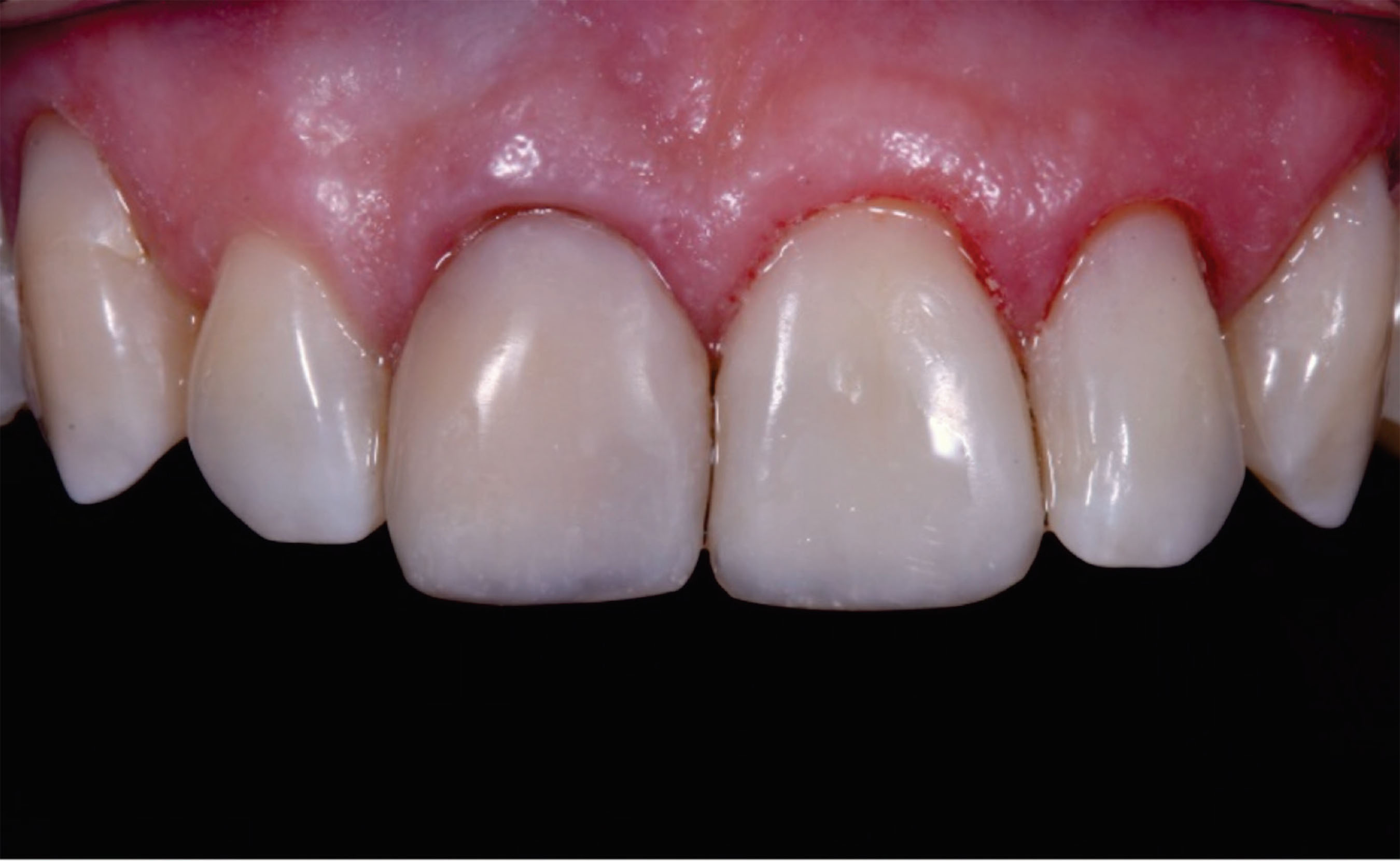
The final clinical outcome demonstrated adequate marginal adaptation and reestablishment of smile harmony (figure 6). The patient reported high satisfaction with the functional comfort and aesthetic results. The 4-week follow-up verified restoration stability, healthy gingival tissues, and maintained surface gloss.

Final clinical outcome. Postoperative aspect after finishing and polishing, demonstrating esthetic integration with the adjacent dentition at follow-up.
DISCUSSION
The choice of the direct-indirect composite resin veneer technique was justified by the clinical characteristics of the case, specifically the presence of a darkened substrate resulting from previous endodontic treatment and dental trauma. In these contexts, achieving a predictable aesthetic result can be challenging when using only the direct technique, as the substrate color significantly influences the outcome of the resin restoration [13]. The direct-indirect technique, in turn, allows for improved mechanical properties provided by the tempering process, more precise marginal adaptation, superior finishing, and the possibility of a previous try-in of the veneer, which ensures greater aesthetic predictability [14]. Additionally, it is a single-session procedure with less wear of the dental structure and reduced costs compared to ceramic veneers; it is easily repairable, making it more convenient for the patient [15].
The analysis of the clinical results revealed adequate gingival adaptation, without signs of inflammation or recession, indicating that the direct-indirect technique provided well-adjusted margins and respected the soft tissues. The patient reported satisfaction with both the comfort during the procedure and the final aesthetic result, highlighting the natural color and shape of the restored teeth. From a functional standpoint, the restoration maintained occlusal integrity and masticatory function, evidencing that the technique allowed for the simultaneous achievement of aesthetic and functional goals, confirming its effectiveness in cases of previously darkened teeth and high aesthetic demand [14,15].
A key advantage of this protocol, as demonstrated in this case, is the possibility of completing the treatment in a single appointment. This “same-day” workflow offers a more conservative and cost-effective alternative to ceramic veneers while being more convenient for the patient [15]. By fabricating the veneer chairside, the clinician maintains full control over the morphology and color masking without the need for multiple laboratory phases.
The results obtained in this case are consistent with the literature, which demonstrates that composite resin veneers, especially using the direct-indirect technique, present good longevity, mechanical resistance, color stability, and satisfactory maintenance of marginal adaptation over time [14]. The literature also indicates that extraoral finishing and the possibility of a prior try-in favor the aesthetic result, providing greater predictability compared to the conventional direct technique. Divergences may arise regarding durability in highly discolored teeth, where the composite thickness and the quality of adhesion may affect color maintenance and marginal integrity [16].
Despite the positive results, it is important to highlight some limitations. The clinical follow-up was relatively short, not allowing for a complete evaluation of the restoration’s longevity and resistance. Furthermore, there is a risk of color change and wear of the composite resin in the long term. Periodic maintenance, including professional polishing and monitoring, is essential to preserve the aesthetics, functionality, and marginal adaptation of the restoration [17].
The prognosis of the case is favorable, considering the technique used, the type of composite resin, and the care with periodic maintenance. The direct-indirect technique proved effective for similar situations involving teeth darkened by previous endodontic treatment or trauma, allowing for predictable aesthetic and functional results. Clinically, the approach proves to be accessible, conservative, low-cost, and capable of meeting patients’ aesthetic demands, making it a viable option in contexts where restoring aesthetics in a minimally invasive manner is desired [14].
The presented treatment demonstrated clinical success in terms of marginal adaptation, aesthetics, and patient satisfaction. The case reinforces the importance of individualizing the treatment plan, considering tooth characteristics, substrate, patient expectations, and technical limitations. This report evidences that the direct-indirect composite resin veneer technique is a reliable and predictable alternative, serving as a reference for decision-making in similar clinical situations.
CONCLUSION
The restorative procedure using composite resin veneers with the direct-indirect technique proved to be an effective and predictable alternative for the aesthetic and functional rehabilitation of cases involving darkened substrates. This technique achieved excellent results regarding marginal adaptation, anatomy, and final aesthetics, resulting in high patient satisfaction.
Furthermore, the extraoral polymerization stage contributed to the enhancement of the physical and mechanical properties of the composite resin, such as a higher degree of conversion, surface resistance, and color stability, granting greater longevity to the clinical outcome.
Thus, the reported case highlights that the direct-indirect technique represents a viable and conservative option for aesthetic anterior restorations, especially when properly planned and executed according to appropriate adhesive and restorative principles.
-
Article aligned with the Good Health and well-being goal of the Sustainable Development Goals (SDGs).
-
How to cite this article
Ribas HZ, Müller BF, Leyton BS. Restorative approach for an anterior tooth with a discolored substrate: a case report. RGO, Rev Gaúch Odontol. 2026;74:e20260026. http://dx.doi.org/10.1590/1981-8637202600262026
Data Availability
The research data are available in the body of the document.
REFERENCES
- 1 Zimmerli B, Jeger F, Lussi A. Bleaching of nonvital teeth. A clinically relevant literature review. Schweiz Monatsschr Zahnmed. 2010;120(4):306-20.
- 2 Şişmanoğlu S. Bleaching of nonvital teeth: a review. Aurum J Health Sci. 2020;2(2):91-114.
-
3 Moccelini BS, de Alencar NA, Bolan M, Magno MB, Maia LC, Cardoso M. Pulp necrosis and crown discoloration: a systematic review and meta-analysis. Int J Paediatr Dent. 2018. doi: http://dx.doi.org/10.1111/ipd.12372
» https://doi.org/10.1111/ipd.12372 - 4 Georges I, Rita G, Zogheib C. Guided endodontics in managing severely calcified teeth: a review. Endodontology. 2023;35(3):195-201.
-
5 Dias PC, Franco FBMJ, Silva RCP, Farani JJ. Aesthetic rehabilitation of discolored nonvital anterior teeth. RGO, Rev Gaúch Odontol. 2020;68:e20200035 doi: http://dx.doi.org/10.1590/1981-863720200003920190174
» https://doi.org/10.1590/1981-863720200003920190174 -
6 Bersezio C, Ledezma P, Mayer C, Rivera O, Junior OBO, Fernández E. Effectiveness and effect of non-vital bleaching on the quality of life of patients up to 6 months post-treatment: a randomized clinical trial. Clin Oral Investig. 2018;22(9):3013-9. doi: http://dx.doi.org/10.1007/s00784-018-2389-y
» https://doi.org/10.1007/s00784-018-2389-y -
7 Dahl JE, Pallesen U. Tooth bleaching--a critical review of the biological aspects. Crit Rev Oral Biol Med. 2003;14(4):292-304. doi: http://dx.doi/10.1177/154411130301400406
» https://doi.org/10.1177/154411130301400406 -
8 Knezevic N, Obradovic M, Dolic O, Veselinovic V, Kojic Z, Josipovic R, et al. Clinical testing of walking bleach, in-office, and combined bleaching of endodontically treated teeth. Medicina (Kaunas). 2022;59(1):18. doi: http://dx.doi/10.3390/medicina59010018
» https://doi.org/10.3390/medicina59010018 -
9 Newton R, Hayes J. The association of external cervical resorption with modern internal bleaching protocols: what is the current evidence? Br Dent J. 2020;228(5):333-7. doi: http://dx.doi/10.1038/s41415-020-1317-0
» https://doi.org/10.1038/s41415-020-1317-0 -
10 Chen Y, Huang Y, Deng X. External cervical resorption-a review of pathogenesis and potential predisposing factors. Int J Oral Sci. 2021;13(1):19. doi: https://dx.doi/10.1038/s41368-021-00121-9
» https://doi.org/10.1038/s41368-021-00121-9 -
11 Kahler B. Present status and future directions: managing discoloured teeth. Int Endod J. 2022;55 Suppl 4(Suppl 4):922-50. doi: https://dx.doi/10.1111/iej.13711
» https://doi.org/10.1111/iej.13711 - 12 Fahl Júnior N. The direct/indirect composite resin veneers: a case report. Pract Periodontics Aesthet Dent. 1996;8(7):627-38.
-
13 Miotti LL, Santos IS, Nicoloso GF, Pozzobon RT, Susin AH, Durand LB. The Use of resin composite layering technique to mask discolored background: a CIELAB/CIEDE2000 analysis. Oper Dent. 2017;42(2):165-74. doi: https://dx.doi/10.2341/15-368-L
» https://doi.org/10.2341/15-368-L -
14 Fahl N Jr, Ritter AV. Composite veneers: the direct-indirect technique revisited. J Esthet Restor Dent. 2021;33(1):7-19. doi: https://dx.doi/10.1111/jerd.12696
» https://doi.org/10.1111/jerd.12696 -
15 Altamimi Y, Fahl N Jr, Rocha MG, Oliveira D, Neiva GF, Maia RR. Shade modulation ability of direct vs. direct-indirect prepless composite veneers. J Esthet Restor Dent. 2025;37(1):48-55. doi: https://dx.doi/10.1111/jerd.13390
» https://doi.org/10.1111/jerd.13390 -
16 Abdulrahman MS. Evaluation of the sealing ability of direct versus direct-indirect veneer techniques: an in vitro study. Biomed Res Int. 2021;2021:1118728. doi: https://dx.doi/10.1155/2021/1118728
» https://doi.org/10.1155/2021/1118728 -
17 Zimmer R, Amorim AH, Portella FF, Arossi GA. Clinical longevity of direct resin-based composite veneers on anterior teeth. Rev Fac Odontol UPF. 2024;29(1):1-13. doi: https://dx.doi/10.5335/rfo.v29i1.15953
» https://doi.org/10.5335/rfo.v29i1.15953
Edited by
-
Assistant editor
Luciana Butini Oliveira