 Fatal cerebral myiasis secondary to squamous cell carcinoma: case report and scoping review
ABSTRACT
Cerebral myiasis is an exceptionally rare condition caused by infestation with dipteran larvae, with only 20 cases reported in the literature to date. A 78-year-old man presented with anorexia, vomiting, and fever. Physical examination revealed a 7 × 8 cm ulcerated scalp lesion with a necrotic base, purulent discharge, a foul odor, and numerous larvae. Computed tomography demonstrated an osteolytic frontal bone defect accompanied by pneumocephalus and subcutaneous emphysema. The larvae were manually removed, an iodoform dressing was applied, and intravenous ceftriaxone therapy was initiated. Progressive neurological decline prompted repeat imaging, which revealed frontal and parietal cerebritis with abscess formation. Surgical debridement was performed to remove necrotic tissue. Histopathological analysis showed moderately differentiated squamous cell carcinoma with acute osteomyelitis, and cultures yielded multidrug-resistant Pseudomonas aeruginosa. Despite targeted antibiotic therapy and intensive supportive care, the patient died. This case highlights the significant morbidity and mortality associated with cerebral myiasis, particularly when complicated by underlying malignancy and multidrug-resistant infection. Early recognition, prompt surgical intervention, and pathogen-directed antimicrobial therapy are crucial, while comprehensive multidisciplinary management remains essential to optimize outcomes in this life-threatening condition.
KEYWORDS:
Myiasis; Infection; Brain; Osteomyelitis; Abscess
INTRODUCTION
Myiasis is defined as the infestation of living vertebrates by larvae of the order Diptera and can be classified according to parasitological or clinical criteria1,2. Alternatively, it may also be categorized based on ecological or anatomical characteristics1,2. Patton3,4 divided myiasis-causing flies into three parasitological groups: obligate, facultative, and accidental. Obligate parasites require living tissue for larval development, whereas facultative parasites typically develop in decaying organic matter but may occasionally invade living tissue. In accidental parasitism, eggs or larvae are inadvertently ingested or inhaled with contaminated food1. Clinical classification is based on the anatomical site of infestation, first proposed by Bishopp5 and later modified by James6 and Zumpt7, encompassing categories such as blood-sucking, tissue-destroying, subdermal, gastrointestinal, and urogenital myiasis, as well as infestations of the head passages (e.g., ear, nose, and throat)1,2,4.
Cerebral myiasis is exceptionally rare, with the first case reported in 1939 by Froomin and Kaznelson8. Since then, only 20 cases have been previously documented in the literature. Despite its rarity, cerebral myiasis poses a severe clinical challenge, often resulting in significant neurological impairment, high morbidity, and, in some cases, mortality9.
Although individual case reports provide valuable insights into the clinical presentation and management of cerebral myiasis, no comprehensive synthesis of the available evidence exists to support clinical decision-making. Important questions remain regarding its epidemiology, risk factors, and optimal therapeutic approaches.
Thus, a scoping review was conducted to systematically map the available research on intracerebral myiasis, identify trends in reported cases, and highlight existing knowledge gaps. This review was guided by the following research question: What does the literature reveal about the clinical presentation, management, and outcomes of patients diagnosed with intracerebral myiasis?
Despite its rarity, the published reports of intracerebral myiasis span a broad temporal range—from 1939 to the present—and encompass diverse etiologic contexts and clinical presentations, including post-traumatic, postoperative, cutaneous head-and-neck malignancy, and pediatric cases. This review covers both historical and contemporary records, which collectively consolidate the clinical spectrum observed to date8–27.
Ethics
This study was approved by the Research Ethics Committee (CEP) of the Padre Albino University Center, under CAAE Nº 68395723.7.0000.5430, and Substantiated Opinion Nº 6.000.608.
MATERIALS AND METHODS
The protocol for this scoping review was developed a priori following the Preferred Reporting Items for Systematic Reviews and Meta-Analyses for Scoping Reviews (PRISMA-ScR) guidelines28. The initial draft was reviewed by the research team and revised accordingly before registration. The final protocol was prospectively registered with the Open Science Framework (OSF) in March 202529. Any deviations from the protocol during the review process are documented and justified in the Results section, ensuring transparency and reproducibility.
Literature search
A comprehensive literature search was conducted across PubMed, SciELO, and BVS on January 26, 2025, with no language or publication date restrictions, to maximize inclusivity of all relevant studies on cerebral myiasis (CM). The search strategy was developed by an experienced librarian and further refined with team discussion, using a combination of controlled vocabulary terms (MeSH) and non-controlled keywords in multiple languages.
The PubMed search strategy included the following combination of MeSH terms and keywords: ("Cerebral Myiasis" OR "intracerebral" OR "Brain Myiasis" OR "Intracranial Myiasis" OR "NeuroMyiasis" OR "Central Nervous System Myiasis") AND ("Myiasis"[MeSH] OR "Parasitic Infestation"[MeSH] OR "Fly Larvae Infestation" OR "Maggot Infestation" OR "Diptera Infestation" OR "Larval Infection" OR "Infection by Fly Larvae" OR "Parasitic Larvae" OR "Myiasis Infection" OR "Grubs Infestation" OR "Grubs" OR "Myiasis").
Additionally, manual reference checks of the included studies were performed to identify potentially overlooked articles and relevant gray literature.
Selection process
Eligibility criteria were established using the PCC framework: population (P): patients diagnosed with intracerebral myiasis; concept (C): clinical presentation, management, and outcomes; context (C): published case reports, case series, and observational studies.
All records were imported into Rayyan (Rayyan Systems Inc., Cambridge, MA, USA) for screening30. Two independent reviewers assessed titles and abstracts, with discrepancies resolved by means of discussion and consensus.
Exclusion criteria comprised non-cerebral myiasis (e.g., ophthalmic, optic, nasopharyngeal), non-dipteran infestations (e.g., gnathostomiasis, nematode larval migration; two papers excluded for this reason), duplicates, and records lacking sufficient case-level information.
Data extraction was performed using a pre-piloted form capturing study identifiers, patient demographics, clinical presentation, underlying conditions, characteristics of the infestation, diagnostic methods, therapeutic interventions, outcomes, and study design features. The extracted variables were informed by, and subsequently compared with, those reported by Cuestas et al.31, enabling identification of methodological similarities and differences and aligning this review with established reporting standards for myiasis in malignant wounds.
Despite the use of a standardized data extraction form, several studies lacked complete information for key variables (e.g., larval species identification, patients’ functional baseline), which constrained the comprehensiveness of the synthesized findings.
RESULTS
The initial search on the SciELO platform yielded a low number of results, which, upon further analysis, suggested an artificial restriction in article retrieval. To address this limitation, search terms were refined by incorporating additional focused descriptors. In PubMed, the addition of the ‘[tiab:∼0]’ operator after certain keywords reduced the number of retrieved records from 65 to 14, underscoring the importance of maintaining a broader initial search to ensure comprehensive coverage.
The final SciELO strategy was therefore simplified to a core set of terms: ‘Myiasis’ OR ‘Cerebral Myiasis’ OR ‘Brain Myiasis.’
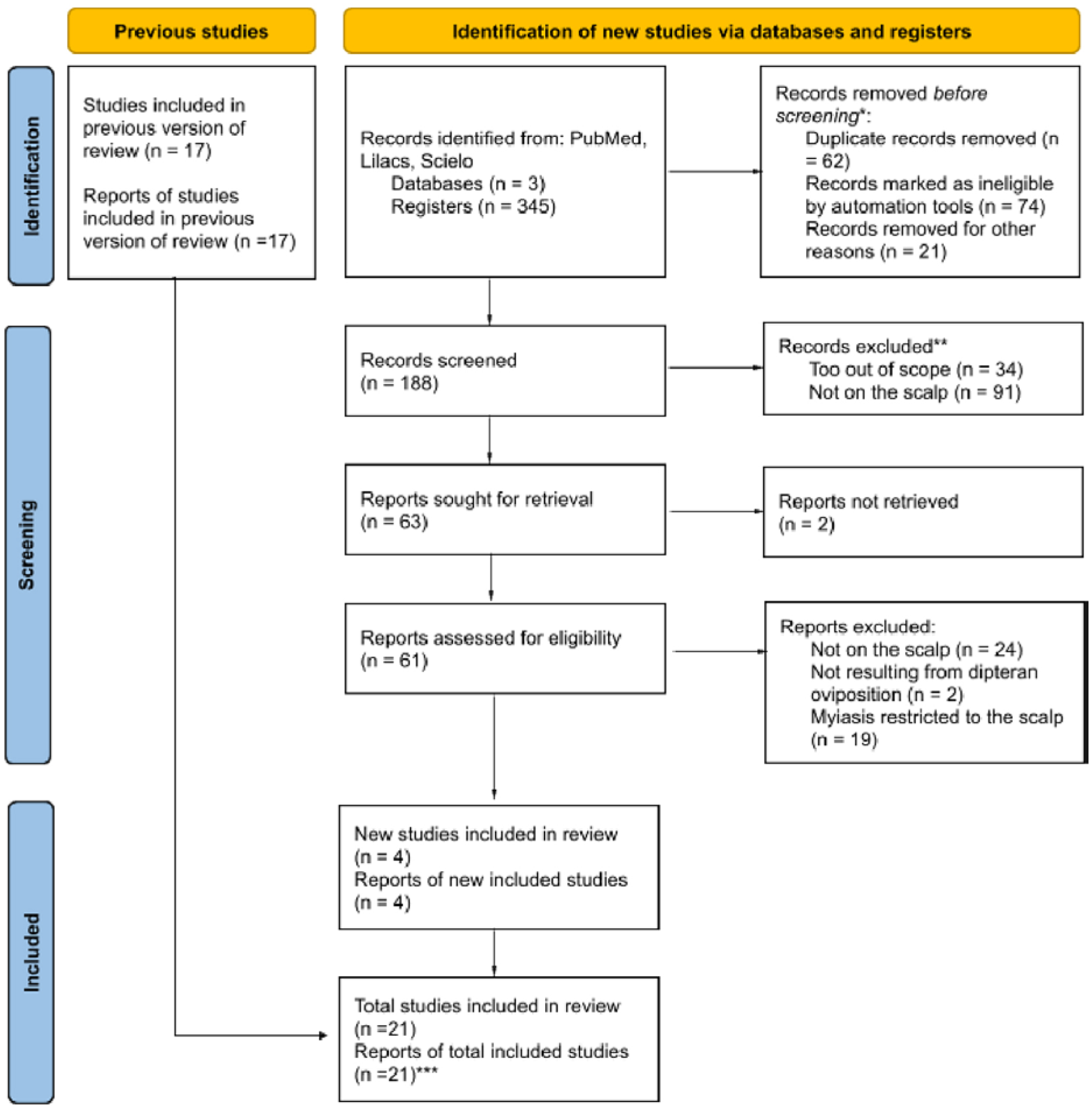
To further supplement the electronic searches, an exploratory search was conducted on the Research Rabbit platform32. In total, 20 studies were deemed pertinent and included for full-text review and data extraction (Figure 1).
PRISMA flow diagram illustrating the selection process of the included studies; *automation tools labeled "keywords" were used in Rayyan to identify articles containing the words "myiasis," "neuromeningeal," or "larvae" that did not refer to humans; despite the use of automation, human analysis was always performed before any exclusion; **the same automation tool was used to help identify articles in which the myiasis did not occur in the brain; other reports that substantially deviated from the central topic were also excluded (essentially those addressing therapies, diagnostic keys, or epidemiological analysis rather than myiasis cases) and categorized as "not related/out of scope"; ***present case included.
CASE REPORT
A 78-year-old Caucasian male was admitted to the emergency department, accompanied by a family member, with complaints of anorexia, vomiting, and fever that had begun earlier that day. He reported living alone and being independent in both basic and instrumental activities of daily living. His only known medical condition was systemic arterial hypertension—confirmed by his daughter—and was taking 50mg of Losartan once every morning.
On physical examination, the patient appeared confused and uncommunicative, with a mildly pale complexion and clinical signs of dehydration, but was afebrile. Inspection of the scalp revealed an ulcerated lesion in the left frontal region extending to the parietal border, measuring approximately 7.0 cm × 8.0 cm. The lesion exhibited irregular, raised, hyperemic margins and a necrotic base with purulent discharge and a strong fetid odor. Numerous fly larvae (myiasis) were observed within the wound, actively consuming necrotic tissue—an observation consistent with the patient's report that the lesion had been progressively enlarging over the past year.
Initially, larvae were manually removed using anatomical forceps and irrigation with 0.9% saline solution. Upon identifying bone exposure, an occlusive iodoform dressing was applied. Based on the initial presentation, the primary hypothesis was cutaneous myiasis. However, after larval removal and identification of bone exposure and involvement, the possibility of extension into the brain parenchyma was considered. No alternative differential diagnoses were proposed. Laboratory tests and a computed tomography (CT) scan of the skull were requested for further evaluation.
The complete blood count (CBC) revealed a hemoglobin level of 11.3 g/dL (reference range for older adult males: 13.0–17.0 g/dL), a hematocrit of 32.7% (reference: 38.8%–50.0%), and leukocytosis with a total leukocyte count of 19,400/µL (reference: 4,000–11,000/µL), with marked neutrophilic predominance (89.8%, reference: 40%–70%; absolute neutrophils count: 17,400/ µL). Serum sodium was 133 mmol/L (reference: 135–145 mmol/L), indicating mild hyponatremia, and blood urea nitrogen was elevated at 72 mg/dL (reference: 7–20 mg/dL).
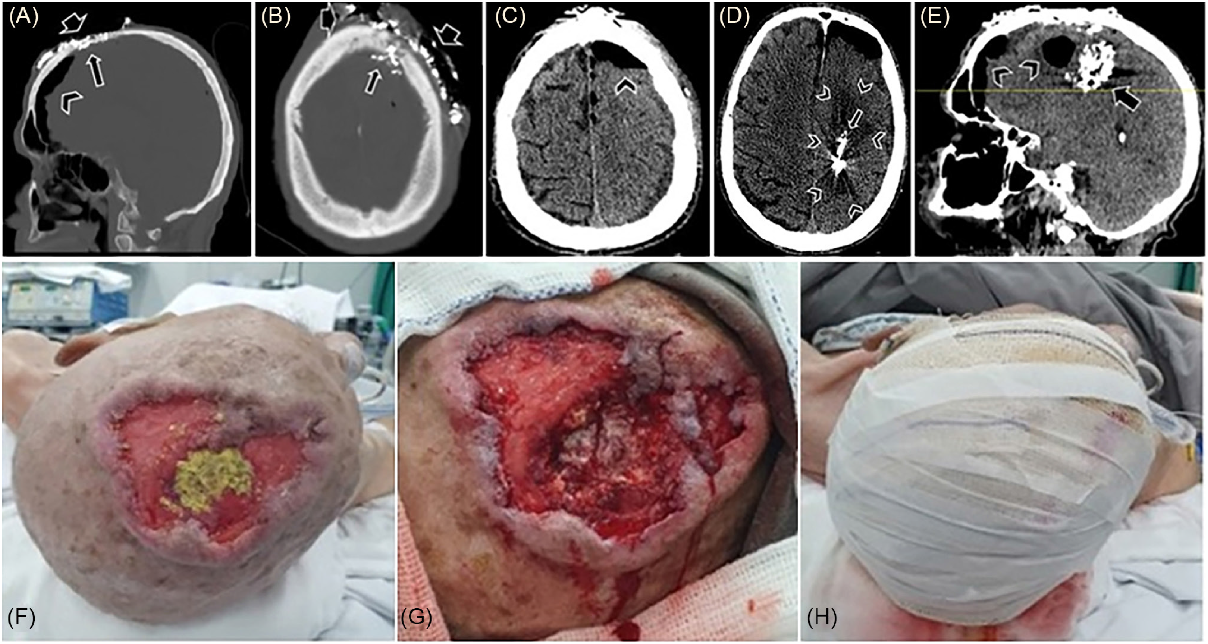
The CT scan revealed an osteolytic defect in the left frontal bone, establishing communication between the meningeal membranes and the external environment through an ulcerated lesion involving the skin and underlying tissues in the frontal region, predominantly left of the midline. Additional findings included pneumocephalus, subcutaneous emphysema, and multiple metallic-density particles interspersing the nervous tissue, consistent with iodoform (Figure 2). The patient was subsequently admitted to the neurosurgery ward for continued care, and intravenous antibiotic therapy with ceftriaxone (1 g every 12 h) was initiated.
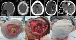
First CT scan: bone window, sagittal (A) and axial (B) planes – osteolytic lesion in the frontal bone (long arrow) establishing communication between the meningeal membranes and the external environment through an extensive ulcerated lesion (short arrow) in the skin and subcutaneous tissue. Pneumocephalus (arrowhead); (C) parenchymal window, pneumocephalus (arrowhead). Second CT scan: parenchymal window: (D) axial plane – hypodense lesions in the left frontal and parietal lobes, suggesting organized cerebritis/abscess with possible areas of necrosis (arrowhead); multiple metallic-density particles intermingling these tissues, representing iodoform (long arrow); (E) sagittal plane – iodoform particles (arrow) and pneumocephalus (arrowhead); (F) extensive irregular wound measuring 7.0 cm × 8.0 cm, with raised and erythematous edges; presence of gauze with Iodoform in the region of greatest excavation, corresponding to the osteolytic lesion; (G) post-debridement local appearance showing exposed meningeal membranes; (H) local occlusive dressing with Rayon, Rifocin, and Dersani gauze.
The patient initially received general care, including respiratory physical therapy and routine wound care with regular dressing changes. On the fifth day of hospitalization, the patient developed drowsiness, asthenia, anorexia, constipation, and urinary retention. Laboratory tests were ordered, along with a chest radiograph, nasoenteral tube placement, and delayed bladder catheterization. Results showed a total leukocyte count of 14,600/µL with 82.1% neutrophils (absolute neutrophils count: 11,986/µL), urea of 94 mg/dL, and a chest radiograph without significant abnormalities.
On the ninth day of hospitalization, despite ongoing supportive measures, the patient exhibited a continued decline in mental status, prompting a repeat cranial CT for preoperative evaluation (Figure 2). Compared to the previous scan, new hypodense lesions were observed in the left frontal and parietal lobes, accompanied by erasure of the ipsilateral cortical sulci, suggesting cerebritis with an organizing abscess and possible necrotic areas. Consequently, the patient underwent surgery, during which the lesion was debrided and irrigated with Hexamidine and 0.9% saline solution. The necrotic tissue, heavily colonized by Iodoform and putrefying myiasis, was thoroughly removed, resulting in complete abscess evacuation. Hemostasis was achieved, followed by the application of an occlusive dressing using Rayon, Rifocin, and Dersani gauze (Figure 2). Samples were submitted for histopathological analysis, bacterioscopy, and antibiogram.
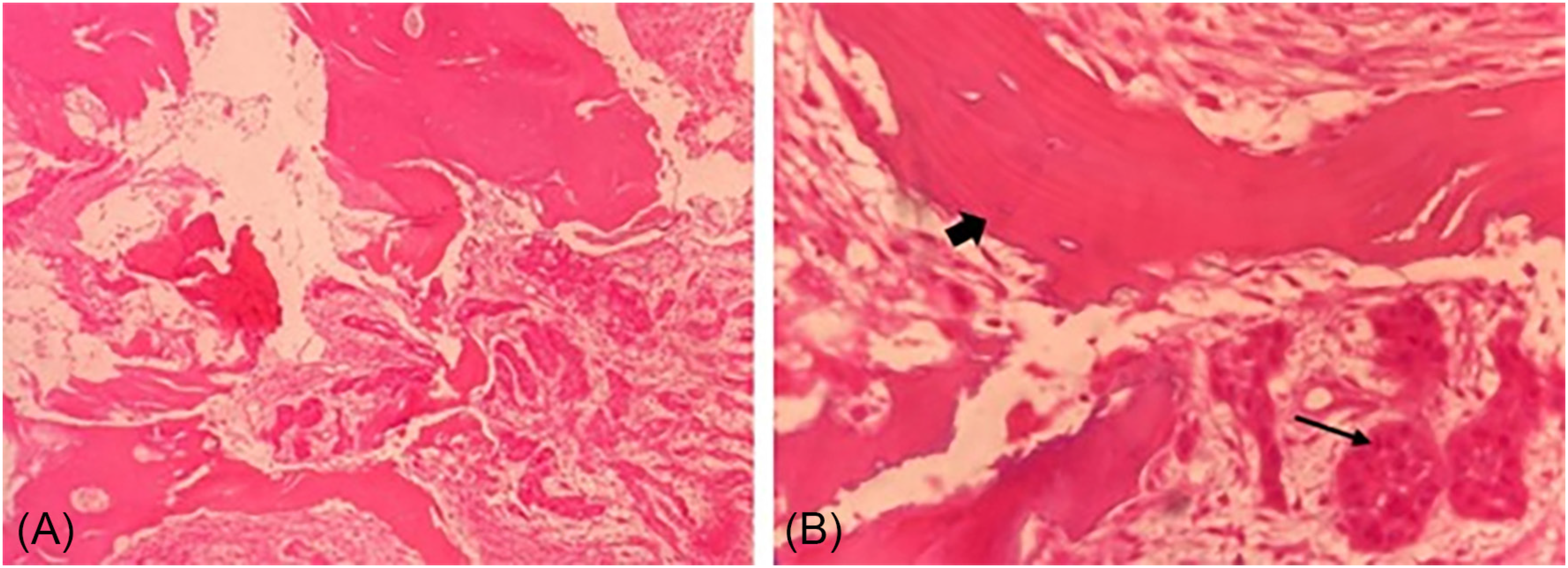
Pathology showed a moderately differentiated and invasive squamous cell carcinoma in the bone tissue, along with acute exudative osteomyelitis (Figure 3). The microbiological culture identified Pseudomonas aeruginosa, with its antimicrobial susceptibility profile determined by automated microdilution.
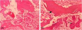
Anatomopathology: (A) bone fragment showing invasion by a moderately differentiated squamous cell carcinoma; neoplastic cells exhibit pleomorphism with intercellular bridges and infiltrate the bone tissue; (B) bone tissue (thick arrow) and nests of moderately differentiated malignant squamous cells (thin arrow).
The isolate was resistant to cefepime (MIC >8 µg/mL), ceftazidime (MIC >8 µg/mL), piperacillin-tazobactam (MIC >16 µg/mL), ciprofloxacin (MIC >1 µg/mL), and levofloxacin (MIC >1 µg/mL). Resistance to broad-spectrum β-lactams, combined with fluoroquinolone resistance, suggests the possible expression of mechanisms such as extended-spectrum β-lactamases (ESBL) or carbapenemases.
Carbapenems showed an intermediate profile with imipenem (MIC undetermined) and meropenem (MIC 8 µg/mL), indicating the need for dose optimization to enhance clinical efficacy. In contrast, the isolate remained susceptible to colistin (MIC ≤2 µg/mL), amikacin (MIC ≤8 µg/mL), and tobramycin (MIC ≤2 µg/mL), suggesting potential treatment options with aminoglycosides or polymyxins, depending on pharmacokinetic and pharmacodynamic parameters at the infection site.
On the first postoperative day (Table 1), the patient's level of consciousness remained depressed. Antibiotic therapy was switched from Ceftriaxone (1g IV every 12 h) to Vancomycin (500mg IV every 12 h) after 12 days of treatment. On the fifth postoperative day, the patient developed an episode of desaturation, which was stabilized with an oxygen mask. However, the patient's sensory decline persisted, prompting a palliative care consultation.
The patient was transferred to the palliative care team at a secondary hospital, where Meropenem was added to the antibiotic regimen. Sodium levels were corrected, and a high-flow oxygen mask (10 L/min) was maintained, resulting in a 96% oxygen saturation (SpO2). Although some transient improvements were observed, such as mild stabilization of diuresis and wound healing, the overall prognosis persisted. The patient remained non-communicative, exhibiting limited responsiveness to painful stimuli, and continued to require intensive supportive care, including nutritional supplementation and secretion management over the subsequent 10 days. Ultimately, the patient died.
DISCUSSION
Intracerebral myiasis is an extremely rare entity, with only 20 cases reported worldwide to date, according to the most recent literature review conducted in 2024 by Ramón-Cuellar et al.10. This is the fifth documented case in Brazil.
Human myiasis is globally distributed, with higher prevalence in developing countries—particularly in humid tropical and subtropical regions—where greater species diversity and abundance are observed1,2,33. The most significant risk factors for myiasis include poor hygiene and low socioeconomic status. Additionally, immunosuppression, diabetes, psychiatric disorders, advanced age, delayed access to specialized medical care, and alcoholism are also recognized as predisposing factors1,2,11,31,33 (Table 2).
Based on our review, the age of patients affected by cerebral myiasis varied from five months to 85 years, with a mean of 37.7 years. Regarding gender, 18 out of 21 cases (85.7%) occurred among men. Reported symptoms include fever, chills, varying degrees of consciousness impairment, intracranial hematoma, motor deficits, convulsions, extrapyramidal effects, secondary infections, and intracranial hypertension with hydrocephalus1,2,11,15,17,25,27. Local symptoms may include pruritus, pain, bloody discharge, and the characteristic sensation of crawling or movement beneath the skin1,11,31. Symptom onset can be delayed for days to years, particularly among patients with dementia11. Additionally, advanced age alone may contribute to delayed clinical manifestation, as seen in this case, in which local symptoms were neglected.
Cutaneous involvement is the most common presentation of myiasis. In this case, the condition originated from a neglected scalp lesion that, upon histopathological examination, was diagnosed as a squamous cell carcinoma with bone tissue infiltration19,25,27,31,34. Notably, only Curzi et al.11 in 2021 have reported squamous cell carcinoma as a risk factor for cerebral myiasis31. Sesterhenn et al.35 reported 20 cases of cutaneous myiasis associated with malignant head-and-neck lesions, seven (35.0%) of which involved preexisting squamous cell carcinoma. No single larval species predominated in those case, although Lucilia sericata was the most frequently identified.
According to the literature, the frontal lobe is the most frequently affected site, followed by the temporal lobe11,16,19,20,23,24. Deep parenchymal involvement and advanced age have been consistently associated with a poor prognosis11. The mortality so far reached 47.6%, representing a 61.9% relative increase compared with the mortality rate previously reported by Ramón-Cuellar (29.4%)10.
Despite prolonged exposure of nervous tissue, secondary infections, whether encephalitis (with or without abscess formation) or meningitis, were rare, with only four cases reported19,20,27,35. Although larvae are known to harbor a broad spectrum of bacteria capable of intensifying the local inflammatory response35, it is assumed that maggot infestation may exert a protective effect on exposed tissues. This occurs by means of mechanical coverage of the wound surface and digestion of necrotic tissue1,21,23,26,35. During the First World War, larvae of the Calliphoridae family, including Lucilia sericata and Phormia regina, were used for debriding wounds and osteomyelitis due to their saprophagous habits35. Pneumocephalus was another exceptional finding, described only in three cases23,27,35. No reports of osteomyelitis were found in the literature.
Various approaches have been described for larval removal35. Except for Algahtany et al.27, who opted for conservative management, all other cases underwent surgical intervention involving larval removal, debridement of necrotic tissue, wound irrigation, and, when feasible, reconstruction of the affected area. These procedures were combined with broad-spectrum intravenous antibiotic therapy, with or without antifungal, antiparasitic, or anticonvulsant medications9,11,19,21,23,26,27. Furthermore, one of the most critical components of treatment is the consistent daily maintenance of dressings and wounds, along with maintaining overall patient hygiene, as these measures are key determinants in reducing infection risk.
Unfortunately, the exact larval species could not be identified in this case. However, considering the endemic species in the patient's geographical region and the known predisposing factors—such as advanced age, an exposed wound, and poor hygiene—it is most likely that Cochliomyia hominivorax was involved, as it is the primary causative agent of myiasis in Brazil1. Identifying the species, and consequently its life cycle, is crucial for understanding the mode of infestation, estimating its duration, predicting its progression, and implementing effective preventive measures35.
Limitations
This scoping review has some inherent limitations. First, the inclusion of only published case reports and series raises the possibility of publication bias, since unusual or severe presentations are more likely to be reported, potentially limiting generalizability. Second, the scarcity and heterogeneity of available data— particularly regarding larval species identification, patients’ functional baseline, and standardized outcomes—restricted the comprehensiveness of the synthesis. Finally, as with all scoping reviews, the methodology prioritizes breadth over depth, aiming to map the existing literature rather than to generate quantitative effect estimates.
Moreover, this study did not receive external funding, which eliminates potential funding-related bias but also limits the availability of resources that might have enhanced data collection and analysis. Similarly, the authors declare no conflicts of interest, ensuring impartiality in the review process.
Despite these limitations, the systematic approach employed, adherence to PRISMA-ScR guidelines, and prospective protocol registration strengthen the transparency and reproducibility of our findings.
CONCLUSION
Cerebral myiasis, although rare, presents significant clinical challenges and can result in fatal outcomes, as demonstrated in this case. Given its rarity, often underestimated and occasionally mismanaged, there remains no clear consensus on optimal management. However, early diagnosis, prompt surgical debridement, targeted antibiotic therapy, and daily wound care are essential. Maintaining strict wound and patient hygiene is also a critical parameter for favorable clinical evolution. The treatment strategy employed highlights the need for a multidisciplinary approach and further research to establish standardized management protocols and improve patient outcomes.
DATA AVAILABILITY
The complete anonymized dataset supporting the findings of this study is included within the article itself.
REFERENCES
- 1 McGraw TA, Turiansky GW. Cutaneous myiasis. J Am Acad Dermatol. 2008;58:907-26.
- 2 Francesconi F, Lupi O. Myiasis. Clin Microbiol Rev. 2012;25:79-105.
- 3 Patton WS. Some notes on Indian Calliphorinae. Part I. Chrysomya bezziana Villeneuve, the common Indian Calliphorinae whose larvae cause cutaneous myiasis in man and animals. Indian J Med Res. 1920;8:17-29.
- 4 Patton WS. Some notes on Indian Calliphorinae. Part VII. Additional cases of myiasis caused by larvae of Chrysomyia bezziana Vill, together with some notes on the Diptera which cause myiasis in man and animals. Indian J Med Res. 1922;9:654-82.
- 5 Bishopp FC. Flies which cause myiasis in man and animals: some aspects of the problem. J Econ Entomol. 1915;8:317-29.
-
6 James MT. The flies that cause myiasis in man. Washington: U.S. Department of Agriculture; 1947. [cited 2025 Nov 21]. Available from: https://doi.org/10.5962/bhl.title.65688
» https://doi.org/10.5962/bhl.title.65688 - 7 Zumpt F. Myiasis in man and animals in the Old World: a textbook for physicians, veterinarians, and zoologists. London: Butterworths; 1965.
- 8 Froomin LL, Kaznelson AB. Intradural cyst of parasitic origin (myiasis clinic). Zh Ushn Nos Gorl Bolezn. 1939;16:427-33.
- 9 Mariottiz Acuña S, Cardona Castellanos DP, Corredor Quintero AB. Miasis intracerebral, generalidades y limitantes en el tratamiento, reporte de caso. Rev Cuarzo. 2017;23:26-34.
- 10 Ramón-Cuellar JF, Mejía-Cordovez JA, Quiñones-Ossa GA, Leal-Isaza JP, Vargas-Osorio MP, Ramirez-Muñoz JD, et al. Cerebral myiasis, an unexpected intraoperative finding: a case report and systematic review. World Neurosurg. 2024;186:138-44.
- 11 Curzi C, Bartoletti V, Canova G, Giordan E. A severe case of brain myiasis: treatment rationale and review of literature. Asian J Neurosurg. 2021;16:582-6.
- 12 Semenov P V. A case of penetration of Hypoderma lineatum de Villers larva into the human brain. Med Parazitol (Mosk). 1969;38:612-3.
- 13 Zucoloto S, Rossi MA. Miíase facial com extensão para a caixa craniana. Rev Bras Med. 1971;28:13-6.
- 14 Rossi MA, Zucoloto S. Fatal cerebral myiasis caused by the tropical warble fly, Dermatobia hominis. Am J Trop Med Hyg. 1973;22:267-9.
- 15 Gilly R, Lapras C, Mamelle JC, Challamel MJ, Ghilhot JH, Nicolas A, et al. Myase migrante du genre hypoderma avec hematome intracerebral: a propos d'une observation chez un enfant de 7 ans. Pediatrie. 1976;31:67-75.
- 16 Pouillaude JM, Dupont J, Gilly R, Lapras Cl. Intracerebral myiasis in a child. Pediatr Radiol. 1980;10:121-3.
- 17 Francois P, Martin G, Goullier A, Plasse M, Beaudoing A. Hypodermose neuromeningee compliquee d'hydrocephalie: interet de l'imagerie par resonance magnetique nucleaire. Presse Med. 1987;16:1231-3.
- 18 Kalelioglu M, Akturk G, Akturk F, Komsuoglu SS, Kuzeyli K, Tigin Y, et al. Intracerebral myiasis from Hypoderma bovis larva in a child: case report. J Neurosurg. 1989;71:929-31.
- 19 Cheshier SH, Bababeygy SR, Higgins D, Parsonnet J, Huhn SL. Cerebral myiasis associated with angiosarcoma of the scalp: case report. Neurosurgery. 2007;61:E167.
- 20 Marco de Lucas E, Díez C, Gutiérrez A, Montiaga F, Arnáiz J, Mandly AG, et al. Unusual MRI findings in a patient with history of frontal fracture and skin infestation by fly larvae, as a possible sign of intracerebral myiasis. Clin Neurol Neurosurg. 2008;110:725-8.
- 21 Terterov S, Taghva A, MacDougall M, Giannotta S. Posttraumatic human cerebral myiasis. World Neurosurg. 2010;73:557-9.
- 22 Holanda LF, Pereira BJ, Holanda CV, Oliveira JG. Cerebral myiasis. Neurology. 2015;84:434-5.
- 23 Giri SA, Kotecha N, Giri D, Diyora B, Nayak N, Sharma A. Cerebral myiasis associated with artificial cranioplasty flap: a case report. World Neurosurg. 2016;87:661.e13-16.
- 24 Navarro JN, Alves RV. Postoperative cerebral myiasis: a rare cause of wound dehiscence in developing countries. Surg Neurol Int. 2016;7:69.
- 25 Aggarwal A, Maskara P. Maggots in the brain: sequelae of ignored scalp wound. World Neurosurg. 2018;109:115-6.
- 26 Deo RC, Acharya A, Panigrahi S, Senapati SB, Mahapatra AK, Puppala S. Cerebral myiasis secondary to burr hole evacuation: a rare illustrative case report. Asian J Neurosurg. 2023;18:410-3.
- 27 Algahtany MA, Alzailaie AK, Khoudir MA, Alqahtani FM, Osman TA, Shobowale EO. Conservative management of post-traumatic cerebral myiasis: report of a unique case and literature review. J Pak Med Assoc. 2022;72:164-7.
- 28 Tricco AC, Lillie E, Zarin W, O’Brien KK, Colquhoun H, Levac D, et al. PRISMA extension for scoping reviews (PRISMA-ScR): checklist and explanation. Ann Intern Med. 2018;169:467-73.
-
29 Open Science Framework. Research protocol: intracerebral myiasis. [cited 2025 Nov 24]. Available from: https://osf.io/u24g9/?view_only=09a48b4a800545f0a5366380d64097f3
» https://osf.io/u24g9/?view_only=09a48b4a800545f0a5366380d64097f3 - 30 Ouzzani M, Hammady H, Fedorowicz Z, Elmagarmid A. Rayyan: a web and mobile app for systematic reviews. Syst Rev. 2016;5:210.
- 31 Cuestas D, Pedraza J, Herrera H, Motta A, Cuestas A, Forero Y, et al. Cutaneous myiasis in skin cancer and malignant wounds: a systematic review. Int J Dermatol. 2021;60:1529-46.
- 32 Cole V, Boutet M. Product: ResearchRabbit. J Can Health Libr Assoc. 2023;44:43-7.
- 33 Tauziede‐Espariat A. Brain myiasis. In: Chrétien F, Wong KT, Sharer LR, editors. Infections of the central nervous system: pathology and genetics. Hoboken: Wiley Blackwell; 2020. p.503-4.
- 34 Arbit E, Varon RE, Brem SS. Myiatic scalp and skull infection with diptera sarcophaga: case report. Neurosurgery. 1986;18:361-2.
- 35 Sesterhenn AM, Pfützner W, Braulke DM, Wiegand S, Werner JA, Taubert A. Cutaneous manifestation of myiasis in malignant wounds of the head and neck. Eur J Dermatol. 2009;19:64-8.
Edited by
-
Editor:
Fabiana Martins de Paula https://orcid.org/0000-0003-3343-0149