 Coating of Ti-6Al-4V sheets with nano-HA-based materials by electrophoretic deposition (EPD) method and investigation of surface properties
ABSTRACT
Titanium alloys, particularly Ti-6Al-4V, are among the most widely used materials in biomedical implant applications due to their superior mechanical strength, corrosion resistance, and compatibility with bone tissue. In this study, Ti-6Al-4V substrates were coated with Hydroxyapatite (HA) and Hydroxyapatite reinforced with 1% Graphene (Gr) using electrophoretic deposition under 40V and 80V. The influence of applied voltage, deposition time, and solution composition on coating morphology, thickness, elemental distribution, and structural properties was systematically evaluated. The most favorable results were obtained at 80V, where homogeneous and adherent coatings were formed. Morphological examinations revealed coating thicknesses ranging from 11.25 µm to 87.5 µm. XRD analyses confirmed the presence of Tiα, Tiβ, HA, and Gr phases, demonstrating successful composite coating. Surface characterization showed that HA coatings exhibited hydrophilic behavior, while HA-Gr coatings demonstrated super-hydrophilic properties, which are highly advantageous for cellular attachment and osseointegration. Contact angle and roughness measurements further indicated improved biocompatibility of the composite coatings. These findings suggest that HA-Gr coated Ti-6Al-4V alloys possess enhanced surface properties and hold significant potential as advanced biomaterials for implant applications.
Keywords:
Implant; Hydrophilic; Ti-6Al-4V; Hydroxyapatite; Graphene
1. INTRODUCTION
Titanium alloys have a wide range of uses in medical and orthopedic fields due to their superior chemical, physical, and biological properties. However, titanium and its alloys used in implants cannot form direct chemical bonds with bone tissue because they are not biologically active, and this can cause deficiencies in implants [1,2,3]. To eliminate this deficiency, coating metal implants with HA based biocompatible materials is of vital importance for implants. HA is the main inorganic component that forms human bone with biocompatible and bioactive properties [4,5,6]. As a result of coating only with HA, the coatings made on the implant surfaces may not be suitable for the shape. This situation causes failure and adhesion deterioration in implants coated with HA. Therefore, by coating implants such as Ti-6Al-4V with biocompatible materials such as nano-sized HA-based Gr, the harmful effects that may occur on implant surfaces are eliminated, in addition to increasing coating quality. Gr prevents implant materials from deteriorating over time thanks to its high biocompatibility and antibacterial properties [7, 8].
Different methods, such as dip coating, plasma spray, electrochemical deposition, electrophoretic deposition, and photoelectrochemical coating, are used for the coating of metallic materials [9]. The electrophoretic deposition method can be applied to materials with very complex geometries because it is relatively easy and fast to implement [10,11,12]. The electrophoretic deposition method (EPD) is assumed to be one of the most effective processes for modifying the titanium surface. One of the most essential features of electrophoretic coating is that it can coat particles suspended in a liquid medium [13, 14]. The coating film thickness in EPD coating is homogeneous, and the bonding force is strong. Here, it is easy to achieve the targeted homogeneous film thickness by adjusting the force on the surfaces. In addition to high film hardness and high conductivity, corrosion prevention performance is also quite good [15,16,17]. With the EPD method, it is also possible to obtain homogeneous thickness in HA-based coatings by good surface preparation, appropriate voltage, time selection, and sintering after coating [18, 19].
Gr-based materials have been widely used in various biological fields because of their special structures, outstanding mechanical properties, good biocompatibility, high specific surface area, electrical conductivity, unparalleled flexibility, and high transparency [20]. Thanks to graphene’s superior properties, HA-based Gr coatings also change the properties of bone-compatible HA materials. Among these coatings, HA-Gr coatings, applied using the EPD method, exhibit biocompatibility, good hydrophilicity, large surface area, adsorption, antibacterial, and physical properties. Thus, by coating implant materials such as Ti-6Al-4V used in the body structure with HA-based Gr-reinforced materials, there will be significant improvements in their durability and longevity [21, 22].
This study aims to introduce the general properties of titanium alloys and to examine in detail the benefits of HA-based Gr coatings on these materials and the effects of electrophoretic coating on titanium alloys. It also aims to protect the surface properties of titanium and its alloys against external factors. HA coatings with 1% graphene (HA-Gr) were successfully applied using the EPD method. The superior mechanical and biocompatibility properties of Gr have enhanced the characteristics of the HA material, making it compatible with bone structure. After coating at different voltages, the coating thickness and morphology of the surfaces, tape-test, contact angle, and surface roughness properties were investigated. Therefore, the coating of HA-based materials on Ti-6Al-4V and the biocompatibility properties of these materials were investigated.
2. MATERIALS AND METHODS
2.1. Materials
This study selected Ti-6Al-4V alloy metal sheets with ASTM Grade 5 properties (Timed, Türkiye) as the substrate material due to their widespread use in biomedical implant applications. The chemical composition of the Ti-6Al-4V alloy sheets (Nanografi, Türkiye) is presented in Table 1. Hydroxyapatite (HA) powder with a purity of 99%, an average particle size of 100 µm, and Graphene (Gr) possessing 99% purity and an average thickness of 3 nm were used as coating materials.
The experimental studies consisted of four main stages: surface preparation of the samples, preparation of coating suspensions, heat treatment of the coated specimens, and characterization analyses. Ti-6Al-4V alloy sheets were cut into 20 × 20 mm dimensions and subsequently subjected to grinding and polishing processes to obtain smooth and uniform surfaces. The prepared samples were then immersed in ethanol or acetone for 30 minutes to remove surface contaminants and oils. After chemical cleaning, the samples were thoroughly rinsed with distilled water to ensure complete removal of residues and were dried before the coating process.
2.2. Preparation of coating suspension
HA-based coating suspensions were prepared using 100 mL of ethanol, 1 g of HA powder, 10 mL of Dimethylformamide (DMF), 1 g of glycerin, and 0.0001 g of Sodium Lauryl Ether Sulfate (SLES). The mixture was stirred for 30 minutes at 500 rpm using a heated magnetic stirrer with adjustable speed control, ensuring a homogeneous solution. The pH value of the suspension was carefully adjusted to 4 during the mixing stage. In this formulation, DMF acted as a dispersing agent to enhance the adhesion of HA particles to the Ti-6Al-4V substrate surfaces. At the same time, glycerin contributed to pH stabilization and improved powder suspension stability. SLES was incorporated as a surfactant to promote smooth and uniform coating layers (Table 2). Finally, the suspensions were ultrasonically dispersed at 30 kHz for 30 minutes using a Kudos Ultrasonic Cleaner to achieve complete homogenization through vibrational energy.
2.3. Electrophoretic deposition (EPD) and heat treatment
Electrophoretic deposition (EPD) of the prepared HA and HA-Gr based suspensions was carried out using an adjustable DC power supply (DefneLab, Türkiye). Ti-6Al-4V plates were used as cathodes for the deposition process, while Ag, Pt, or Pb electrodes were employed as anodes, ensuring appropriate electrode spacing. Metal clamps were used to secure the connections and prevent accidental contact between cables. A schematic representation of the EPD setup is shown in Figure 1.
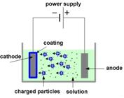
The suspension pH was carefully adjusted to 4 using nitric acid (HNO3) after verification with a calibrated pH meter (WTW, Inolab Level 1). Dimethylformamide (DMF, 10% vol., Merck) was added to enhance coating strength and to promote the formation of a homogeneous and adherent layer on the substrate surface. Coating experiments were conducted at 40 V and 80 V for a deposition time of 90 s. Although 60 V was considered an intermediate parameter, two representative voltages (40 V and 80 V) were selected for comparative evaluation, based on literature data and preliminary experiments.
After deposition, the coated specimens were dried under ambient air conditions and subjected to a heat treatment at 800 °C for two hours in a protective gas atmosphere furnace to improve coating crystallinity, adhesion, and stability.
2.4. Characterization test of coating
After the coating processes, analyses, and tests were performed to evaluate the surface properties and structural characteristics of the Ti-6Al-4V implant materials. We assessed biocompatibility-related features of the coatings through measurements of surface roughness, contact angle, and tape tests. Morphological and elemental analyses were carried out using a Scanning Electron Microscope equipped with Energy Dispersive X-ray Spectroscopy (SEM-EDX, LEO 1430 VP). The measurements were conducted at a magnification of 1,000X, with elemental mapping performed on regions approximately 10 µm in size. For the structural analysis of the coatings, we used X-ray Diffraction (XRD) with a Shimadzu XRD-6000 instrument. The diffraction patterns were obtained in the range of 2θ = 10–80°, allowing us to identify crystalline phases such as Tiα, Tiβ, HA, and Gr.
Contact angle measurements were conducted using a One Attension Theta Lite instrument to evaluate the wettability of the coated surfaces. This technique involves assessing the droplet’s shape, which is influenced by the liquid’s surface tension and the density difference between the liquid and the surrounding medium. This analysis provides insights into the hydrophilic or hydrophobic characteristics of the coatings. Surface roughness tests were performed by calculating the arithmetic mean roughness (Ra) values from five measurements obtained on the coated plate surfaces. The average values were analyzed to assess the impact of coating type and deposition parameters on the surface topography. Tape testing was conducted with a cross-hatch tester to evaluate the mechanical adhesion strength of the coatings. This test followed the procedures outlined in ISO 2409 and ASTM D3359 standards. During the test, vertical and horizontal cuts were made in a cross-hatch pattern on the coated surfaces. The performance of the coating adhesion was assessed based on the classifications defined in the relevant standards, specifically regarding the degree of coating detachment along the cross-hatch areas.
3. RESULTS AND DISCUSSION
3.1. Structural analysis
After coating the Ti-6Al-4V sheets at 40 V and 80 V for 90s, structural characterization was performed using XRD. The most consistent and uniform results were achieved at 80 V for 90s, which provided the optimal coating thickness. Before the analysis, the coated surfaces were treated with a carbon layer, and measurements were conducted within the 2θ range of 10–80°, comparing the results against standard literature data.
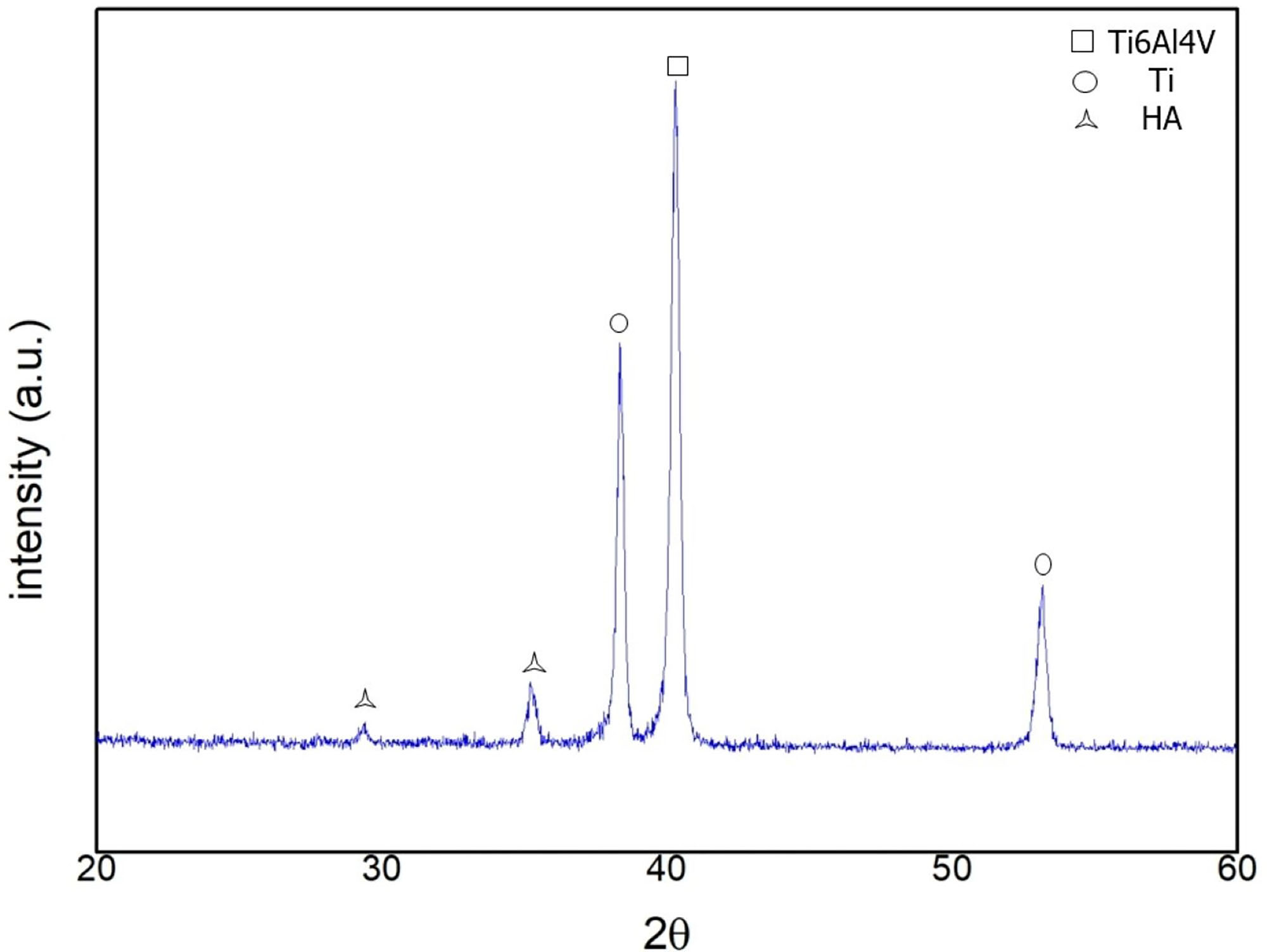
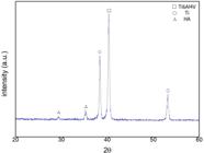
The HA-based coating layers on the implant materials were confirmed using Cu-Kα radiation. The representative XRD spectrum for the coating performed at 80 V for 90s, using a 100% HA suspension, is illustrated in Figure 2. The strongest diffraction peak was observed at 2θ = 41°, followed by notable Tiβ peaks at 2θ = 38° and 53°. Additionally, HA phases were identified, demonstrating the successful deposition and crystallization of the hydroxyapatite layer under the specified conditions. The dominant Tiα and Tiβ peaks are attributed to the inherent alloy composition of the Ti-6Al-4V substrate, which is also supported by previous studies [23].
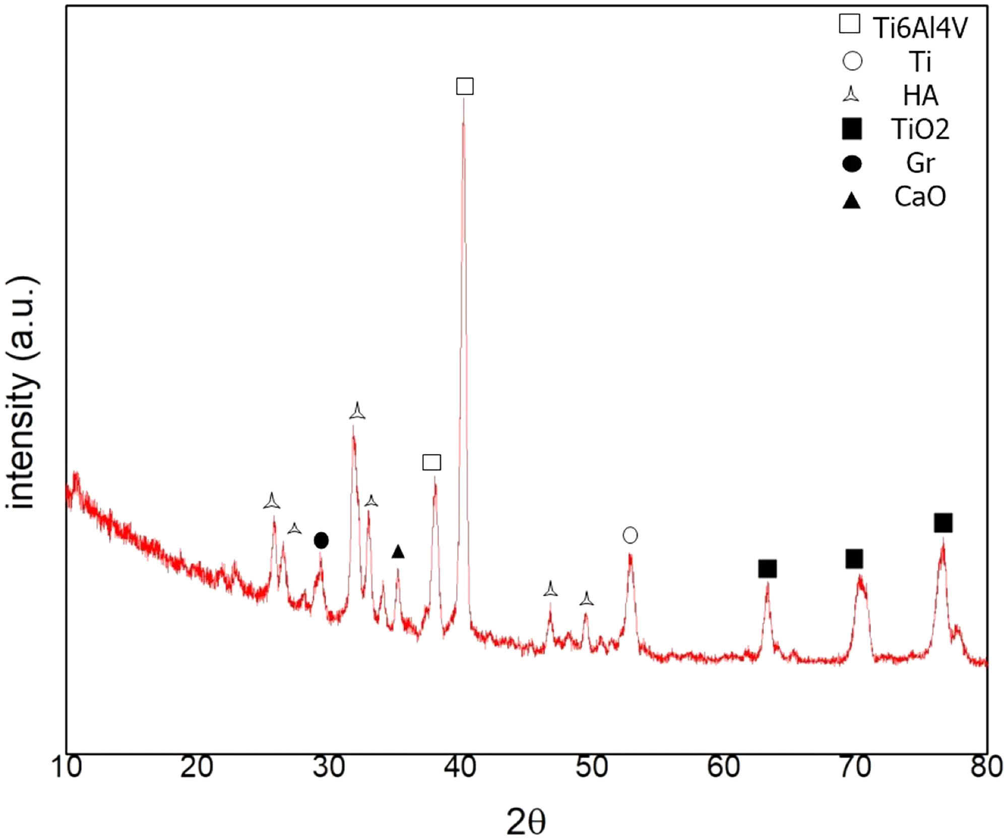
The XRD patterns of Ti-6Al-4V samples coated with HA–1%Gr suspensions under the 80 V-90s regime are presented in Figure 3. The analysis confirmed the presence of Tiα, Tiβ, HA, and Gr phases. Significant Tiα peaks were observed at 2θ = 41°, 63°, 70°, and 77°, while Tiβ peaks were detected at 2θ = 38° and 53°. The formation of Tiα and Tiβ phases was attributed to the addition of Gr and the influence of the applied voltage, with thermal effects during heat treatment also playing a role in their crystallization.
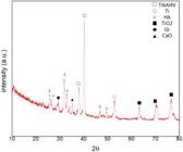
Characteristic peaks for HA were identified at 2θ = 26°, 27°, 32°, 33°, 47°, and 49°, confirming the successful incorporation of hydroxyapatite into the coating structure. Furthermore, the presence of Gr was evident through a diffraction peak at 2θ = 28°, which arose from the inclusion of Gr in the suspension formulation [24]. These findings indicate that the addition of Gr modifies the crystallographic structure and enhances the composite nature of the HA-Ti-6Al-4V system, supporting its potential for biomedical applications.
3.2. Morphology-elemental analysis
The coating experiments revealed that the optimal deposition conditions were achieved at voltages between 40 and 80 V for a duration of 90s. Coatings produced under these conditions exhibited relatively uniform coverage but did not adhere completely to the substrate surface. At voltages exceeding 80V, the coatings showed an increase in fissures and microcracks, likely due to excessive particle deposition and localized stress accumulation during the electrophoretic process. These findings suggest that while 40 to 80V is an ideal operational range, higher voltages undermine coating integrity and mechanical stability, thereby limiting their suitability for use in implant materials.
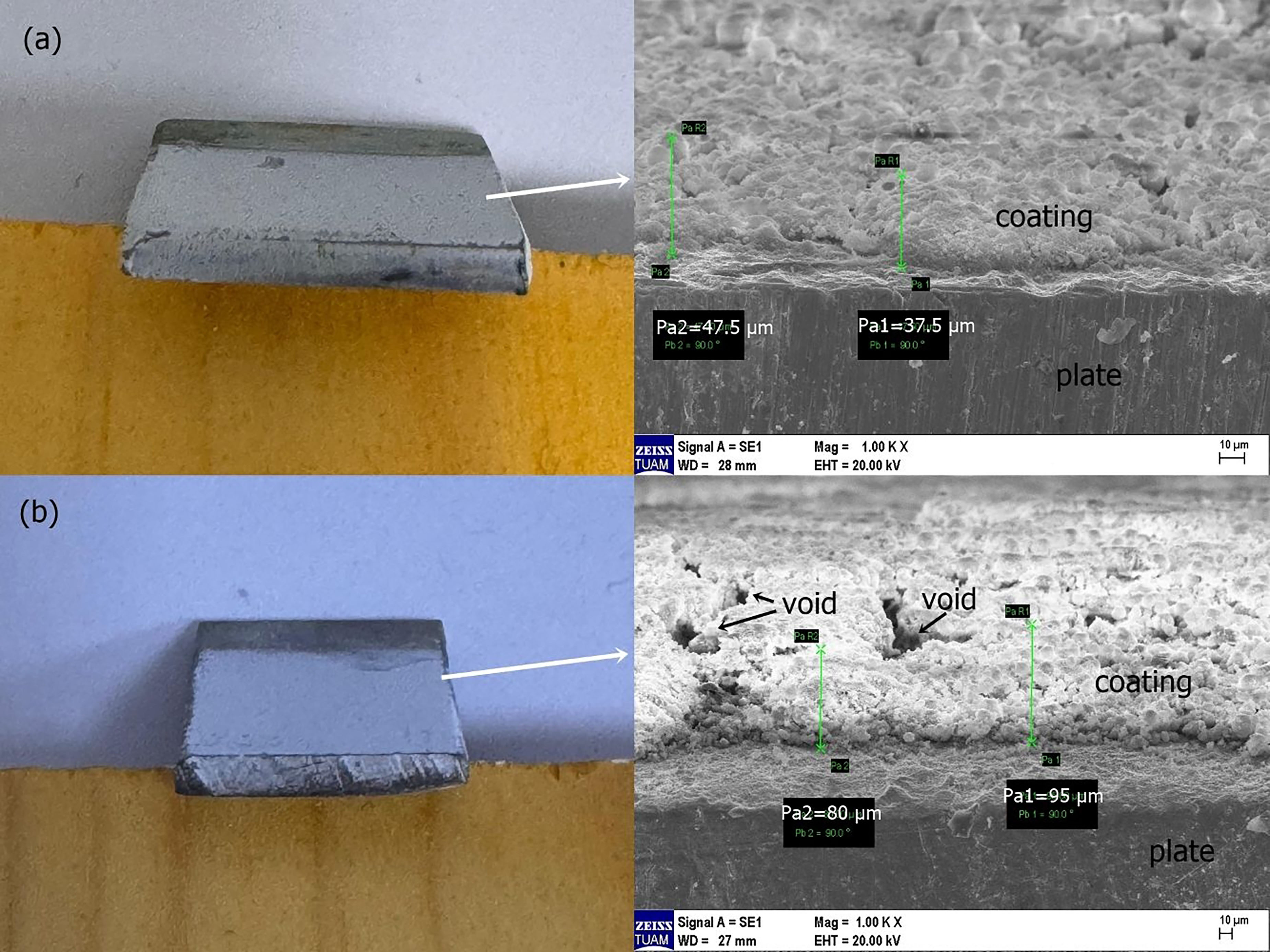
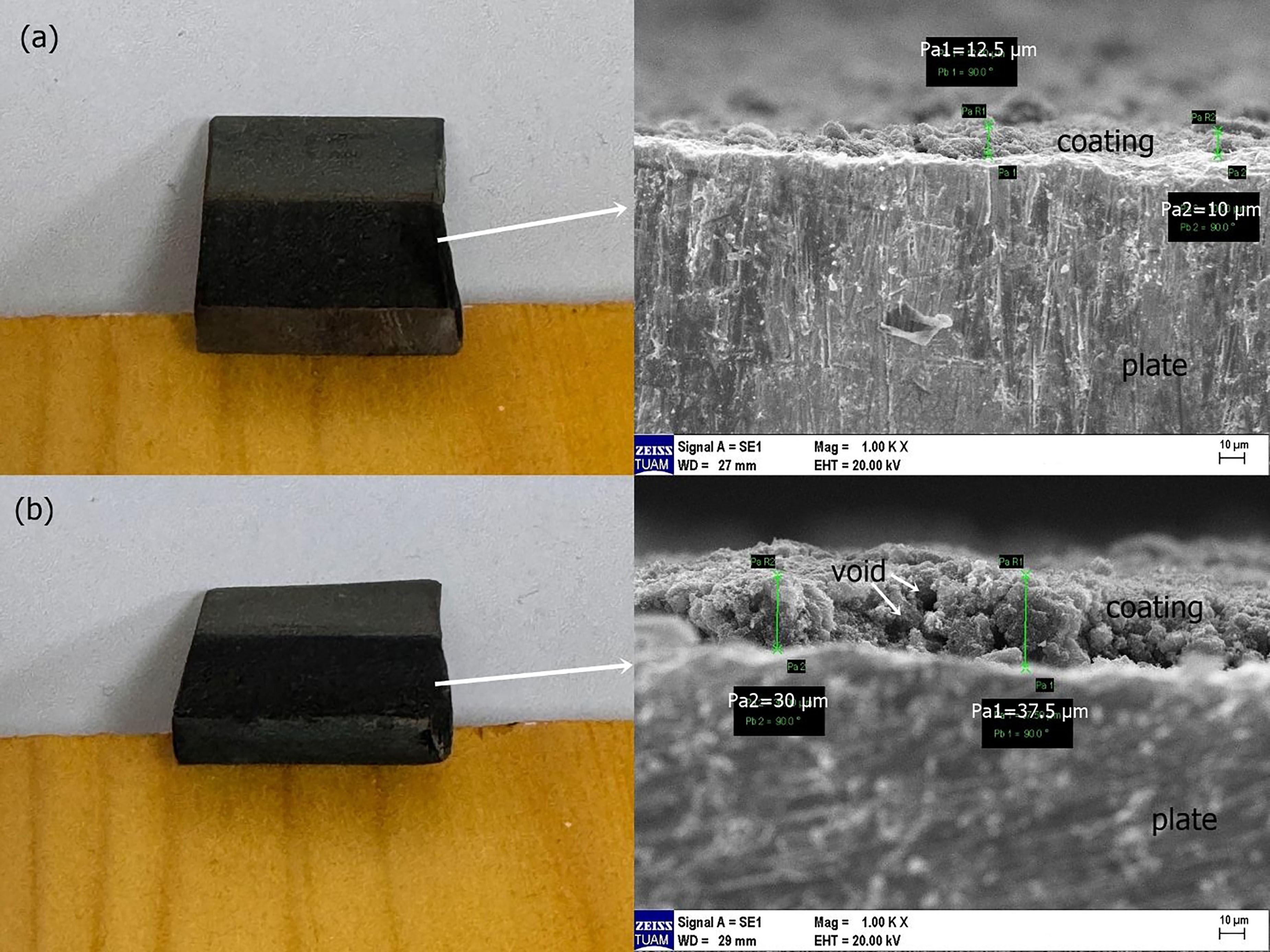
The surface morphologies of the Ti-6Al-4V specimens coated with HA-based suspensions were examined using SEM, as depicted in Figure 4. Figures 4a and 4b show the front-edge regions of the panels that were sanded and then carbon-coated. Cross-sectional SEM measurements indicated that the coating thickness varied between 37 µm and 87.5 µm, depending on the applied voltage.
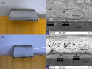
These results are comparable to the findings of NARANJO et al. [25], who coated TiNi shape memory alloys with HA using the electrophoretic deposition (EPD) method at voltages ranging from 20 to 40V and deposition times from 30s to 5 min. They reported coating thicknesses in the range of 42 to 57 µm at 40V, which aligns with the findings of this study. Such comparisons highlight the importance of both deposition voltage and time in controlling coating thickness.
In addition to variations in thickness, localized voids were observed within the coatings. These voids are thought to be related to increases in surface tension and the higher voltage values used during deposition, which can lead to heterogeneous particle distribution and microstructural irregularities [26]. Additionally, the uniformity of powder dispersion within the suspension was a critical factor influencing the quality and uniformity of the coatings.
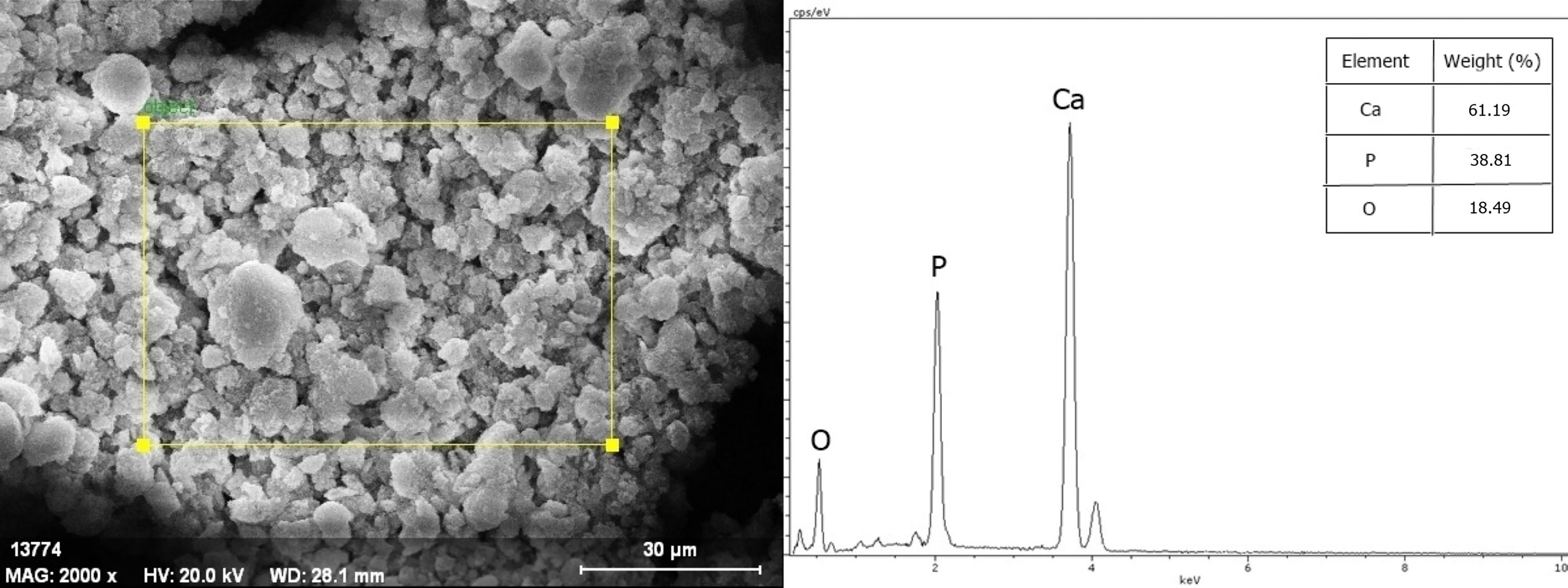
The elemental composition of the HA-coated Ti-6Al-4V samples was analyzed using regional EDX analysis, with a representative spectrum shown in Figure 5. The study confirmed the presence of calcium (Ca), phosphorus (P), and oxygen (O) in the coating layer. The detection of Ca and P is directly associated with the HA powders used in the suspension. Additionally, the oxygen content is believed to have originated both from the HA structure and from oxide formation during the heat treatment process.
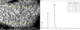
Quantitative EDX results revealed that the coating contained 61.19 wt.% Ca and 38.81 wt.% P. Using these values, the calculated Ca/P molar ratio was 1.6, which is very close to the stoichiometric value of 1.67 reported in the literature for hydroxyapatite. This result indicates that the prepared coatings-maintained a near-stoichiometric HA structure, confirming the effectiveness of the deposition process and the thermal stability of the coatings [27].
After applying the voltage and time, the coating’s homogeneity and surface qualities were improved through heat treatment. The heat treatment was done at 800 ˚C for 2h. The objective is to ensure the coating’s adherence to the implant material while optimizing its thickness and durability (Table 3).
In addition to pure HA suspensions, coatings were performed using HA-based suspensions doped with Gr on Ti-6Al-4V sheets. SEM analysis revealed that the average coating thicknesses varied between 11.25 µm and 33.75 µm depending on the applied voltage (Figure 6). These results demonstrate that Gr incorporation affects the deposition kinetics and final coating morphology.
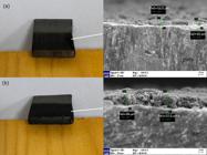
Comparable results have been reported by LIU et al. [28], who developed silver–graphene coatings with enhanced electrical conductivity. By doping suspensions with 5–30 g/L of graphene at 3 V for 2 min, they obtained coating thicknesses up to 23.5 µm. The present study’s SEM measurements performed across multiple regions confirmed coating thickness variations. They revealed the presence of regional and superficial cavities, which are attributed to voltage changes during the EPD process.
Despite the occurrence of voids, both coating thickness and adhesion to the substrate improved with Gr addition. Furthermore, post-deposition heat treatment at 800 °C for two hours enhanced coating homogeneity and surface quality. This treatment was particularly effective in improving coating adherence to the Ti-6Al-4V implant substrates while also optimizing the thickness and durability of the HA–Gr layers.
The formation of voids within the HA–Gr coatings is due to the combined effects of increased voltage and surface tension during the deposition process. Additionally, the diffusion zones between the Ti-6Al-4V substrate and the coating layer became more pronounced, indicating improved interfacial bonding. As shown in Figure 6, the gray regions represent Gr structures, while the lighter areas correspond to HA phases, confirming the successful incorporation of graphene within the composite layer. The observed increase in coating thickness with higher voltage values further suggests that the suspension mixture reached a more homogeneous and balanced state, enhancing the overall quality of the coating [29].
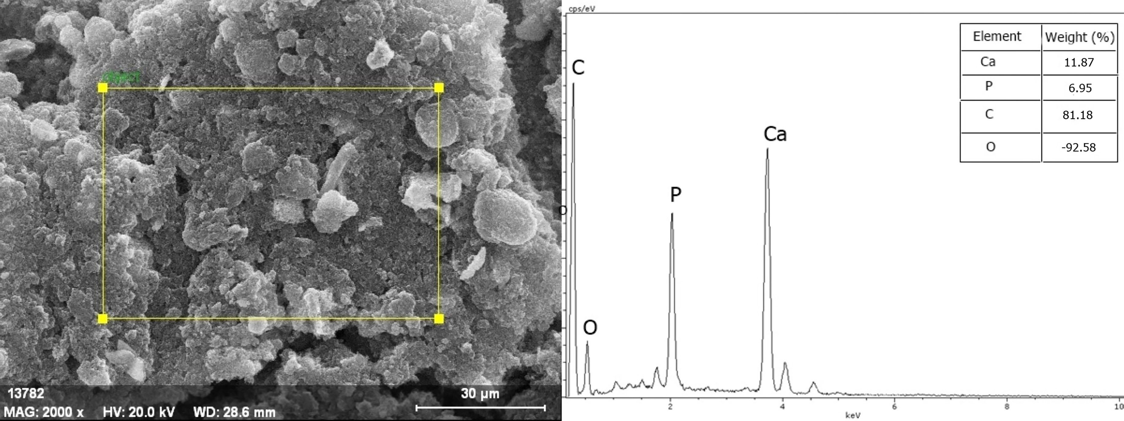
The elemental composition of the coatings obtained with HA–Gr suspensions was investigated by EDX, and the representative spectrum is shown in Figure 7. The analysis revealed the presence of Ca, P, C, and O elements within the coating layer. Quantitative results showed Ca at 11.87wt.% and P at 6.95wt.%, corresponding to a Ca/P molar ratio of approximately 1.7. This value is in close agreement with the stoichiometric ratio of 1.67 reported in the literature, indicating that HA powders were homogeneously distributed within the suspension and successfully incorporated into the coating layer.
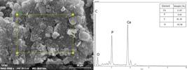
The presence of carbon was attributed to the graphene addition in the suspension, although minor contributions from surface contamination during the coating process cannot be excluded. Moreover, variations in applied voltage may have influenced the incorporation of carbon and the elemental distribution observed in the coating [30, 31]. These findings confirm the effective integration of Gr within the HA-based structure, while maintaining the near-stoichiometric composition essential for biocompatibility.
3.3. Contact angle results
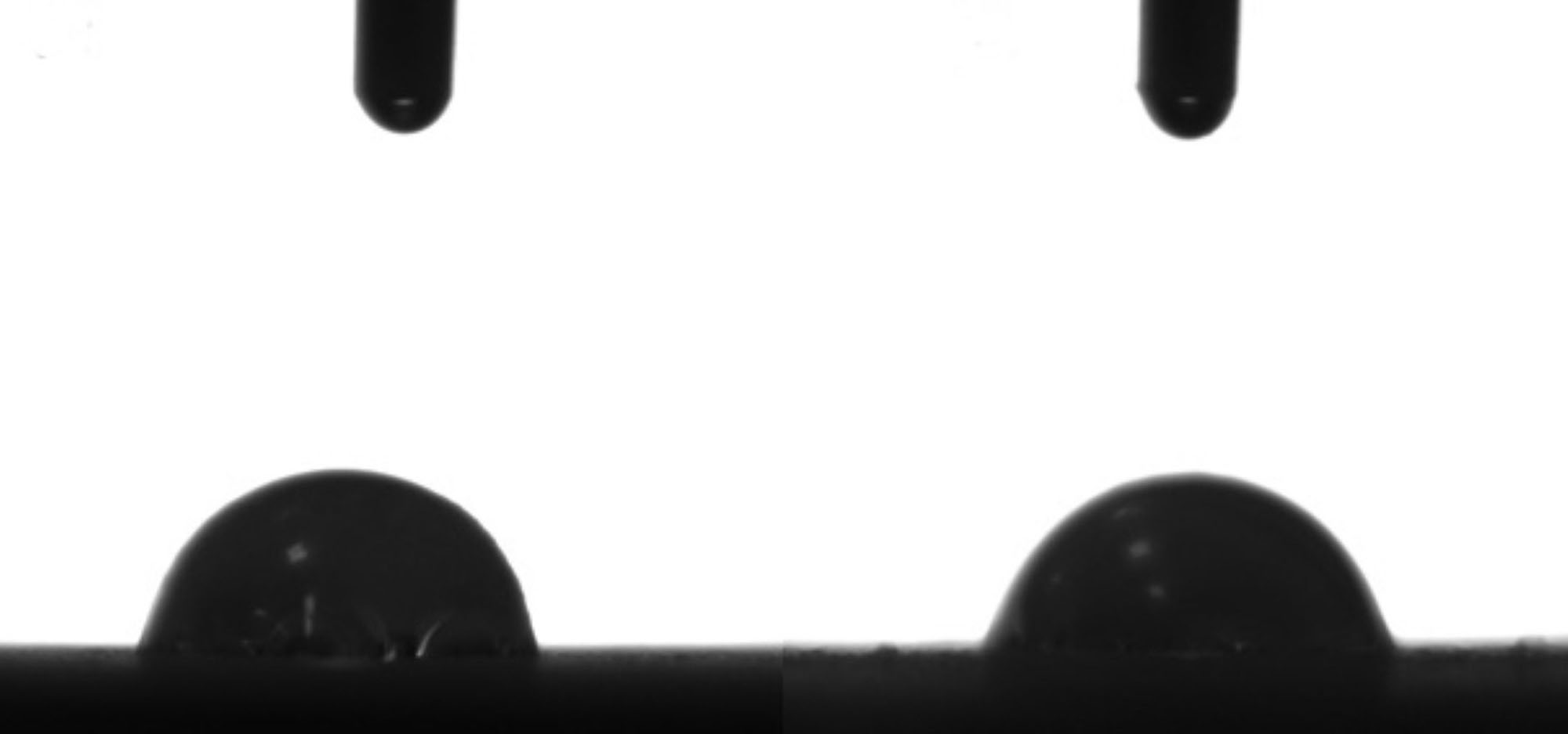
The wettability of the coated Ti-6Al-4V surfaces was evaluated by contact angle (θ) measurements, which quantify the extent of liquid wetting on a solid surface. Geometrically, the contact angle is formed at the three-phase boundary between liquid, gas, and solid. For the coatings prepared under the 80 V–90s regime using the HA suspension, an average contact angle value of 73.32 ± 0.02° was obtained (Figure 8, Table 4). According to these results, the HA-coated surfaces exhibit a hydrophilic character, indicating favorable wetting behavior for initial cell attachment and osseointegration [32].
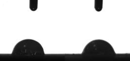
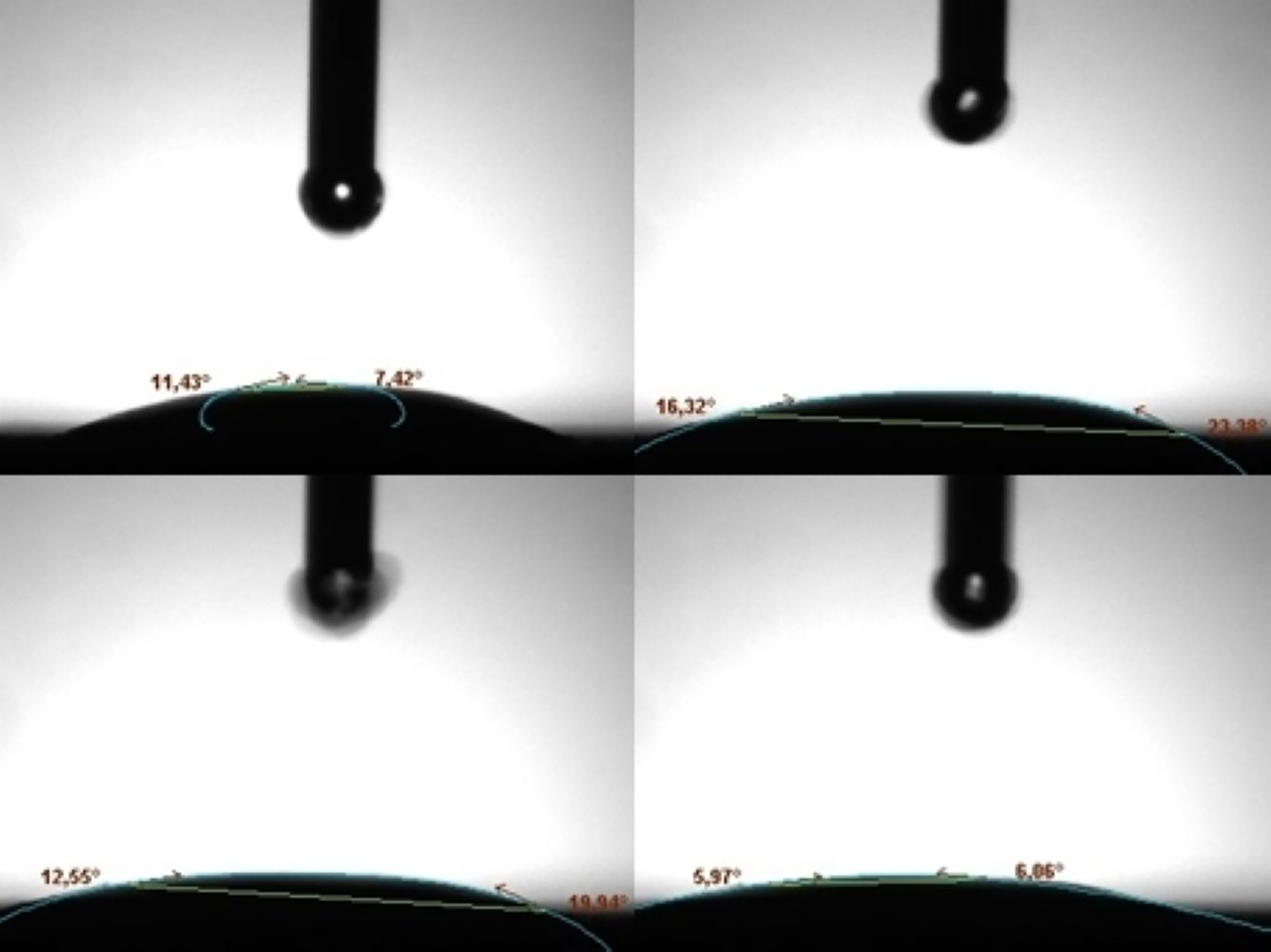
The wettability of the Ti-6Al-4V surfaces coated with HA–Gr suspensions under the 80 V–90s regime was also investigated. The average contact angle value was determined to be approximately 19° (Figure 9, Table 5). This result indicates that the HA–Gr coatings exhibit super-hydrophilic properties, which are expected to enhance bioactivity and promote osseointegration. The super-hydrophilic behavior may be attributed to increased surface tension on the coated surfaces as well as incomplete suspension of Gr powders during the coating process. Similar findings have been reported by WEIWEI et al. [33], who coated graphene oxide/hydroxyapatite (GO/HA) composites using the EPD method. They reported contact angle values of 20.6°, 25.5°, 32°, and 38.1° for HA, 5GO/HA, 8GO/HA, and 12GO/HA coatings, respectively, with an average contact angle of 26° for GO-containing structures. The present results are consistent with these values, further supporting the positive influence of graphene incorporation on surface wettability.

Contact angle measurement after coating on Ti-6Al-4V plates in HA-Gr suspension at 80V-90s.
3.4. Surface roughness test results
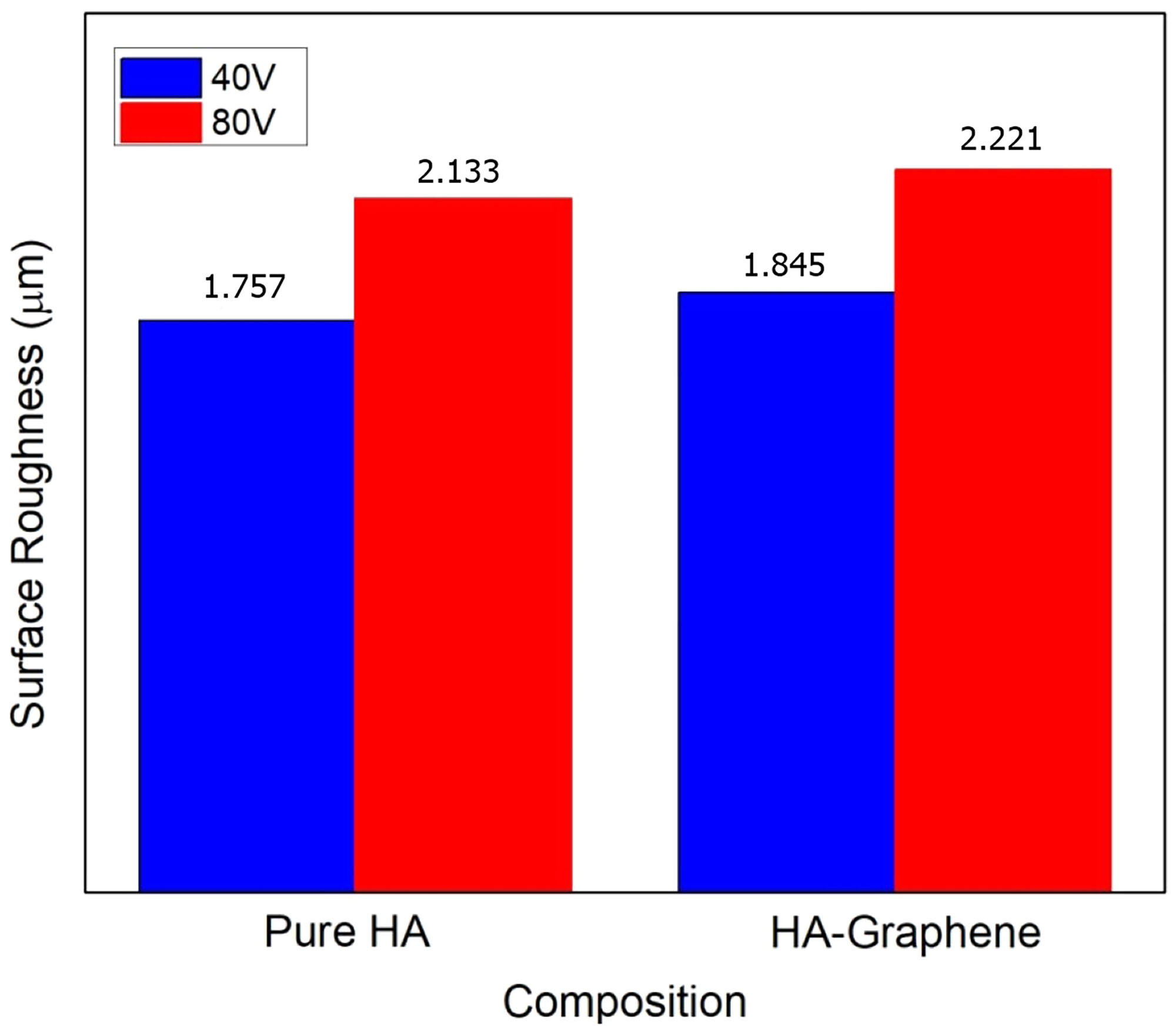
Surface roughness measurements were performed on the coated and uncoated Ti-6Al-4V surfaces, and the arithmetic mean roughness (Ra) values were calculated by averaging at least five independent measurements. The results, expressed in micrometers (µm), are presented in Figure 10 and Table 6.
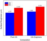
Graph of surface roughness results for sheets coated with pure hydroxyapatite (HA) and HA-Graphene.
The analysis revealed that surface roughness values increased with applied voltage. This increase may be attributed to residual stresses generated on the metal surfaces during deposition or partial disintegration of the coating layer at higher voltages. Nevertheless, the surface profiles appeared relatively smooth, indicating that the coatings adhered uniformly to the substrate.
The type of powder material used in the suspension also influenced surface roughness. For example, coatings produced under the 80 V–90s regime exhibited Ra values of 2.133 µm and 2.221 µm, respectively. As observed in the SEM images, localized microcracks and surface discontinuities contributed to these increases in roughness. Similar findings have been reported in the literature, where roughness values were closely linked to voltage and coating parameters [34, 35].
3.5. Tape-test results
The adhesion strength between the coating and the Ti-6Al-4V substrate is a critical factor that directly influences implant materials’ long-term durability and reliability under physiological conditions. The adhesion behavior of the coatings was evaluated using the cross-hatch tape test per the ASTM D3359 standard method.
For this test, vertical and horizontal incisions were applied to the coated surfaces to create a cross-hatch pattern. Subsequently, an adhesive tape (3M Scotch) was firmly applied to the scratched regions and rapidly peeled off. The extent of coating removal and the resulting surface damage were examined using an optical microscope to determine the degree of adhesion.
The adhesion strength between the coating and the Ti-6Al-4V substrate was evaluated using the cross-hatch tape test (ASTM D3359 standard). For this purpose, vertical and horizontal incisions were applied to the coated surfaces to form a cross-hatch pattern, after which adhesive tape (3M Scotch) was used and rapidly peeled off. The degree of coating detachment was then examined under an optical microscope.
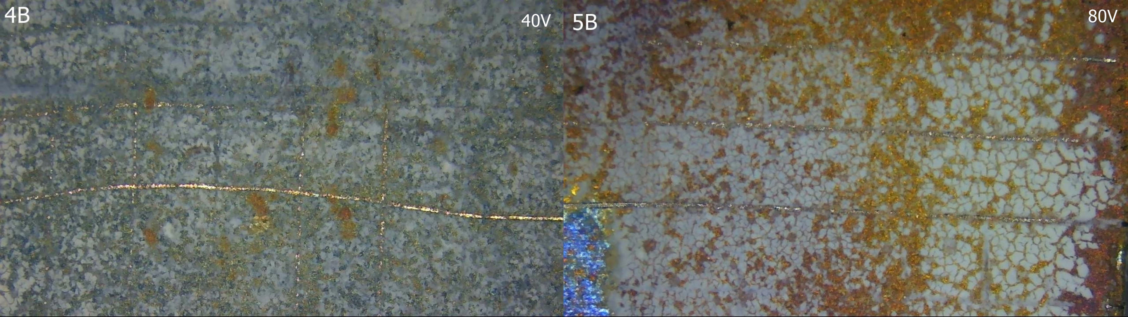
As shown in Figure 11, regional variations in line thickness were observed after the test. For samples coated under 40 V, the adhesion was classified as 4B, indicating minor coating removal in localized regions. With increasing deposition time and voltage, the adhesion performance improved significantly. The best adhesion was obtained under the 80V and 90s condition, where the coatings were classified as 5B, demonstrating excellent adhesion with no visible coating detachment. These results suggest that higher deposition voltages promoted stronger bonding of HA powders to the Ti-6Al-4V surfaces, confirming the effectiveness of the optimized coating parameters [36].
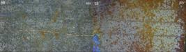
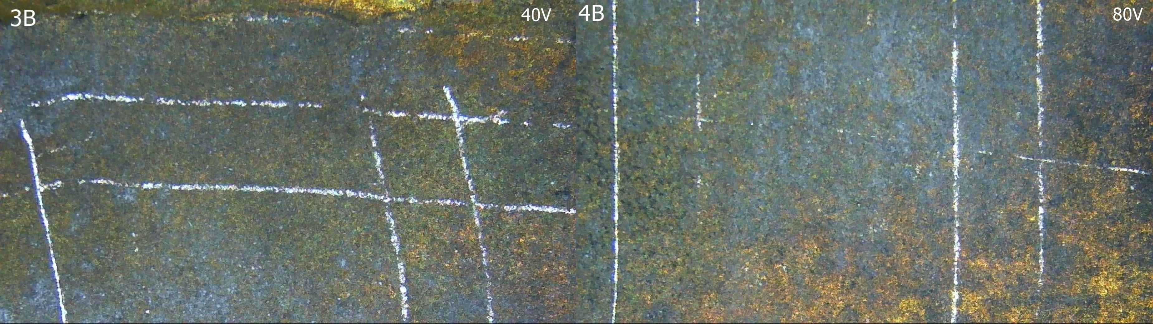
The adhesion strength of the HA–Gr coatings was examined using the tape test under deposition conditions of 40 V and 80 V (Figure 12). According to ASTM classification, the adhesion levels of the coated samples were determined to be 3B and 4B, respectively. These results indicate moderate to good adhesion performance, with minimal coating removal observed after tape application.

Variations in scratch morphology were influenced by the content of graphene in the suspension and the applied deposition parameters. It is believed that changes in voltage directly affected coating thickness, which in turn impacted the adhesion behavior observed in the tape test results. While incorporating Gr improved wettability and surface characteristics, the adhesion strength was lower than that of pure HA coatings. This suggests that further optimization of suspension stability and deposition conditions may be necessary to maximize mechanical performance.
4. CONCLUSION
In this study, the results of HA and HA-based graphene-coated Ti-6Al-4V sheets at different voltages were investigated. As a result of the study:
-
Coating Parameters: This study aimed to identify the most suitable voltage and deposition time for producing HA-based coatings on Ti-6Al-4V implant substrates. Among the investigated parameters, the optimal results were obtained at 80V for 90s, which provided the most homogeneous and adherent coatings. Under these conditions, the maximum coating thickness reached 87.5 µm. Despite internal stresses and localized effects of voltage variation, the coatings exhibited uniform morphology and stable structural properties. These findings confirm that the 80V–90s regime represents the most effective deposition condition for enhancing the surface characteristics of Ti-6Al-4V implant materials.
-
Surface Wettability and Adhesion: The type of coating material significantly influenced the wettability and adhesion characteristics of the Ti-6Al-4V surfaces. Pure HA coatings demonstrated hydrophilic behavior and were favorable for cell attachment and biocompatibility. In contrast, HA-Gr coatings exhibited super-hydrophilic properties, further enhancing surface energy and potentially promoting superior biointegration. Regarding adhesion performance, HA coatings achieved stronger bonding with the substrate (up to 5B classification). In contrast, HA–Gr coatings showed moderate to good adhesion (3B–4B), influenced by voltage variations and graphene content.
-
Mechanical strength properties: Significant improvements were observed in surface roughness, adhesion, and overall coating strength of the Ti-6Al-4V implants after applying HA-based coatings. In particular, the tape test results demonstrated strong adhesion, with 4B and 5B classifications, indicating excellent bonding between the coating and the substrate. The increase in deposition voltage contributed to enhanced adhesion performance, which is expected to improve the long-term durability of the coated implant materials. These findings confirm that optimized EPD parameters not only enhance coating uniformity but also extend the service life of Ti-6Al-4V implants by strengthening the mechanical stability of the surface layers.
-
Future studies: Future investigations will evaluate in vitro and in vivo biocompatibility and corrosion resistance, among the most critical parameters in assessing biomaterial coatings. Due to financial and laboratory limitations, these studies could not be performed within the present work. Planned in vitro and in vivo biocompatibility tests will provide essential insights into the biological response and integration of the coatings with bone and surrounding tissues. Furthermore, corrosion testing will be conducted to determine the degradation behavior of the coated Ti-6Al-4V implants under physiological conditions. The results of these studies will be used to optimize coating properties further, ensuring both long-term stability and functional performance of the implants in clinical applications.
This study showed that incorporating advanced materials like Gr into HA-based suspensions significantly enhance the coating performance on Ti-6Al-4V substrates. Due to its superior physicochemical properties, graphene helps optimize suspension stability, improve coating homogeneity, and refine surface characteristics. The resulting HA–Gr composite coatings demonstrated better wettability, favorable roughness values, and strong adhesion, all of which are essential for biomedical implant applications. These findings indicate that graphene-reinforced coatings are highly promising for enhancing both the surface and mechanical properties of Ti-6Al-4V implants, thereby extending their potential use and functional lifespan within the human body.
5. ACKNOWLEDGMENTS
This study was supported by Afyon Kocatepe University BAPK 21.MYO.02. Many thanks to the Scientific Research Projects Commission for their support.
6. BIBLIOGRAPHY
-
[1] HOMA, K., ZAKRZEWSKI, W., DOBRZYŃSKI, W., et al., “Surface functionalization of titanium-based implants with a nanohydroxyapatite layer and its impact on osteoblasts: a systematic review”, Journal of Functional Biomaterials, v. 15, n. 2, pp. 45, 2024. doi: http://doi.org/10.3390/jfb15020045. PubMed PMID: 38391898.
» https://doi.org/10.3390/jfb15020045 -
[2] YU, Y.T., LEE, W.H., HSIEH, S.F., et al., “Effects of hydroxyapatite size on the drug release characteristics of drug-containing coatings”, Progress in Organic Coatings, v. 195, pp. 108600, 2024. doi: http://doi.org/10.1016/j.porgcoat.2024.108600.
» https://doi.org/10.1016/j.porgcoat.2024.108600 -
[3] MA, H.Y., WANG, J.C., QIN, P., et al., “Advances in additively manufactured titanium alloys by powder bed fusion and directed energy deposition: microstructure, defects, and mechanical behavior”, Journal of Materials Science and Technology, v. 183, pp. 32–62, 2024. doi: http://doi.org/10.1016/j.jmst.2023.11.003.
» https://doi.org/10.1016/j.jmst.2023.11.003 -
[4] SHANKAR, D., JAMBAGI, S.C., “Improvements in bioactivity, blood compatibility, and wear resistance of thermally sprayed carbon nanotube reinforced hydroxyapatite-based orthopedic implants”, Tribology International, v. 197, pp. 109809, 2024. doi: http://doi.org/10.1016/j.triboint.2024.109809.
» https://doi.org/10.1016/j.triboint.2024.109809 -
[5] GUO, Y., YAN, X., YANG, Y., et al., “Graphene oxide/ hydroxyapatite composite coatings on additively manufactured NiTi alloy for biomedical applications”, Ceramics International, v. 50, n. 1, pp. 2479–2489, 2024. doi: http://doi.org/10.1016/j.ceramint.2023.11.041.
» https://doi.org/10.1016/j.ceramint.2023.11.041 -
[6] ANNUR, D., HASANAH, L.N., KARTIKA, I., et al., “Effect of hydroxyapatite concentration and electrophoretic deposition (EPD) parameter process on hydroxyapatite/chitosan coating quality for porous titanium as an orthopedic implant”, The Canadian Journal of Metallurgy and Materials Science, v. 64, n. 1, pp. 35–42, 2024. doi: https://doi.org/10.1080/00084433.2024.2310339.
» https://doi.org/10.1080/00084433.2024.2310339 -
[7] SUN, M., CHEN, X., WU, Z., et al., “Numerical simulation and experimental study of deposition behavior for cold sprayed dual nano HA/30 wt.% Ti composite particle”, Coatings, v. 14, n. 10, pp. 1330, 2024. doi: http://doi.org/10.3390/coatings14101330.
» https://doi.org/10.3390/coatings14101330 -
[8] JALALI BIDAKHAVIDI, M., OMIDVAR, H., ZAMANIAN, A., et al., “Characterization, wear behavior and biocompatibility of HA/Ti composite and functionally graded coatings deposited on Ti−6Al−4V substrate by mechanical coating technique”, The Nonferrous Metals Society of China, v. 34, n. 2, pp. 547–559, 2024. doi: http://doi.org/10.1016/S1003-6326(23)66416-7.
» https://doi.org/10.1016/S1003-6326(23)66416-7 - [9] MANPREET, K., PRASHANT, K., HARJIT, S.G., “A review on advances in photoelectrochemical (PEC-type) photodetectors: a trending thrust research area”, International Journal of Hydrogen Energy, v. 49, n. Pt C, pp. 1095–1112, 2024. doi.http://doi.org/10.1016/j.ijhydene.2023.11.018.
-
[10] GUNDUZ, D.O., KUÇUKTURK, G., PUL, M., et al., “Focus on advanced materials and processes on sustainability”, Materials Research Express, v. 11, n. 6, pp. 066522, 2024. doi: http://doi.org/10.1088/2053-1591/ad59ed.
» https://doi.org/10.1088/2053-1591/ad59ed -
[11] GUTIÉRREZ-MEJÍA, F.A., VÁSQUEZ-LÓPEZ, C., VARGAS-CORONADO, R.F., et al., “Electrophoretic deposition of TiO2 nanotubes and antibiotics on polyurethane coated stainless steel for improved antibacterial response and cell viability”, Materials Today. Communications, v. 40, pp. 109428. doi: http://doi.org/10.1016/j.mtcomm.2024.109428.
» https://doi.org/10.1016/j.mtcomm.2024.109428 -
[12] PAWŁOWSKI, Ł., MANIA, S., BANACH-KOPEĆ, A., et al., “The influence of chitosan’s molecular weight, concentration, and is suspension method on the properties of electrophoretically deposited coatings on the Ti13Nb13Zr alloy surface”, Materials Chemistry and Physics, v. 331, pp. 130174, 2025. doi: http://doi.org:10.1016/j.matchemphys.2024.130174.
» https://doi.org/10.1016/j.matchemphys.2024.130174 -
[13] SOHRABPOOR, H., SALARVAND, V., YAZDI, M.S., et al., “Enhancing bioactivity and corrosion resistance of laser powder bed-fused Ti-6Al-4V using titania-reinforced hydroxyapatite nanocomposite coatings through electrophoretic deposition”, SSRN, 2024. Preprint. doi: http://doi.org/10.2139/ssrn.4942457.
» https://doi.org/10.2139/ssrn.4942457 -
[14] MAKURAT-KASPROLEWICZ, B., OSSOWSKA, B.A., “Electrophoretically deposited titanium and its alloys in biomedical engineering: recent progress and remaining challenges”, Journal of Biomedical Materials Research. Part B, Applied Biomaterials, v. 112, n. 1, pp. e35342, 2024. doi: http://doi.org/10.1002/jbm.b.35342. PubMed PMID: 37905698.
» https://doi.org/10.1002/jbm.b.35342 -
[15] PAWŁOWSKI, Ł., BARTMAŃSKI, M., STRUGAŁA, G.A., et al., “Electrophoretic deposition and characterization of chitosan/eudragit E 100 coatings on titanium substrate”, Coatings, v. 10, n. 7, pp. 607, 2020. doi: http://doi.org/10.3390/coatings10070607.
» https://doi.org/10.3390/coatings10070607 -
[16] KIRMAN, M., DOGAN, H., DIKICI, T., “Deposition of HA-GO on ASO modified Ti surface by EPD method and characterization of surface and biological properties of the coating”, Ceramics International, v. 51, n. 3, pp. 3703–3713, 2024. doi: http://doi.org/10.1016/j.ceramint.2024.11.346.
» https://doi.org/10.1016/j.ceramint.2024.11.346 -
[17] HADZHIEVA, Z., BOCCACCINI, A.R., “Recent developments in electrophoretic deposition (EPD) of antibacterial coatings for biomedical applications – a review,” Current Opinion in Biomedical Engineering, v. 21, pp. 100367, 2022. doi: http://doi.org/10.1016/j.cobme.2021.100367.
» https://doi.org/10.1016/j.cobme.2021.100367 -
[18] OKUNIEWSKI, W., WALCZAK, M., SZALA, M., “Effects of shot peening and electropolishing treatment on the properties of additively and conventionally manufactured Ti-6Al-4V alloy: a review”, Materials (Basel), v. 17, n. 4, pp. 934, 2024. doi: http://doi.org/10.3390/ma17040934. PubMed PMID: 38399186.
» https://doi.org/10.3390/ma17040934 -
[19] BANSAL, G., GAUTAM, R.K., MISRA, J.P., et al., “Synthesis and characterization of poly (methyl methacrylate)/silver-doped hydroxyapatite dip coating on Ti-6Al-4V”, Colloids and Surfaces A: Physicochemical and Engineering Aspects, v. 689, pp. 133662, 2024. doi: http://doi.org/10.1016/j.colsurfa. 2024.133662.
» https://doi.org/10.1016/j.colsurfa.2024.133662 -
[20] BAHETI, W., LV, S., MILA., et al., ”Graphene/hydroxyapatite coating deposit on titanium alloys for implant application”, Journal of Applied Biomaterials & Functional Materials, v. 21, pp. 22808000221148104, 2023. doi: http://doi.org/10.1177/22808000221148104.
» https://doi.org/10.1177/22808000221148104 -
[21] IATSENKO, A., SYCH, O., NIKOLENKO, A., et al., “Structure Investigation of highly porous bioceramics based on biogenic hydroxyapatite with graphene oxide coating”, Results in Surfaces and Interfaces, v. 6, pp. 100265, 2024. doi: https://doi.org/10.1016/j.rsurfi.2024.100265.
» https://doi.org/10.1016/j.rsurfi.2024.100265 -
[22] HU, B., CAO, J., WANG, K., et al., “PI/PAI-E44 synthesis-inspired composite coating with graphene- reinforced hydroxyapatite: enhanced wear resistance, antibacterial properties, biological activity, and corrosion resistance”, Surface and Coatings Technology, v. 497, pp. 131786, 2025. doi: http://doi.org/10.1016/j.surfcoat.2025.131786.
» https://doi.org/10.1016/j.surfcoat.2025.131786 -
[23] DREVET, R., JABER, N.B., FAURÉ, J., et al., “Electrophoretic deposition (EPD) of nano-hydroxyapatite coatings with improved mechanical properties on prosthetic Ti-6Al-4V substrates”, Surface and Coating Technology, v. 301, pp. 94–99, 2016. doi: http://doi.org/10.1016/j.surfcoat.2015.12.058.
» https://doi.org/10.1016/j.surfcoat.2015.12.058 -
[24] LI, B., ZHAO, P., LIU, S., et al., “Preparation and properties of hydroxyapatite/chitosan coatings on Ti surface by different electrochemical methods”, Journal of Materials Engineering and Performance, v. 34, n. 9, pp. 7946–7954, 2025. doi: http://doi.org/10.1007/s11665-024-09741-8.
» https://doi.org/10.1007/s11665-024-09741-8 -
[25] NARANJO, E., QUINCHUELA PAUCAR, J.C., ULLOA, N., et al., “Hydroxyapatite coating of TiNi shape memory alloy via Electrophoretic Deposition (EPD)”, Materials Research Innovations, v. 29, n. 2, pp. 57–63, 2024. doi: http://doi.org/10.1080/14328917.2024.2372897.
» https://doi.org/10.1080/14328917.2024.2372897 -
[26] CHENG, X., LIU, Y., LIU, O., et al., “Electrophoretic deposition of coatings for local delivery of therapeutic agents”, Progress in Materials Science, v. 136, pp. 101111, 2023. doi: http://doi.org/10.1016/j.pmatsci.2023.101111.
» https://doi.org/10.1016/j.pmatsci.2023.101111 -
[27] BALAEI, H., GHASEMI, H.M., AGHDAM, R.M., et al., “The effect of silver nanoparticles on biological and corrosion behavior of electrophoretically deposited hydroxyapatite film on Ti-6Al-4V”, Journal of Materials Science: Materials in Medicine, v. 35, pp. 18, 2024. doi: http://doi.org/10.1007/s10856-024-06784-0.
» https://doi.org/10.1007/s10856-024-06784-0 -
[28] YONG, L., XIJUN, W., DAPENG, L., et al., “Brush plating process and study on corrosion resistance and hardness of modified graphene on copper-based silver-graphene composite coating”, Revista Matéria, v. 29, n. 4, pp. e20240493, 2024. doi: https://doi.org/10.1590/1517-7076-RMAT-2024-0493.
» https://doi.org/10.1590/1517-7076-RMAT-2024-0493 -
[29] YI, J., LI, M., ZHU, J., et al. “Recent development and applications of electrodeposition bio-coatings on medical titanium for bone repair”, Journal of Materials Chemistry B, v. 39, n. 12, pp. 9863–9893, 2024. doi: http://doi.org/10.1039/D4TB01081G.
» https://doi.org/10.1039/D4TB01081G -
[30] MA, Y., HAN, J., WANG, M., et al., “Electrophoretic deposition of graphene-based materials: a review of materials and their applications”, Journal of Materiomics, v. 4, n. 2, pp. 108–120, 2018. doi: https://doi.org/10.1016/j.jmat.2018.02.004.
» https://doi.org/10.1016/j.jmat.2018.02.004 -
[31] CHEN, X., CHEN, S., LIANG, L., et al., “Electrochemical behavior of EPD synthesized graphene coating on titanium alloys for orthopedic implant application”, Procedia CIRP, v. 71, pp. 322–328, 2018. doi: http://doi.org/10.1016/j.procir.2018.05.035.
» https://doi.org/10.1016/j.procir.2018.05.035 -
[32] YU, Y.T., LEE, W. H., HSIEH, S. F., et al., “Effects of hydroxyapatite size on the drug release characteristics of drug-containing coatings”, Progress in Organic Coatings, v. 195, pp. 108600, 2024. doi: http://doi.org/10.1016/j.porgcoat.2024.108600.
» https://doi.org/10.1016/j.porgcoat.2024.108600 -
[33] WEIWEI, H., LI, L., BENRUI, Y., et al., “Electrodeposition of graphene oxide-hydroxyapatite composite coating on titanium substrate”, Ceramics International, v. 49, n. 6, pp. 9647–9656, 2023. doi: https://doi.org/10.1016/j.ceramint.2022.11.135.
» https://doi.org/10.1016/j.ceramint.2022.11.135 -
[34] PAN, M., WENG, Z., LIU, J., “Effect of positive bias on properties of chitosan coating prepared on micro-arc oxidation surface of Ti–6Al–4V alloy by electrophoretic deposition”, Materials Chemistry and Physics, v. 275, pp. 125257, 2022. doi: http://doi.org/10.1016/j.matchemphys.2021.125257.
» https://doi.org/10.1016/j.matchemphys.2021.125257 -
[35] MALISZ, K., ŚWIECZKO-ŻUREK, B., OLIVE, J.M., et al., “Research of chitosan coatings deposited by electrophoretic deposition method at various voltage and time parameters”, Materials Chemistry and Physics, v. 315, pp. 128984, 2024. doi: http://doi.org/10.1016/j.matchemphys.2024.128984.
» https://doi.org/10.1016/j.matchemphys.2024.128984 -
[36] VARLIK, O., GONCU, Y., AY, N., “Electrophoretic deposition of composite titanium diboride-chitosan coating”, Materials Chemistry and Physics, v. 282, pp. 125927, 2022. doi: http://doi.org/10.1016/j.matchemphys.2022.125927.
» https://doi.org/10.1016/j.matchemphys.2022.125927