
 Non-radioactive strontium as a supplement to enhance osseointegration
Abstract
Introduction Due to its positive results on bone tissue, strontium also began to be used as an adjuvant in bone neoformation processes, mainly maxillofacial surgical procedures.
Objective To assess if the non-radioactive strontium (Sr) supplementation enhances the osseointegration of titanium implants in rats.
Material and method Seventy male rats (Rattus Norvegicus) were randomly divided into 5 groups, according to the systemic supplementation: Control - saline solution; SRAN50 - strontium ranelate (SRAN) 50mg/kg/day; SRAN625 - SRAN 625mg/kg/day; SCAR/SCHL30 - strontium carbonate and strontium chloride (SCAR/SCHL) 30mg/kg/day; SCAR/SCHL365 - SCAR/SCHL 365mg/kg/day. The drugs were administered via gavage, once a day, starting 15 days before surgery (1 titanium implant in each tibia), and persisted for 15 or 60 days. The right tibiae were used for biomechanical (removal torque) and immunohistochemical (Osteocalcin – OCN, and bone morphogenetic protein - BMP-2) evaluation. The left were used for microtomographic, and histomorphometric evaluation.
Result Increased removal torque for SRAN625 and SCAR/SCHL365 were observed when compared with the Control, in 15 days. However, no differences were found in the 60-days period among the groups. Microtomographic evaluation showed larger bone volume at 60 days, compared to 15 days, for all groups but SCAR/SCHL30. When all groups were compared, no differences were seen in the 15-days period, while in the 60-days SRAN625 and SCAR/SCHL365 were statistically higher than the Control. In the immunohistochemical analysis, higher doses (SRAN625 and SCAR/SCHL365) led to an increase of BMP-2 in 15 days. Histomorphometric analysis revealed no differences among the groups regarding bone-to-implant-contact and bone area around the implant threads.
Conclusion This study suggests that higher concentrations of systemic Sr lead to variably improved osseointegration-related parameters regarding the biomechanical and microtomographic evaluation.
Descriptors:
Strontium; systemic use; osseointegration; bone remodeling; implants
Resumo
Introdução Devido a seus resultados positivos sobre o tecido ósseo o estrôncio passou a ser utilizado também como coadjuvante de processos de neoformação óssea, principalmente procedimentos cirúrgicos maxilo-faciais.
Objetivo Avaliar se a suplementação de estrôncio (Sr) não radioativo melhora a osseointegração de implantes de titânio em ratos.
Material e método Setenta ratos machos (Rattus Norvegicus) foram divididos aleatoriamente em 5 grupos, de acordo com a suplementação sistêmica: Controle - solução salina; SRAN50 - ranelato de estrôncio (SRAN) 50mg/kg/dia; SRAN625 - SRAN 625mg/kg/dia; SCAR/SCHL30 - carbonato de estrôncio e cloreto de estrôncio (SCAR/SCHL) 30mg/kg/dia; SCAR/SCHL365 - SCAR/SCHL 365mg/kg/dia. Os medicamentos foram administrados por gavagem, uma vez ao dia, iniciando 15 dias antes da cirurgia (1 implante de titânio em cada tíbia), e persistiram por 15 ou 60 dias. As tíbias direitas foram utilizadas para avaliação biomecânica (torque de remoção) e imuno-histoquímica (Osteocalcina – OCN e proteína morfogenética óssea - BMP-2). As esquerdas foram utilizadas para avaliação microtomográfica e histomorfométrica.
Resultado Aumento do torque de remoção para SRAN625 e SCAR/SCHL365 foi observado quando comparado ao Controle, em 15 dias. Entretanto, não foram encontradas diferenças no período de 60 dias entre os grupos. A avaliação microtomográfica mostrou maior volume ósseo em 60 dias, comparado a 15 dias, para todos os grupos, exceto SCAR/SCHL30. Quando todos os grupos foram comparados, não foram observadas diferenças no período de 15 dias, enquanto no período de 60 dias SRAN625 e SCAR/SCHL365 foram estatisticamente maiores que o Controle. Na análise imuno-histoquímica, doses maiores (SRAN625 e SCAR/SCHL365) levaram a um aumento de BMP-2 em 15 dias. A análise histomorfométrica não revelou diferenças entre os grupos quanto ao contato osso-implante e área óssea ao redor das roscas do implante.
Conclusão Este estudo sugere que concentrações mais altas de Sr sistêmico levam a parâmetros relacionados à osseointegração melhorados de forma variável quanto à avaliação biomecânica e microtomográfica.
Descritores:
Estrôncio; uso sistêmico; osseointegração; remodelação óssea; implantes
INTRODUCTION
Strontium (Sr) is a chemical element that belongs to the group of alkaline earth metals, which presents similarities to Calcium, such as incorporation with the bone tissue and high affinity for hydroxyapatite1. Sr is easily absorbed in bone and teeth1 and can exert a “dual action” by stimulating the production of osteoblasts and inducing the apoptosis of osteoclasts, simultaneously2. However, the mechanisms for this dual action are not totally understood. A theory suggests that these mechanisms are associated with the interaction of Sr and calcium-sensing receptors (CaR)3. Another theory is that Sr downregulates the nuclear factor-kappa B (RANK), preventing the ligation of ligand (RANKL) and therefore leading to enhanced production of osteoprotegerin (OPG)4. Both mechanisms suggest that Sr could interfere favorably towards bone formation2.
Emerging Sr-based medications for the treatment/prevention of bone-related pathologies, such as osteoporosis and osteoarthritis, showed good initial outcomes in human trials5-7. Currently, the most known representative of this class of medicaments is Sr ranelate (SRAN). This drug is usually administered in humans daily, in a concentration of 2 g/day8. When this drug was tested in animals, such as rats, the tested concentration was 625 mg/kg/day9, which would, based on the diminished intestinal absorption of the medication by the rats´, leading to plasmatic levels which would be similar to that tested in humans9. Studies testing such concentrations showed promising results regarding in healthy and osteoporosis animals10.
In face of these findings, Sr-based therapies could also benefit in others research areas which also depend on new bone formation, with increased quality and quantity of newly formed bone, such as implant-dentistry11. As an example, one could speculate on possible implant-osseointegration enhancement effects, which would allow faster and more efficient bone formation surrounding the implants, allowing the rehabilitation of the patients to be concluded in in less time12.
Not only SRAN, but also diverse forms of strontium supplementation could be tested for such application. One example would be a new formula based on Sr carbonate and Sr chloride. The protocol for using this new formula was based on the molecular weight of strontium ranelate, being the quantity of the Sr equivalent for the new supplement. For this creation two molecules were mixed in distilled water, being one substance with acid pH (strontium chloride) and one substance with basic pH (strontium carbonate), providing a neutral salt, which would, in theory, keep the pH equilibrium, leading to a better strontium intestinal absorption.
The aim of study was to develop a new Sr-based supplement which could substitute strontium ranelate as an adjunct therapy following the placement of implants, and assess if it enhances the osseointegration of titanium implants inserted in rats’ tibiae by means of biomechanical, histomorphometric, immunohistochemical, and microtomographic evaluation.
MATERIAL AND METHOD
Animals and Diet
This experimental protocol was approved by the Animal Ethics Committee of the University of São Paulo – UNESP - Araraquara-SP (04/2012). Seventy male rats (3 month-old Rattus Norvegicus, albinus variation) were maintained housed in a room with controlled temperature, and access to water and food ad libitum. The animals were randomly divided into five groups(n=14): Control, treated with saline solution; SRAN50, treated with strontium ranelate in the concentration of 50mg/kg/day (Protos; Laboratory Servier, São Paulo, Brasil); SRAN625, treated with strontium ranelate in the concentration of 625mg/kg/day; SCAR/SCHL30, treated with strontium carbonate and strontium chloride (50/50% proportion) in the concentration of 30mg/kg/day (Cromoline Química Fina, Diadema, São Paulo, Brasil); SCAR/SCHL365, treated with strontium carbonate and strontium chloride (50/50% proportion) in the concentration of 365mg/kg/day. All animals received the supplement via gavage, starting 15 days before the installation of implants on their tibiae. The period of 15 days was selected to allow the supplement reaching adequate plasmatic levels before the surgery took place13. Supplementation was always provided in the morning, fitting the rats´ circadian cycle14.
During treatment, the animals were weighted weekly, and their respective supplement dose was adjusted. The adjustment of the dose for the SRAN groups was based on a high dose recommended for rats (625mg/kg/day SRAN)(9) and on a low dose-equivalent used in humans (50mg/kg/day SRAN). After the two doses were defined for the ranelate group, the new supplement (strontium carbonate and strontium chloride) dose was defined based on the number of molecules of SRAN, 365mg/kg/day and 30mg/kg/day.
Implantation Technique
In this study, specially produced titanium implants (2.2 mm Ø and 4 mm long, Neodent, Curitiba, Brasil), with an acid-etched surface were used.
The animals were anesthetized by an intramuscular injection of ketamine (hydrochloride of ketamine 10%) (Francotar; Virbac, São Paulo, Brasil) and xylazine (hydrochloride of xylazine 2%) (Virbaxil; Virbac, São Paulo, Brasil). The bilateral tibiae were cleaned using a solution containing 0.2% chlorexidine. An incision of 1.5 centimeters was made exposing the bone tissue. Using a hand-piece (Mont Blanc Anthogyr, São Paulo, Brasil), attached to an electrical motor BLM 600 (Driller, Carapicuíba, Brasil). A progressive sequence of drills (1.4 mm, KG Sorensen Implant; 2 mm Neodent, Curitiba, Brasil) was used to prepared the bone tissue, always accompanied by saline irrigation, and not exceeding the speed of 900 rpm. The implants were inserted into the cavity using a specific key-set (Neodent, Curitiba, Brasil), connected to the same hand-piece used to prepare the bone cavity at 15 rpm. After of the insertion of the implants, the tissues were sutured in planes (Vycril; Ethicon, Spreintenbach, Switzerland; and Seda; Ethicon, Spreintenbach, Switzerland). After surgery, the animals were medicated by a subcutaneous injection of penicillin associated to streptomycin 0.1mg/kg (Pentabiotic; FortDodge, Campinas, Brasil), and an intramuscular injection of sodic dipirone 5mg/kg (Febrax; Lema Injex Biologic, São Paulo, Brasil) twice a day for 2 days.
After 15 or 60 days the animals were euthanized by means of prolonged general anesthesia, and the tibias were retrieved and stored in 70% alcohol. Biomechanical evaluation was performed for the implants placed in the right tibiae, assessing their removal torque. The bone piece from which the implant was removed was also used for immunohistochemical evaluation. The implants in the left tibiae were used for microtomographic, histological, and histomorphometric evaluation.
Biomechanical Analysis
The tibias were mounted on a stabilization table and a hexagonal key (Neodent, Curitiba, Brasil) was connected to the implant and to a torque wrench (Tohnichi ATG24CN-S, Tokyo, Japan), graduated on a scale of 0.05 Ncm. The maximum value needed to remove the implant was assessed.
Imunnohistochemical Analysis
The same specimens used for the biomechanical evaluation (without the implant) were then used for the immunohistochemical evaluation. The specimens were decalcified in 7% buffered EDTA, dehydrated and then embedded in paraffin. Histological serial sagittal sections, with a thickness of 4 μm were mounted on silanized slides (Fisher Scientific UK Ltd., Loughborough, Leicestershire, UK).
The tissues were treated with an avidin-biotin-peroxidase (ABC) complex using an ABC staining system kit (Santa Cruz Biotechnology, Santa Cruz, CA, USA), followed by the detection of two antibodies associated to bone formation, Osteocalcin (OCN) and Bone Morphogenetic Protein (BMP-2). The analysis was made in bone region around the first 2-3 threads and with the use of the optical microscope (DIASTAR Microscope, Leica Reichert & Jung products, Wetzlar, Hessen, Germany) for make the counting. The intensity of the proteins markers was evaluated by one blinded examiner for the experimental groups according a four-grade scale: negative (–), positive (+), superpositive (++), and hyperpositive (+++)15.
Microtomography Analysis
The tibiae area containing the implant were imaged using a micro CT scanner (SkyScan 1176 Bruker, Aatselaar, Belgium), in sections of 9 μm, rotation step 0.3 mm, and aluminum and coper filters. The obtained images were reconstructed using dedicated software (NRecon,SkyScan, Aatselaar, Belgium), properly positioned using a dedicated data viewer (SkyScan, Aatselaar, Belgium), and evaluated for the bone volume around the implant (region of interest - ROI) also using dedicated software (CTAnalyser, SkyScan, Aatselaar, Belgium). The ROI was defined as a rectangular (4.5 x 3.2 mm) region extending 0.5 millimeters around the implant. Using a fixed, manually determined threshold (25-90), the area inside the ROI was transformed to a binary image, in which the bone volume around the implants was measured. As sometimes there was bone formation inside the in the region of the implant used to fit the placement key, this areas were later subtracted from the final results.
Histological and Histomorphometric Evaluation
The same samples that were scanned for the micro CT evaluation were later prepared for non-decalcified histological evaluation, according to the protocol previously described by Donath, Breuner16. The sections were stained using Stevenel’s Blue, and photographed using light microscopy (Diastar microscope, Leica Reichert & Jung products, Germany), connected to a camera (Leica Microsystems DFC-300-FX, Leica Reichert & Jung Products, Germany). Obtained images were used for descriptive and quantitative evaluation of the tissues formed around the implants.
For the quantitative evaluation, osseointegration was assessed by means of the percentage of bone-to-implant-contact (BIC) and the bone area (BA) around the implants threads. The three first threads of the implants were analyzed, showing representative amounts of both cortical and cancellous bone. Dedicated software (Image J 1.42q, Bethesda, Maryland, USA) was used to make these assessments in the histological images.
Statistical Methods
All assessments were conducted by the same trained observer, blinded to the groups being evaluated. Results were always expressed as means and standard deviation. As the sample is rather small, data was treated as non-parametrical and the Kruskall-Wallis followed by Dunn´s post-test, for the evaluation among the groups in the same period. For the comparison between the periods of evaluation, the Mann-Whitney test was applied.
RESULT
Biomechanical Evaluation
In the short period (15 days), the animals of SRAN625 and SCAR/SCHL365 groups showed higher torque values when compared to Control. However in the longer period of evaluation (60 days) all groups were statistically equal.
When the two evaluation periods were compared, there was a significant increase of removal torque from 15 days to 60 days for all evaluated (Figure 1).

Mean and stardant deviation of the values assessed during the biomechanical analysis, for all studied groups and periods (*p<0.05; ***p<0.01).
Imunnohistochemical Evaluation
The groups SRAN625 and SCAR/SCHL365 presented higher expression of BMP-2 at 15 days than the groups SRAN50 and SCAR/SCHL30, however these statistical differences were not detected at 60 days. There were no statistical differences in the expression of the OCN between the groups in the both periods of evaluation. The expression of the BMP-2 and OCN were reduced at 60 days in the SCAR/SCHL365 (Figure 2).

Mean and standard deviation of the values assessed during the BMP-2 (figure A) and OCN (figure B) immunohistochemical analysis, for all studied groups and periods (*p<0.05; ##p=0.0079, #p=0.04).
Microtomography Evaluation
In the period of 15 days, there was a larger bone formation around the implants in the groups SRAN625 and SCAR/SCHL365 compared to SRAN50. In the period of 60 days, Control and SRAN50 groups showed the lowest values for bone volume percentage compared to SRAN625 and SCAR/SCHL365 groups.
When the two evaluation periods were compared, an increase in the percentage of bone around the implants from 15 to 60 days was verified for all groups except SCAR/SCHL30 (Figure 3).
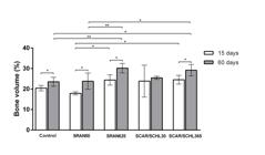
Mean and standard deviation of the values assessed in the microtomography analysis, for all evaluated groups and periods (*p<0.05; **p<0.01).
Histologic Description
For all groups and periods, elements of compact and medullary bone could be seen. Compact bone was composed by circumferential lamellae involving vascular figures, interstitial lamellae, and osteocytes. Further, it was in intimate contact with the titanium (implants), in some regions. The regions presenting medullary bone were morphologically consistent with the anatomical features of the rat’s tibiae17. Representative images of each group are shown in Figure 4.

Microphotographies representing each evaluated group and period. These sections were posteriorly evaluated in for histomorphometry.
Histomorphometric Evaluation
There were no statistical differences among the groups regarding assessed BIC and BA. When the two evaluation periods were compared, only the Control and SCAR/SCHL30 groups had a progressive increase from 15 to 60 days in BIC values. Regarding BA, Control, SCAR/SCHL30, and SRAN625 showed significant increase from 15 to 60 days (Table 1).
Mean and standard deviation of BIC (%) and BA (%), as assessed in the histomorphometric evaluation, for all evaluated groups and periods
DISCUSSION
The use of the Sr in the medical field brought favorable results in the prevention and treatment of the bone metabolism diseases such as osteoporosis1. Following this idea, one could speculate that not only bone-related diseases, but also bone-related surgical procedures could benefit from Sr supplementation11,18. This involves any procedures which depend on new bone formation, like the osseointegration of titanium implants used for oral rehabilitation19. However, to the present, only one medication has been tested for such task, which is Sr ranelate20.
The literature has shown promising, but at same time controversial results from the systemic use of Sr, in the form of SRAN to enhance the osseointegration of titanium implants20. Studies have shown that the systemic administration of SRAN could improve the bone formation around the implants, both in heathy18 and in osteoporotic rats11,12. However, other studies showed that SRAN had a weak effect over heathy rats21. Independent of the controversial results, no variation for SR supplementation have been tested aiming to potentialize Sr absorption and action. A relatively simple way to vary Sr administration and potentialize its action would be to find forms to enhance its absorption. In this study, we suggest a formula containing Sr in the form of carbonate and chloride, which would in theory lead to a better Sr absorption, and which could act as a supplement for bone neoformation, acting for a short period.
Dose choice was defined based on the molecular weight of SRAN (C12H6N2O8SSr2 - 513.49 mg, being 175.24 mg of Sr), which has been the standard drug tested for the same aim to the present. Therefore, the association between Sr carbonate (SrCO3 – 147.63 mg) and Sr chloride (SrCl2 –158.52 mg) adds the same 175.24 mg of Sr. The proportion, in weight, between SRAN and the new test supplement is approximately 1.7, in such a way that 625 mg of SRAN leads to the same amount of Sr as in 365 mg of the test formula, and 50 mg of SRAN compares to 30 mg of the test supplement formula. Therefore, this association between the two molecules was due to prove a neutral salt, which would keep the pH equilibrium, leading to a better Sr intestinal absorption and consequently greater use of the same, once time that generally the Sr is poorly absorbed in the intestinal tract22.
The biomechanical evaluation (removal torque), although seen as an indirect form to measure (assess) osseointegration, is a valid evaluation method23. The present results suggest that only the formulas providing higher Sr doses were able to induce positive effects towards bone formation, based on the results seen in the 15-days period of evaluation. The lack of differences among the groups in the 60-days period is possibly related to the fact that this relates to a period in which the osseointegration of implants is already consolidated in healthy animals15.
In the immunohistochemical analysis, higher doses of Sr (SRAN625 and SCAR/SCHL365) lead to an increase of BMP-2 in 15 days, suggesting the Sr action in initial bone formation. These results are according with Lv et al.24 and Li et al.25, which showed similar results in vitro, for diverse concentrations of SRAN. However, although a tendency of increased OCN for the Sr groups in 15 (SCAR/SCHL365) and 60 days (SRAN50 and SRAN625) was seen, the results showed no difference among groups. Differently, the study of Li et al.11 and Ibrahim et al.26 showed increased values for OCN, but also considering a differente dosage of SRAN.
The microtomographic analysis allows quantitative evaluation, assessing the bone quantity and quality around the implants, as means of the bone volume in the region around the implant. Assessing BIC based on microtomographies is not suggested, due to the artefacts related to metal which could lead to false results regarding the bone surrounding the implants27. In an attempt to lead to a better standardization of the results, in the present study we used, for all animals, the same (fixed) threshold for bone detection, making the results comparable within the study.
The results of the microtomographic analysis were in agreement with those of the biomechanical analysis in the period of the 15 days. The groups with high Sr concentration (SRAN625 and SCAR/SCHL365) showed a superior bone formation in the early periods of evaluation, in comparison to SRAN50. In the period of 60 days, the same groups showed larger values for bone volume in comparison to Control and SRAN50. These larger values found both in 15 and 60 days might suggest that the Sr acts both in early and late stages of bone healing/formation as suggested by Dahl et al.13. In addition, there was a significate increase in the assessed bone volume from 15 to 60 days in all groups, except for SCAR/SCHL30, suggesting that the effects of Sr might exist all through the dynamic process of bone formation. These findings are compatible with those showed by Li et al.11, who, however, found larger values than these reported in the present study. This difference can be explained by the fact that the used thresholds were not the same, added to the fact that Sr administration occurred for a longer period, and that the sample consisted of ovariectomized rats, simulating the osteoporotic state, that could accentuate the effect of the tested supplement.
Thus, Sr can act both in the early and late stages of bone maturation through two diverse mechanisms of action, according to the cellular intake dynamics. First, a mechanism of fast intake, which is directly associated to the osteoblastic activity, in which Sr is absorbed by ionic exchanges with Calcium (Ca) or by binding to osteoid proteins and/or both. The slower mechanism occurs through of the incorporation of Sr ions into the bone mineral crystals, which takes place gradually, due to exchanges in the structure of the mature bone. In the end, these processes are related both to bone neoformation and bone maturation bone, being able to exert its effect in both osseointegration stages13.
In the histological evaluation, bone tissue showed normal morphology, with characteristics of healthy tissue. This suggests that the process of bone formation around the implants was not only a response to their primary stability28, but a true osseointegration process, in which secondary (away from the implant) bone formation takes place. For the quantitative evaluation of osseointegration, the bone tissue was measured by means of BIC and BA assessment. The results did not show any statistical differences among the groups, and only a suggestive tendency indicating larger values for the groups with higher Sr concentration (625mg and 365mg). These controversial results, when compared with the other assessment methods used in the present study can be explain by the small number of sections (only the most central one) used for the histomorphometrical evaluation. This happened due to methodological limitations which cannot be overcome when such small implants are used16. In this way, we believe that this method does not fully represent the real condition of the bone around the implants, for the present study.
High Sr concentrations can lead to many benefits when incorporated into the bone tissue. However, Sr favorable results depends on the use of ideal concentrations, the correct period of administration even before the surgical procedure (allowing proper plasmatic levels of the drug to be reached), and also which are not so high to induce toxicity13. The low concentrations tested in the present study did not lead to any side effects, but higher concentrations could lead to adverse effects, not only locally, to the bone. Other studies showed that high concentrations can cause, at the more severe cases, cardiac diseases29. However, other studies showed controversial results, not being able to estabilish the link between Sr administration and systemic alterations30. In any case, finding the parameters for Sr administration which could lead to bone tissue enhanced formation, with no (or reduced) side effects, is the next challenge. This would be the only way in which the suggested new formulas could be used as a supplement, in substitution of SRAN, in those cases in which bone formation needs to be enhanced.
CONCLUSION
This study shows that higher concentrations of systemic Sr lead to variably improved osseointegration-related parameters regarding the biomechanical and microtomographic evaluation. No differences were found on the other assessment levels.
ACKNOWLEDGMENTS
This study was supported by the Coordination for the Improvement of Higher Education Personnel (CAPES-001), and National Council for Scientific and Technological Development – CNPq.
-
How to cite:
Scardueli CR, Spin-Neto R, Oliveira GJLP, Stavropoulos A, Marcantonio RAC, Marcantonio Jr E. Non-radioactive strontium as a supplement to enhance osseointegration. Rev Odontol UNESP. 2024;53:e20240026. https://doi.org/10.1590/1807-2577.02624
REFERENCES
-
1 Blake GM, Fogelman I. Strontium ranelate: a novel treatment for postmenopausal osteoporosis: a review of safety and efficacy. Clin Interv Aging. 2006;1(4):367-75. http://doi.org/10.2147/ciia.2006.1.4.367 PMid:18046914.
» http://doi.org/10.2147/ciia.2006.1.4.367 -
2 Reginster JY, Deroisy R, Neuprez A, Hiligsmann M, Zegels B, Bruyere O. Strontium ranelate: new data on fracture prevention and mechanisms of action. Curr Osteoporos Rep. 2009 Sep;7(3):96-102. http://doi.org/10.1007/s11914-009-0016-1 PMid:19723468.
» http://doi.org/10.1007/s11914-009-0016-1 -
3 Chattopadhyay N, Quinn SJ, Kifor O, Ye C, Brown EM. The calcium-sensing receptor (CaR) is involved in strontium ranelate-induced osteoblast proliferation. Biochem Pharmacol. 2007 Aug;74(3):438-47. http://doi.org/10.1016/j.bcp.2007.04.020 PMid:17531955.
» http://doi.org/10.1016/j.bcp.2007.04.020 -
4 Brennan TC, Rybchyn MS, Green W, Atwa S, Conigrave AD, Mason RS. Osteoblasts play key roles in the mechanisms of action of strontium ranelate. Br J Pharmacol. 2009 Aug;157(7):1291-300. http://doi.org/10.1111/j.1476-5381.2009.00305.x PMid:19563530.
» http://doi.org/10.1111/j.1476-5381.2009.00305.x -
5 Reginster JY, Seeman E, De Vernejoul MC, Adami S, Compston J, Phenekos C, et al. Strontium ranelate reduces the risk of nonvertebral fractures in postmenopausal women with osteoporosis: Treatment of Peripheral Osteoporosis (TROPOS) study. J Clin Endocrinol Metab. 2005 May;90(5):2816-22. http://doi.org/10.1210/jc.2004-1774 PMid:15728210.
» http://doi.org/10.1210/jc.2004-1774 -
6 Kaufman JM, Audran M, Bianchi G, Braga V, Diaz-Curiel M, Francis RM, et al. Efficacy and safety of strontium ranelate in the treatment of osteoporosis in men. J Clin Endocrinol Metab. 2013 Feb;98(2):592-601. http://doi.org/10.1210/jc.2012-3048 PMid:23341486.
» http://doi.org/10.1210/jc.2012-3048 -
7 Reginster JY, Beaudart C, Neuprez A, Bruyère O. Strontium ranelate in the treatment of knee osteoarthritis: new insights and emerging clinical evidence. Ther Adv Musculoskelet Dis. 2013 Oct;5(5):268-76. http://doi.org/10.1177/1759720X13500862 PMid:24101948.
» http://doi.org/10.1177/1759720X13500862 -
8 Meunier PJ, Roux C, Ortolani S, Diaz-Curiel M, Compston J, Marquis P, et al. Effects of long-term strontium ranelate treatment on vertebral fracture risk in postmenopausal women with osteoporosis. Osteoporos Int. 2009 Oct;20(10):1663-73. http://doi.org/10.1007/s00198-008-0825-6 PMid:19153678.
» http://doi.org/10.1007/s00198-008-0825-6 -
9 Ammann P. Strontium ranelate: a physiological approach for an improved bone quality. Bone. 2006 Feb;38(2 Suppl 1):15-8. http://doi.org/10.1016/j.bone.2005.09.023 PMid:16455318.
» http://doi.org/10.1016/j.bone.2005.09.023 -
10 Li YF, Luo E, Feng G, Zhu SS, Li JH, Hu J. Systemic treatment with strontium ranelate promotes tibial fracture healing in ovariectomized rats. Osteoporos Int. 2010 Nov;21(11):1889-97. http://doi.org/10.1007/s00198-009-1140-6 PMid:19957162.
» http://doi.org/10.1007/s00198-009-1140-6 -
11 Li Y, Li X, Song G, Chen K, Yin G, Hu J. Effects of strontium ranelate on osseointegration of titanium implant in osteoporotic rats. Clin Oral Implants Res. 2012 Sep;23(9):1038-44. http://doi.org/10.1111/j.1600-0501.2011.02252.x PMid:22117625.
» http://doi.org/10.1111/j.1600-0501.2011.02252.x -
12 Li Y, Feng G, Gao Y, Luo E, Liu X, Hu J. Strontium ranelate treatment enhances hydroxyapatite-coated titanium screws fixation in osteoporotic rats. J Orthop Res. 2010 May;28(5):578-82. http://doi.org/10.1002/jor.21050 PMid:20014319.
» http://doi.org/10.1002/jor.21050 -
13 Dahl SG, Allain P, Marie PJ, Mauras Y, Boivin G, Ammann P, et al. Incorporation and distribution of strontium in bone. Bone. 2001 Apr;28(4):446-53. http://doi.org/10.1016/S8756-3282(01)00419-7 PMid:11336927.
» http://doi.org/10.1016/S8756-3282(01)00419-7 -
14 Almon RR, Yang E, Lai W, Androulakis IP, DuBois DC, Jusko WJ. Circadian variations in rat liver gene expression: relationships to drug actions. J Pharmacol Exp Ther. 2008 Sep;326(3):700-16. http://doi.org/10.1124/jpet.108.140186 PMid:18562560.
» http://doi.org/10.1124/jpet.108.140186 -
15 de Oliveira GJ, de Paula LG, Spin-Neto R, Stavropoulos A, Spolidório LC, Marcantonio E Jr, et al. Effect of avocado/soybean unsaponifiables on osseointegration: a proof-of-principle preclinical in vivo study. Int J Oral Maxillofac Implants. 2014 Jul-Aug;29(4):949-57. http://doi.org/10.11607/jomi.3498 PMid:25032777.
» http://doi.org/10.11607/jomi.3498 -
16 Donath K, Breuner G. A method for the study of undecalcified bones and teeth with attached soft tissues. The Säge-Schliff (sawing and grinding) technique. J Oral Pathol. 1982 Aug;11(4):318-26. http://doi.org/10.1111/j.1600-0714.1982.tb00172.x PMid:6809919.
» http://doi.org/10.1111/j.1600-0714.1982.tb00172.x -
17 Conte N No, de Andrade CR, Spolidorio LC, Planeta Cda S, Cruz FC, de Souza Bastos A, et al. Effects of chronic stress and alendronate therapy on the osseointegration of titanium implants. Clin Implant Dent Relat Res. 2014 Oct;16(5):762-71. http://doi.org/10.1111/cid.12046 PMid:23448531.
» http://doi.org/10.1111/cid.12046 -
18 Maïmoun L, Brennan TC, Badoud I, Dubois-Ferriere V, Rizzoli R, Ammann P. Strontium ranelate improves implant osseointegration. Bone. 2010 May;46(5):1436-41. http://doi.org/10.1016/j.bone.2010.01.379 PMid:20116464.
» http://doi.org/10.1016/j.bone.2010.01.379 -
19 Brånemark PI, Adell R, Albrektsson T, Lekholm U, Lundkvist S, Rockler B. Osseointegrated titanium fixtures in the treatment of edentulousness. Biomaterials. 1983 Jan;4(1):25-8. http://doi.org/10.1016/0142-9612(83)90065-0 PMid:6838955.
» http://doi.org/10.1016/0142-9612(83)90065-0 -
20 Scardueli CR, Bizelli-Silveira C, Marcantonio RAC, Marcantonio-Jr E, Stavropoulos A, Spin-Neto R. Systemic strontium to the osseointegration of titanium implants in animals: systematic review of the literature. Int J Implant Dent. 2018 Jul;4(1):21. http://doi.org/10.1186/s40729-018-0132-8 PMid:30014305.
» http://doi.org/10.1186/s40729-018-0132-8 -
21 Linderbäck P, Agholme F, Wermelin K, Närhi T, Tengvall P, Aspenberg P. Weak effect of strontium on early implant fixation in rat tibia. Bone. 2012 Jan;50(1):350-6. http://doi.org/10.1016/j.bone.2011.10.034 PMid:22108138.
» http://doi.org/10.1016/j.bone.2011.10.034 -
22 Pors Nielsen S. The biological role of strontium. Bone. 2004 Sep;35(3):583-8. http://doi.org/10.1016/j.bone.2004.04.026 PMid:15336592.
» http://doi.org/10.1016/j.bone.2004.04.026 -
23 Sakakura CE, Margonar R, Holzhausen M, Nociti FH Jr, Alba RC Jr, Marcantonio E Jr. Influence of cyclosporin A therapy on bone healing around titanium implants: a histometric and biomechanic study in rabbits. J Periodontol. 2003 Jul;74(7):976-81. http://doi.org/10.1902/jop.2003.74.7.976 PMid:12931759.
» http://doi.org/10.1902/jop.2003.74.7.976 - 24 Lv H, Huang X, Jin S, Guo R, Wu W. [Strontium ranelate promotes osteogenic differentiation of rat bone mesenchymal stem cells through bone morphogenetic protein-2/Smad signaling pathway]. Nan Fang Yi Ke Da Xue Xue Bao. 2013 Mar;33(3):376-81. Chinese. PMid: 23529235.
- 25 Li Z, Wang Y, Wang XN, Lan AP, Wu W. [Strontium ranelate promotes osteogenic differentiation of rat bone marrow mesenchymal stem cells by increasing bone morphogenetic protein-7 expression]. Nan Fang Yi Ke Da Xue Xue Bao. 2011 Nov;31(11):1949-53. Chinese. PMid: 22126789.
-
26 Ibrahim MRM, Singh S, Merican AM, Raghavendran HR, Murali MR, Naveen SV, et al. The effect of strontium ranelate on the healing of a fractured ulna with bone gap in rabbit. BMC Vet Res. 2016 Jun;12(1):112. http://doi.org/10.1186/s12917-016-0724-6 PMid:27307015.
» http://doi.org/10.1186/s12917-016-0724-6 -
27 Ejima K, Omasa S, Motoyoshi M, Arai Y, Kai Y, Amemiya T, et al. Influence of metal artifacts on in vivo micro-CT for orthodontic mini-implants. J Oral Sci. 2012 Mar;54(1):55-9. http://doi.org/10.2334/josnusd.54.55 PMid:22466887.
» http://doi.org/10.2334/josnusd.54.55 -
28 Brånemark R, Ohrnell LO, Skalak R, Carlsson L, Brånemark PI. Biomechanical characterization of osseointegration: an experimental in vivo investigation in the beagle dog. J Orthop Res. 1998 Jan;16(1):61-9. http://doi.org/10.1002/jor.1100160111 PMid:9565075.
» http://doi.org/10.1002/jor.1100160111 -
29 Reginster JY. Cardiac concerns associated with strontium ranelate. Expert Opin Drug Saf. 2014 Sep;13(9):1209-13. http://doi.org/10.1517/14740338.2014.939169 PMid:25020233.
» http://doi.org/10.1517/14740338.2014.939169 -
30 Cooper C, Fox KM, Borer JS. Ischaemic cardiac events and use of strontium ranelate in postmenopausal osteoporosis: a nested case-control study in the CPRD. Osteoporos Int. 2014 Feb;25(2):737-45. http://doi.org/10.1007/s00198-013-2582-4 PMid:24322476.
» http://doi.org/10.1007/s00198-013-2582-4


