Kaposi sarcoma with extensive thoracic involvement
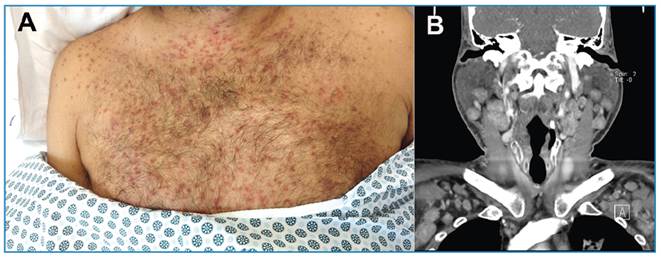
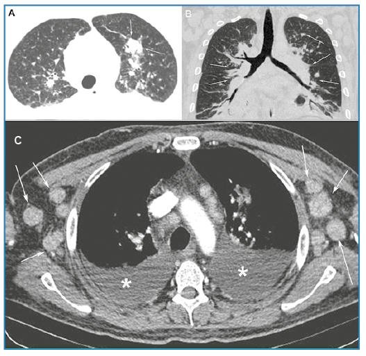
A 45-year-old male diagnosed with acquired immunodeficiency syndrome (AIDS) who was irregularly receiving antiretroviral therapy, reported episodes of fever and general malaise for the previous 30 days, followed by the appearance of disseminated red and purple skin lesions, predominantly on the upper trunk (Figure 1A). Computed tomography (CT) of the neck revealed bilateral lymphadenopathy, mainly affecting the cervical, supraclavicular, and axillary chains (Figure 1B). The CD4 cell count was 145 cells/mm3 and the viral load was 46,000 copies/mL. Panel results for other sexually transmitted diseases were unremarkable. Subsequently, the patient developed dyspnea and a dry cough. CT of the chest revealed irregular thickening of the peribronchovascular interstitium and interlobular septa, parenchymal nodules, mediastinal and axillary lymph node enlargement, and bilateral pleural effusion (Figure 2A-C). Flexible bronchoscopy revealed multiple lesions in the tracheobronchial mucosa, consistent with Kaposi sarcoma (KS). An axillary lymph node biopsy was also diagnostic of KS. Due to complications, the patient died three months later.
A. The upper trunk showing disseminated red and purple skin lesions. B. Computed tomography image of the cervical region showing bilateral and relatively symmetrical lymphadenopathy, affecting mainly the cervical and supraclavicular chains.
Computed tomography (CT) images of the chest with axial (A) and coronal (B) reconstructions showing an irregular parenchymal nodule (arrows in A) and thickening of the peribronchovascular interstitium and interlobular septa (arrows in B). C. Axial CT image showing axillary lymph-node enlargement (arrows) and bilateral pleural effusions (asterisks).
Kaposi sarcoma is a malignant angioproliferative mesenchymal tumor caused by human herpes virus-8. AIDS-related KS is aggressive and frequently fatal. Pulmonary manifestations can involve the tracheobronchial tree, parenchyma, and pleural space. The most frequent CT findings in AIDS-related KS include peribronchovascular and interlobular septal thickening, ill-defined parenchymal nodules, fissural nodules, lymph node enlargement, and pleural effusion. The incidence of KS in the population with AIDS has dramatically decreased in this era of highly active antiretroviral therapy1-4.
In conclusion, KS should be included in the differential diagnosis of disseminated lymphadenopathy-associated skin lesions in patients with AIDS.
REFERENCES
-
1 Gasparetto TD, Marchiori E, Lourenço S, Zanetti G, Vianna AD, Santos AA, et al. Pulmonary involvement in Kaposi sarcoma: correlation between imaging and pathology. Orphanet J Rare Dis. 2009;4:18. doi: 10.1186/1750-1172-4-18.
» https://doi.org/10.1186/1750-1172-4-18 -
2 Guan C, Shi Y, Liu J, Yang Y, Zhang Q, Lu Z, et al. Pulmonary involvement in acquired immunodeficiency syndrome-associated Kaposi's sarcoma: a descriptive analysis of thin-section manifestations in 29 patients. Quant Imaging Med Surg. 2021;11(2):714-24. doi: 10.21037/qims-20-284.
» https://doi.org/10.21037/qims-20-284 -
3 Silva SQ, Frank CHM, Almeida TVR. Extensive pulmonary involvement in Kaposi sarcoma in a patient with human immunodeficiency virus-acquired immunodeficiency syndrome. Rev Soc Bras Med Trop. 2020:54:e20200192. doi: 10.1590/0037-8682-0192-2020.
» https://doi.org/10.1590/0037-8682-0192-2020 -
4 Epelbaum O, Go R, Patel G, Braman S. Pulmonary Kaposi's Sarcoma and Its Complications in the HAART Era: A Contemporary Case-Based Review. Lung. 2016;194(1):163-9. doi: 10.1007/s00408-015-9830-7.
» https://doi.org/10.1007/s00408-015-9830-7
Edited by
-
Associated Editor:
Manoel O. da Costa Rocha Orcid: https://orcid.org/0000-0002-4721-1767