Distribution of yellow fever vectors in a disease-free area of Northeast Brazil
ABSTRACT
Background: Owing to the recent spread of the yellow fever virus in Brazil and imminent risk of its establishment in previously disease-free areas in the northeast of the country, epidemiological surveillance actions are necessary, including knowledge of non-human primate populations and the vectors that inhabit risk areas. The objective of this study was to evaluate the movement of sylvatic and urban yellow fever vectors between an Atlantic Forest fragment and its surrounding areas.
Methods: The study site was the Mata do Junco Wildlife Refuge Conservation Unit (CU) in Capela, Sergipe, Brazil. Immatures were collected using ovitraps in the forested area and in peridomestic environments surrounding the CU.
Results: Fourteen species were recorded, six of which were epidemiologically relevant. The main vectors of yellow fever in Brazil were present in the CU. Aedes albopictus (Skuse, 1894) was the most abundant species and was present in all studied environments. Aedes aegypti (Linnaeus, 1762) was not collected from forested areas. Among the native species, Haemagogus leucocelaenus (Dyar & Shannon, 1924) and Haemagogus janthinomys (Dyar, 1921) were collected only in the forested area of the CU, whereas Haemagogus spegazzinii (Brètes, 1921) was collected both in the forest and in the area around the CU.
Conclusion: Exotic species circulate between wild and urban areas for feeding and oviposition, but circulation to the urban area is limited among native species.
Keywords:
Arboviruses; Vector ecology; Entomological surveillance; Epizootics
INTRODUCTION
The last major urban yellow fever outbreak occurred between 1928 and 1929 in Brazil, with the final urban case reported in 19421. Since then, cases of the sylvatic form continued to occur primarily in the Amazon and Central-West regions. However, during the first two decades of this century, several outbreaks of the virus were recorded outside the Amazon region, culminating in one of the most substantial events in the history of sylvatic yellow fever in Brazil. Between 2016 and 2019, the disease spread through previously unaffected areas in the southeastern states, where more than 4,000 epizootics and 2,237 human cases of yellow fever were confirmed2-4. In the Northeast region, during this reemergence event, yellow fever epizootics were confirmed in the state of Bahia, although no human cases were recorded5.
Although non-human primates of the genus Alouatta are the most affected by the yellow fever virus, during the recent outbreak, many marmosets (Callithrix spp.) were diagnosed with the virus, including animals from urban areas6-8. Marmosets are common in the peripheral areas of several cities, urban parks, and residential condominiums9-12, where they function as epidemiological sentinels but are also victims of the disease.
Mosquito species of the genera Haemagogus and Sabethes, widely distributed across tropical regions, act as vectors of the yellow fever virus in wild environments13-15. In Brazil, Haemagogus leucocelaenus (Dyar & Shannon, 1924) and Haemagogus janthinomys (Dyar, 1921) are considered the primary vectors of the virus16 and are abundant in the Atlantic Forest, particularly during the rainy season17,18. The exotic species Aedes aegypti (Linnaeus, 1762) and Aedes albopictus (Skuse, 1894) remain widely distributed in Brazil despite intensive control efforts and are regarded as epidemiological links with the potential to facilitate the re-establishment of the urban transmition19-21. In the context of yellow fever reemergence, understanding the Culicidae fauna is crucial for surveillance, particularly in regions where Culicidae surveys are sporadic, such as the Northeast of Brazil22-24. A lack of comprehensive data can undermine estimates of species distribution and consequently the assessment of arbovirus transmission risk.
This study aimed to survey the fauna of sylvatic and urban yellow fever vectors in a Conservation Unit (CU) in the Atlantic Forest and its surroundings in the state of Sergipe, in the light of the findings of a study by Li et al. (2022)24, which suggested that the state of Sergipe is not suitable for Hg. janthinomys and only its western part is suitable for Hg. leucocelaenus, despite the presence of Atlantic Forest fragments. Given the proximity between forested and urban areas, and the potential movement of yellow fever vectors between sylvatic and urban environments, we also tested the hypothesis that primary vector species move between these settings in search of oviposition sites, which may facilitate the transfer of the yellow fever virus between anthropogenic and natural areas.
METHODS
Study area
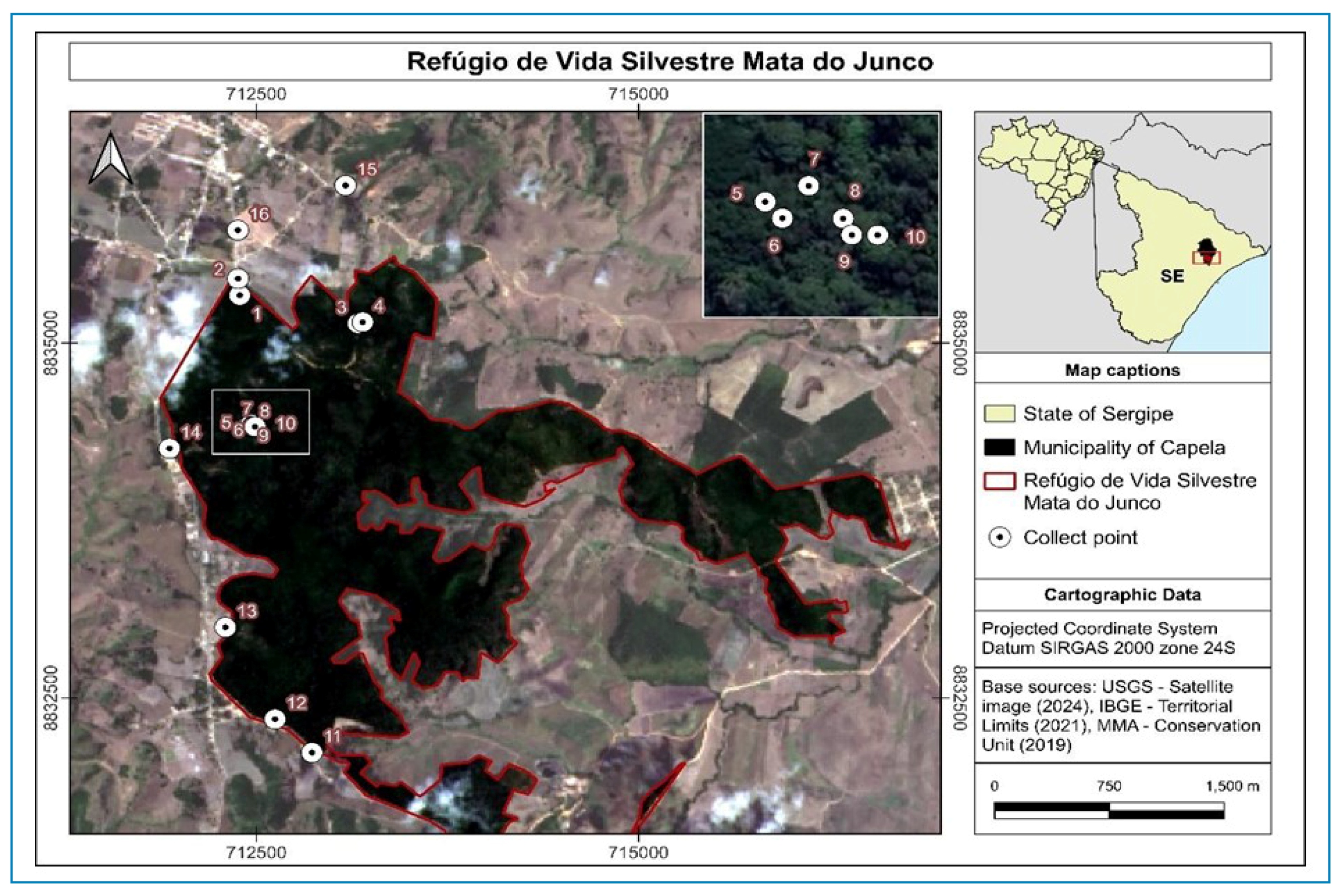
The study area comprised the Refúgio de Vida Silvestre Mata do Junco CU located in the municipality of Capela in the state of Sergipe, 67 km from the capital Aracaju (10°30′35″ S and 37°03′17″ W) (Figure 1). The climate is classified as equatorial dry summer (As), according to the updated Köppen-Geiger climate classification25, with an average annual temperature of 24.9 °C and accumulated annual precipitation of 1372 mm. The rainy season runs from April to July, with monthly average above 100 mm; the dry season runs from October to January, with monthly rainfall average below 50 mm. The CU is composed of fragmented forest patches resulting from intensive logging prior to its consolidation and is classified as a sub-deciduous Atlantic Forest, island type26. The area covers 894.90 ha, of which only 498.61 ha are covered by vegetation, characterized as secondary forest under regeneration27. According to floristic studies conducted in the area, the average canopy height was 12 m, with some trees reaching up to 20 m, which is considered low according to Atlantic Forest standards. Fabaceae had the highest tree species richness, followed by Myrtaceae. The most abundant species belonged to the families Lecythidaceae, Myrtaceae, Sapotaceae, Fabaceae, and Melastomataceae. The canopy structure ranges from closed areas with lianas, epiphytes, and climbers, to more open areas28-30.
Location of the Mata do Junco Wildlife Refuge Conservation Unit, and the 16 collection points in Capela, Sergipe, Brazil.
The CU was created with the aim of protecting fragments of the Atlantic Forest, the endangered species Callicebus coimbrai (Kobayashi & Langguth, 1999) (Coimbra Filho's titi monkey), and preserving the source of Rio Lagartixo, which supplies water to the municipality of Capela26,28. In addition to C. coimbrai, Callithrix jacchus (Linnaeus, 1762) (marmosets) and, occasionally, Sapajus sp. (capuchin monkeys) are found in the area31,32. The CU provides habitats for sylvatic vectors and is located in close to anthropized areas, where artificial containers suitable for oviposition are available.
Sample collection and processing
Ovitraps were used to collect eggs and occasionally larvae and pupae33. Each ovitrap consisted of a black plastic container with a wide opening that was partially filled with water. A fiberboard paddle was positioned as an oviposition substrate with a rough surface facing outward and fixed vertically inside the container using a metal clip. Immatures were collected monthly between March 2019 and February 2020. Traps were installed at 10 points within the CU and six points in its surroundings. Within the CU, four points were located in anthropized areas (Figure 1, 1-4), close to the reserve administration building and water collection station, in places where human activities frequently occur (hereinafter referred to as the open area); the remaining six points within the CU were located in forested areas (Figure 1, 5-10) (hereinafter referred to as the forest area). The six additional points were established in rural areas surrounding the CU (Figure 1, 11-16). At every location, two traps were installed at different vertical strata: one at 1 m from ground level and the other at 3 m, targeting both female mosquitoes that oviposit at low heights and acrodendrophilic species; thus, a total of 32 traps were installed. The maximum height was defined based on the average height of the bifurcations of the main trunks (potential sites for cavity formation) of trees located in rural areas surrounding the forest, where tree height was generally lower than that in the forested area. Ovitraps were installed by securing them to trees and bamboo trunks within the forest and to fruit trees, beneath roof eaves or on water tank platforms in rural areas, positioned near potential mosquito habitats and protected from rain. The paddles and remaining water in the traps were checked once a month and replaced.
The collected contents were sent to the insectarium of the Tropical Entomology and Parasitology Laboratory (LEPaT) at the Federal University of Sergipe. Specimens collected in the pupal form were placed in cages until adults emerged. When present, Toxorhynchites larvae were placed into individual vials to prevent predation. After drying, the paddles were examined under a stereoscopic microscope, and all eggs present were counted regardless of whether they had hatched. Paddles were maintained on a drying rack for one week and subsequently immersed in trays containing 400 mL of mineral water and 0.01 g of fry food granules until larval hatching. The larvae were reared to the fourth-instar stage, after which they were either sacrificed or individually isolated for the collection of larval e pupal exuviae. Immediately after emergence, adults were placed in a freezer for 20 min, transferred to microtubes containing silica, and stored in a refrigerator until use.
Mosquito identification
Taxonomic identification was carried out following the keys available14,34-37. The genus was abbreviated according to Reinert (2001)38. For the genus Aedes, the old taxonomic classification was maintained as recommended by Wilkerson et al. (2015)39.
The identification of some species was also confirmed at the molecular level by DNA barcoding. Depending on the availability, one to five specimens of each species were processed. DNA was extracted from whole adult specimens using the PureLink® Viral RNA/DNA Mini Kit (Life Technologies, Carlsbad, CA, USA). The primers LCOI490 and HCO219840 were used to amplify an approximately 658 bp fragment located at the 5′ end of the Cytochrome Oxidase subunit I sequence, which was trimmed to 609 bp. Both the PCR and sequencing reaction were run using the Phire Tissue Direct PCR Master Mix (Thermo Scientific™, Anjou, QC, CA), according to the manufacturer's instructions. PCR products were electrophoresed on a 2% agarose gel stained with Diamond Nucleic Acid Dye (Promega Corporation). The products were sequenced in both directions with the same set of primers and analyzed in an ABI 3130 DNA Analyzer (PE Applied Biosystems, Warrington, UK); the quality of the chromatograms was verified visually in Chromas v2.6.5 (Technelysium Pty Ltd, Brisbane, AU). Homologies, insertions/deletions, and frameshifts were identified using MUSCLE v3.8.3141. The sequences obtained in FASTA format were aligned using ClustalW42. The sequence from each specimen was compared to barcode sequences from GenBank using BLAST. The Cytochrome Oxidase subunit I sequences were deposited in the National Center for Biotechnology Information nucleotide sequence database. The degree of divergence was determined using the genetic distance matrix in MEGA v6.043. DNA amplification was unsuccessful for specimens preserved in naphthalene.
Statistical analysis
The richness was estimated based on the number of species and morphotypes. Species were characterized according to their relative abundance and constancy44, categorized as constant (c) when recorded in more than 50% of collections, accessory (a) when recorded in 25-50% of collections, and occasional (t) when recorded in fewer than 25% of collections.
The analyses were performed using R version 3.6.145. Normality of the data was tested using the Shapiro-Wilk statistical test. The correlation between distribution and precipitation was assessed using Spearman’s correlation test. Precipitation data from an Agrometeorological Monitoring System were used46. To compare the distribution of Culicidae in terms of collection areas, a multinomial test was used, considering the difference in the number of collection points between the areas. Species abundance was compared according to collection height using a binomial test. For all analyses, a significance level of 0.05 was considered.
Ethical approval
This study did not involve human experimentation. The umbrella project entitled “Investigation of the Circulation of Yellow Fever Virus, Hematozoa and Gastrointestinal Endoparasites in Neotropical Primates in the State of Sergipe” was approved by the UFS Animal Research Ethics Committee, the Chico Mendes Biodiversity Institute (ICMBio), Sergipe State Secretariat for Urban Development and Sustainability, and registered with the National System for the Management of Genetic Heritage and Associated Traditional Knowledge (SISGEN).
RESULTS
Richness of 14 species was observed, distributed across six genera: Aedes, Culex, Haemagogus, Limatus, Toxorhynchites, and Wyeomyia. Among the identified species, Ae. aegypti, Ae. albopictus, Hg. janthinomys, Hg. leucocelaenus, and Haemagogus spegazzinii (Brètes, 1921) are considered of medical importance36. Several species showed homology with sequences deposited in GenBank; however, some showed a divergence greater than 3% (Table 1). The species of the genus Toxorhynchites was morphologically identified as Toxorhynchites theobaldi (Dyar & Knab, 1906), using the available key35; however, because of the divergence found with the sequence of Mexican origin deposited in GenBank (KY782655; identity 94.90%, coverage 99.00%, and a genetic distance of 5.9%), in contrast to the morphological homology with another species collected by our group in the State of Bahia (MF537259; identity 99.84%, coverage 89%, and a genetic distance of 0.1%), we chose to maintain the species as Toxorhynchites sp.23.
Species and accession codes of Cytochrome Oxidase subunit I gene sequences deposited in GenBank.
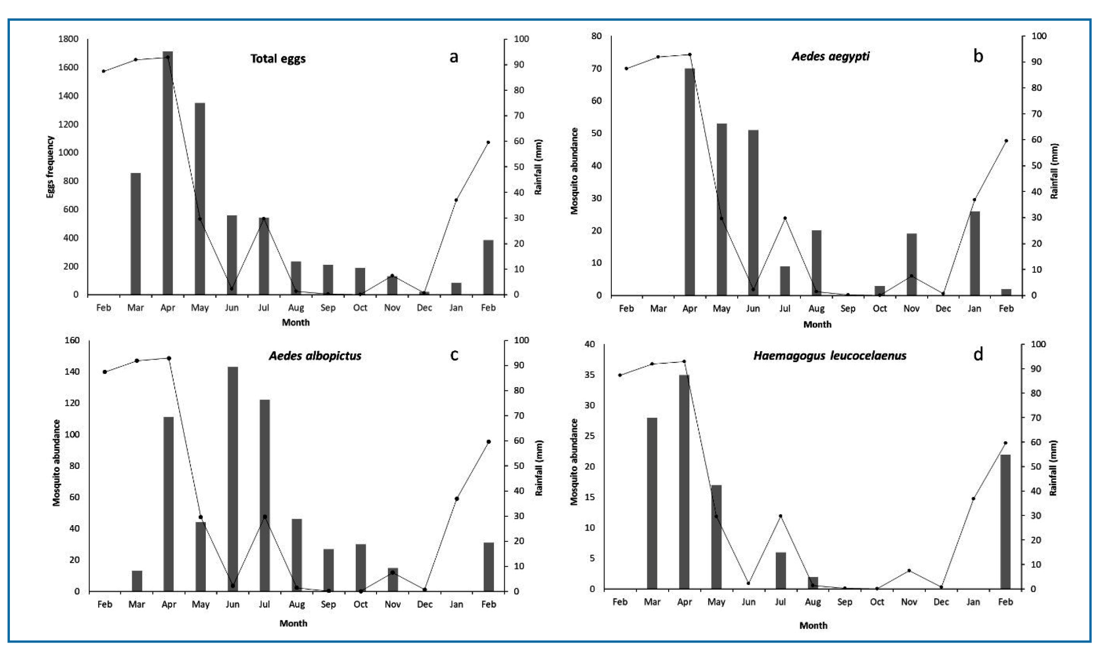
A total of 6,277 eggs, 2,508 larvae, and 42 pupae were collected from the ovitraps. The greatest abundance was observed in April and May 2019, consistently decreasing afterwards before increasing again in February of the following year, following an increase in the precipitation rate in January (Figure 2a ). The pattern observed in the distribution of the number of eggs over time showed a moderate correlation with the accumulated precipitation of the previous 30 d (ρ = 0.56; p = 0.0585) but a strong correlation with the accumulated precipitation in the month prior to collection (ρ = 0.79; p = 0.0002) or in the 60 d prior to collection (ρ = 0.72; p = 0.0082).
Accumulated precipitation in the 30 d prior to collection and the number of eggs collected by month in areas of Mata do Junco, in the municipality of Capela, Sergipe, from 2019 to 2020; a - total eggs; b - Aedes albopictus; c - Aedes aegypti; d - Haemagogus leucocelaenus.
After laboratory rearing, 1,355 specimens were identified: 1,203 were adults and 152 were fourth-stage larvae. The most abundant species was Ae. albopictus (42.9%), followed by Ae. aegypti (18.6%) and Hg. leucocelaenus (8.1%). Haemagogus janthinomys and Hg. spegazzinii had abundances of 1.3% and 0.93%, respectively (Table 2). Of the three most common species, Ae. albopictus (ρ = 0.36; p = 0.23) and Ae. aegypti (ρ = 0.28; p = 0.36) did not show a correlation with precipitation in the month prior to collection; however, Hg. leucocelaenus (ρ = 0.81; p = 0.0012) showed a strong correlation with rainfall (Figure 2b-d ).
The number of mosquitoes identified in traps installed at a height of 1 m (56.6%) was significantly higher than the number identified in traps at 3 m (43.3%) (p < 0.0001). However, considering the total number of eggs collected, the abundance was significantly higher at 3 m (53.5%) (p < 0.0001). Of the 15 species captured, Culex (Microculex) group pleuristriatus was only captured at heights above 3 m. Aedes aegypti, Ae. albopictus, and Limatus durhamii (Theobald, 1901) were significantly more abundant at 1 m, whereas Culex originator (Gordon & Evans, 1922) was most abundant at 3 m. There were no statistically significant differences between heights for any Haemagogus species.
The relative abundance was significantly higher in the rural area surrounding the CU, whereas species richness was greater in the forest. Aedes aegypti was statistically more abundant in rural sites and was absent from the forest, occurring only in open areas of the CU. Aedes albopictus was recorded in all three sampling environments and was most abundant in open CU sites. Haemagogus leucocelaenus and Hg. janthinomys were absent from rural collections and were statistically more abundant in forest and open CU environments respectively. Haemagogus spegazzinii occurred in open and rural sites, where it was significantly more abundant.
Among the species of medical importance, Ae. aegypti, Ae. albopictus, and Hg. leucocelaenus were constant in the traps, whereas Hg. janthinomys was an accessory species. Of the other species, Aedes fulvithorax (Lutz, 1904), Aedes terrens (Walker, 1956), C. originator, Li. durhamii, and Toxorhynchites sp. were constant species; Wyeomyia arthrostigma (Lutz, 1905) was considered accessory; and Culex (Mcx) group pleuristriatus, Hg. spegazzinii, Limatus flavisetosus (Oliveira Castro, 1935), and Wyeomyia (Pho.) pilicauda (Root, 1928) were occasional species (Table 2).
DISCUSSION
To date, there is no evidence of reemergence of the yellow fever virus in the forest areas of the State of Sergipe. The last reported outbreak in Sergipe and neighboring states dates back to 192647. Regarding vector distribution, Li et al. (2022)24 estimated that the presence of Hg. janthinomys and Hg. leucocelaenus was unlikely; however, these primary sylvatic vectors of yellow fever virus were recorded in the present study, in addition to the exotic species Ae. aegypti. Other species that are experimentally competent or have been found naturally infected were also recorded, including Ae. albopictus and Hg. spegazzinii5,48-50.
Taken together, these results, combined with the presence of non-human primates, indicate high local receptivity to the sylvatic yellow fever transmission. The CU area studied here comprises a small fragment of the Atlantic Forest located close to urbanized areas with limited vegetation cover (Figure 1); however, it represents an important refuge for wildlife, including mosquitoes51. This scenario is likely to be repeated across Atlantic Forest remnants along the coastal region of northeastern Brazil52.
In Brazil, Hg. janthinomys has been found from the North to the Southeast, whereas Hg. leucocelaenus extends from the North to the South of the country13,15. The Culicidae fauna of northeastern Brazil remains poorly sampled; there are only occasional and non-systematic records of the distribution of Hg. leucocelaenus and Hg. janthinomys, except for the Atlantic Forest of Bahia, where Hg. janthinomys has been documented14,23,53. In this survey, Hg. leucocelaenus and Hg. janthinomys were recorded exclusively in forested areas, although previous studies indicate that both species can disperse long distances-up to 5.7 km and 11.5 km, respectively54-and that Hg. leucocelaenus may occur in anthropogenic environments, including peri-urban areas55. The forested area of Capela presents slightly lower mean annual air temperature and relative humidity (23.7 °C and 89.5%) than clearing areas (24.3 °C and 82.6%)56, conditions that may favor sylvatic species and may account for their restriction to the forested habitats in the presence of vertebrate hosts.
As studies targeting adult mosquitoes have successfully recorded Haemagogus species in degraded areas57, it is possible that these species forage for blood meals in anthropic environments but return to forest for shelter and oviposition. This behavior may explain their absence in ovitraps from rural areas in our study; however, it does not preclude their circulation in areas surrounding the forest. Only Hg. spegazzinii, a common species in northeastern Brazil occurring in both the Caatinga and Atlantic Forest biomes5,58, was successfully collected as immature in rural areas. Although its vector competence is unknown, Hg. spegazzinii has previously been reported as naturally infected48,59. Its wide distribution, potential vector competence, and ability to circulate between wild and anthropogenic environments suggest that this species may play an important role in arboviruses transmission in northeastern Brazil.
Typically, epizootics and epidemics of diseases transmitted by mosquitoes are associated with the beginning of the rainy season, when vector population densities are higher60. In this study, the abundance of immature forms in ovitraps was correlated with precipitation, although there was an artificial supply of water to the ovitraps. This additional water availability appeared to favor the exotic species Ae. albopictus and Ae. aegypti, whereas native species followed a precipitation pattern (Figure 2).
In this study, Ae. albopictus was the most abundant species and was recorded at all three collection sites. This species has a wide geographic distribution in Brazil61-65, and in Sergipe, it was first detected in 2011, followed by its gradual spread throughout the state65. Dispersal studies have shown that Ae. albopictus females can disperse long distances within a few days, from wild to modified environments66. This dispersal capacity, together with ecological adaptability, enables the species to occupy both wild and urban environments. Its ability to feed on a wide range of mammals and its high susceptibility to infection make Ae. albopictus a potential risk factor for yellow fever transmission in urban areas67-69.
Recent yellow fever outbreaks have been reported in Brazil in forested areas near urban centers, including locations without previous evidence of viral activity or established vaccination policies16,69,70. In the Northeast region, yellow fever vaccination began to be recommended in the second half of 2020; however, implementation was hindered by the prioritization of COVID-19 (SARS-CoV-2) vaccination. As a result, vaccination coverage remains low in many municipalities across the region, increasing vulnerability to human cases of this arbovirus.
Despite their acrodendrophilic behavior, Haemagogus species were recorded at both heights in this study, suggesting that the availability of suitable habitats may outweigh height in determining oviposition sites. However, this finding should be interpreted with caution, as trap placement was limited to a maximum height of 3 m. While no significant differences were reported between 1 m and 3 m by Silva-Inácio et al. (2020)⁵⁹, Dias et al. (2023b) 71 documented greater Haemagogus abundance at higher strata (6-8 m).
Study limitations include discrepancies between the total of immature collected and the number ultimately available for identification. Mortality during rearing may have affected the representativeness of the sample, as more fragile species could have been disproportionately impacted by laboratory handling, leading to an underestimation of their frequencies. The exclusive use of a single trap type (ovitraps) may also have limited the detection of species that do not use this container, for example, species of the genus Sabethes72.
Recent yellow fever epizootics have resulted in population declines of approximately 30% in golden lion tamarins (Leontopithecus) and 10-26% in the muriquis (Brachyteles hypoxanthus (Kuhl, 1920)) in Southeast Brazil73,74. If the virus were introduced into the CU Mata do Junco, it could place the endangered C. coimbrai at critical risk of extinction75. In addition to C. coimbrai, C. jacchus also occurs in the Mata do Junco. During the 2016 and 2017 outbreaks in Brazil, several epizootics were reported among marmosets. Because marmoset groups move between forest fragments and urban and peri-urban environments, they may act as a link in the yellow fever transmission cycle and facilitate virus spread in these areas5-8.
The presence, abundance and constancy of sylvatic (Hg. leucocelaenus, Hg. janthinomys, and Hg. spegazzinii) and urban (Ae. aegypti and Ae. albopictus) yellow fever vectors across forested, rural, and peridomestic environments, suggest mosquito movement between areas and the potential interaction between sylvatic and anthropogenic transmission cycles. In the context of non-human primates, including endangered species, low vaccination coverage, and proximity to areas with reported epizootics, these findings highlight the importance of sustained entomological and epizootic surveillance in this CU and in other Atlantic Forest remnants in northeastern Brazil.
REFERENCES
-
1 Tartaglia TTS, Pacca CC. Febre amarela no Brasil: da história ao risco de reemergência. Rev Med. 2019;98(5):334-40. doi:10.11606/issn.1679-9836.v98i5p334-340.
» https://doi.org/10.11606/issn.1679-9836.v98i5p334-340 -
2 Possas C, Oliveira RL, Tauil PL, Pinheiro FP, Pissinatti A, Cunha RV, et al. Yellow fever outbreak in Brazil: the puzzle of rapid viral spread and challenges for immunisation. Mem Inst Oswaldo Cruz. 2018;113:e180278 . doi:10.1590/0074-02760180278.
» https://doi.org/10.1590/0074-02760180278 -
3 Giovanetti M, Mendonça MCL, Fonseca V, Mares-Guia MA, Fabri A, Xavier J, et al. Yellow fever virus reemergence and spread in Southeast Brazil, 2016-2019. J Virol. 2019;94(16):e01623-19. doi:10.1128/JVI.01623-19.
» https://doi.org/10.1128/JVI.01623-19 -
4 Figueiredo P, Stoffella-Dutra AG, Barbosa GC, Oliveira JS, Amaral CD, Santos JD, et al. Re-Emergence of Yellow Fever in Brazil during 2016-2019: Challenges, Lessons Learned, and Perspectives. Viruses. 2020;12(11):1233. doi:10.3390/v12111233.
» https://doi.org/10.3390/v12111233 -
5 Silva NIO, Sacchetto L, Rezende IM, Trindade GS, LaBeaud AD, Thoisy B, et al. Recent sylvatic yellow fever virus transmission in Brazil: the news from an old disease. Virol J. 2020;17(1):1-12. doi:10.1186/s12985-019-1277-7.
» https://doi.org/10.1186/s12985-019-1277-7 -
6 Domingo C, Charrel RN, Schmidt-Chanasit J, Zeller H, Reupsken C. Yellow fever in the diagnostics laboratory. Emerg Microbes Infect. 2018;7(1):1-15. doi:10.1038/s41426-018-0128-8.
» https://doi.org/10.1038/s41426-018-0128-8 -
7 Paploski IAD, Souza RL, Tauro LB, Cardoso CW, Mugabe VA, Alves ABPS, et al. Epizootic Outbreak of Yellow Fever Virus and Risk for Human Disease in Salvador, Brazil. Ann Intern Med. 2018;168(4):301-2. doi:10.7326/M17-1949.
» https://doi.org/10.7326/M17-1949 -
8 Mares-Guia MAMM, Horta MA, Romano A, Rodrigues CDS, Mendonça MCL, Santos CC, et al. Yellow fever epizootics in non-human primates, Southeast and Northeast Brazil (2017 and 2018). Parasites Vectors. 2020;13(1):1-8. doi:10.1186/s13071-020-3966-x.
» https://doi.org/10.1186/s13071-020-3966-x -
9 Favoretto SR, Mattos CC, Morais NB, Araújo FAA, Mattos CA. Rabies in marmosets (Callithrix jacchus), Ceará, Brazil. Emerg Infect Dis. 2001;7(6):1062-5. doi:10.3201/eid0706.010630.
» https://doi.org/10.3201/eid0706.010630 -
10 Goulart VDLR, Teixeira CP, Young RJ. Analysis of callouts made in relation to wild urban marmosets (Callithrix penicillata) and their implications for urban species management. Eur J Wildl Res. 2010;56(4):641-9. doi:10.1007/s10344-009-0362-4.
» https://doi.org/10.1007/s10344-009-0362-4 -
11 Rodrigues NN, Martinez RA. Wildlife in our backyard: interactions between Wied's marmoset Callithrix kuhlii (Primates: Callithrichidae) and residents of Ilhéus, Bahia, Brazil. Wildl Biol. 2014;20(2):91-6. doi:10.2981/wlb.13057.
» https://doi.org/10.2981/wlb.13057 -
12 Dias JS, Beltrão-Mendes R, Bezerra TL, Lima VFS, Dolabella SS, La Corte R. Gastrointestinal parasites in free-ranging common marmosets (Callithrix jacchus Linnaeus, 1758) in the state of Sergipe, northeastern Brazil. Vet Parasitol Reg Stud Rep. 2023a;38:100822. doi:10.1016/j.vprsr.2022.100822.
» https://doi.org/10.1016/j.vprsr.2022.100822 -
13 Gomes AC, Torres MAN, Paula MB, Fernandes A, Marassá AM, Consales CA, et al. Ecologia de Haemagogus e Sabethes (Diptera: Culicidae) em áreas epizoóticas do vírus da febre amarela, Rio Grande do Sul, Brasil. Epidemiol Serv Saude. 2010;19(2):101-13. doi:10.5123/S1679-49742010000200003.
» https://doi.org/10.5123/S1679-49742010000200003 - 14 Marcondes CB, Alencar J. Revisão de mosquitos Haemagogus Williston (Diptera: Culicidae) do Brasil. Rev Biomed. 2010;21(4):221-38.
-
15 Inácio CLS, Silva JHT, Freire RCM, Gama RA, Marcondes CB, Ximenes MFFM. Checklist of Mosquito Species (Diptera: Culicidae) in the Rio Grande do Norte State, Brazil - Contribution of Entomological Surveillance. J Med Entomol. 2017;54(3):763-73. doi:10.1093/jme/tjw236.
» https://doi.org/10.1093/jme/tjw236 -
16 Abreu FVS, Ribeiro IP, Ferreira-de-Brito A, Santos AAC, Miranda RM, Bonelly IS, et al.Haemagogus leucocelaenus and Haemagogus janthinomys are the primary vectors in the major yellow fever outbreak in Brazil, 2016-2018. Emerg Microbes Infect. 2019;8(1):218-31. doi:10.1080/22221751.2019.1568180.
» https://doi.org/10.1080/22221751.2019.1568180 -
17 Couto-Lima D, Andreazzi CS, Leite PJ, Bersot MIL, Alencar J, Lourenço-de-Oliveira R. Seasonal population dynamics of the primary yellow fever vector Haemagogus leucocelaenus (Dyar & Shannon) (Diptera: Culicidae) is mainly influenced by temperature in the Atlantic Forest, southeast Brazil. Mem Inst Oswaldo Cruz. 2020;115:e200218 . doi:10.1590/0074-02760200218.
» https://doi.org/10.1590/0074-02760200218 -
18 Freitas-Silva OS, Mello CF, Machado SL, Leite PJ, Alencar J. Interaction of Haemagogus leucocelaenus (Diptera: Culicidae) and Other Mosquito Vectors in a Forested Area, Rio de Janeiro, Brazil. Trop Med Infect Dis. 2022;7(6):94. doi:10.3390/tropicalmed7060094.
» https://doi.org/10.3390/tropicalmed7060094 - 19 Santos RLCD. Atualização da distribuição de Aedes albopictus no Brasil (1997-2002). Rev Saude Publica. 2003;37(5):671-3.
-
20 Zara ALDSA, Santos SMD, Fernandes-Oliveira ES, Carvalho RG, Coelho GE.Aedes aegypti control strategies: a review. Epidemiol Serv Saude. 2016;25(2):391-404. doi:10.5123/S1679-49742016000200017.
» https://doi.org/10.5123/S1679-49742016000200017 -
21 Gava C, Silva TCCD, Lyra DGP, Ardisson KS, Marques CS, Almada GL, et al. Prevenção e controle da febre amarela: avaliação de ações de vigilância em área indene no Brasil. Cad Saude Publica. 2022;38(5):e00000521 . doi:10.1590/0102-311X00000521.
» https://doi.org/10.1590/0102-311X00000521 -
22 Marteis LS, Natal D, Sallum MAM, Medeiros-Sousa AR, Oliveira TMP, La Corte R. Mosquitoes of the Caatinga: 1. Adults stage survey and the emerge of seven news species endemic of a dry tropical forest in Brazil. Acta Trop. 2017;166:193-201. doi:10.1016/j.actatropica.2016.11.022.
» https://doi.org/10.1016/j.actatropica.2016.11.022 -
23 Andrade DC, Morais SA, Marteis LS, Gama RA, Freire RCM, Rekowski BS, et al. Diversity of Mosquitoes (Diptera: Culicidae) in the Caatinga Biome, Brazil, from the Widespread to the Endemic. Insects. 2020;11(8):468. doi:10.3390/insects11080468.
» https://doi.org/10.3390/insects11080468 -
24 Li SL, Acosta AL, Hill SC, Brady OJ, de Almeida MAB, Cardoso JDC, et al. Mapping environmental suitability of Haemagogus and Sabethes spp. mosquitoes to understand sylvatic transmission risk of yellow fever virus in Brazil. PLoS Negl Trop Dis. 2022;16(3):e0010019 . doi:10.1371/journal.pntd.0010019.
» https://doi.org/10.1371/journal.pntd.0010019 -
25 Kottek M, Grieser J, Beck C, Rudolf B, Rubel F. World map of the Köppen-Geiger climate classification updated. Meteorol Z. 2006;15(3):259-63. doi:10.1127/0941-2948/2006/0130.
» https://doi.org/10.1127/0941-2948/2006/0130 - 26 Santos MJS, Souza HTR, Souza RM. Biomonitoramento através de indicadores ambientais abióticos - Mata do Junco (Capela-SE). Scientia Plena. 2007;3:142-51.
- 27 Neto JM, Guimarães DV, Gonzaga MIS. Estimativa e análise temporal do uso e ocupação do solo no entorno do refúgio de vida silvestre Mata do Junco com base em processamento digital de imagens orbitais. Sci Plena. 2012;8:1-5.
-
28 Oliveira EVS, Gomes LA, Déda RM, Melo LMS, Silva ACC, Farias MCV, et al. Floristic survey of the Mata do Junco Wildlife Refuge, Capela, Sergipe State, Brazil. Hoehnea. 2016;43(4):645-67. doi:10.1590/2236-8906-28/2016.
» https://doi.org/10.1590/2236-8906-28/2016 -
29 Sousa-Alves JP, Thomas WW, Barbosa MRV. Phytosociology of angiosperms in a highly-fragmented landscape of Coastal Atlantic Forest in Northeastern Brazil. Rev Nordestina Biol. 2018;26(1):124-37. doi:10.22478/ufpb.2236-1480.2018v26n1.18414.
» https://doi.org/10.22478/ufpb.2236-1480.2018v26n1.18414 -
30 Souza-Alves JP, Barbosa MRV, Ferrari SF, Thomas WW. Diversity of trees and lianas in two sites in the coastal Atlantic Forest of Sergipe, Northeastern Brazil. Check List. 2014;10:709-17. doi: 10.15560/10.4.709.
» https://doi.org/10.15560/10.4.709 - 31 Veiga LM, Sousa MC, Jerusalinsky L, Ferrari SF, Oliveira MM, Santos SSD, et al.Callicebus coimbrai In: IUCN Red List of Threatened Species. 2008.
-
32 Hilário RR, Ferrari SF. Dense Understory and Absence of Capuchin Monkeys (Sapajus xanthosternos) Predict Higher Density of Common Marmosets (Callithrix jacchus) in the Brazilian Northeast. Am J Primatol. 2015;77:425-33; https://doi.org/ 10.1002/ajp.22358
» https://doi.org/10.1002/ajp.22358 - 33 Fay RW, Perry AS. Laboratory studies of ovipositional preferences of Aedes aegypti Mosq News. 1965;25(3):276-81.
- 34 Lane J, Cerqueira NL. Os Sabetíneos da América (Diptera: Culicidae). São Paulo: Arquivos de Zoologia do Estado de São Paulo; 1942.
- 35 Lane J. Neotropical Culicidae. Vol. I. São Paulo: University of São Paulo; 1953.
- 36 Forattini OP. Culicidologia médica: identificação, biologia, epidemiologia. São Paulo: Editora da Universidade de São Paulo; 2002.
- 37 Consoli RAGB, Oliveira RL. Principais mosquitos de importância sanitária no Brasil. Rio de Janeiro: Editora Fiocruz; 1994.
- 38 Reinert JF. Revised list of abbreviations for genera and subgenera of Culicidae (Diptera) and notes on generic and subgeneric changes. J Am Mosq Control Assoc. 2001;17(1):51-5.
-
39 Wilkerson RC, Linton YM, Fonseca DM, Schultz TR, Price DC, Strickman DA. Making mosquito taxonomy useful: A stable classification of tribe Aedini that balances utility with current knowledge of evolutionary relationships. PLoS ONE. 2015;10(7):e0133602 . doi:10.1371/journal.pone.0133602.
» https://doi.org/10.1371/journal.pone.0133602 - 40 Folmer O, Black M, Hoeh W, Lutz R, Vrijenhoek R. DNA primers for amplification of mitochondrial cytochrome c oxidase subunit I from diverse metazoan invertebrates. Mol Mar Biol Biotechnol. 1994;3(5):294-9.
-
41 Edgar RC. MUSCLE: Multiple sequence alignment with high accuracy and high throughput. Nucleic Acids Res. 2004;32(5):1792-7. doi:10.1093/nar/gkh340.
» https://doi.org/10.1093/nar/gkh340 -
42 Larkin MA, Blackshields G, Brown NP, Chenna R, McGettigan PA, McWilliam H, et al. Clustal W and Clustal X version 2.0. Bioinformatics. 2007;23(21):2947-8. doi:10.1093/bioinformatics/btm404.
» https://doi.org/10.1093/bioinformatics/btm404 - 43 Tamura K, Stecher G, Peterson D, Filipski A, Kumar S. MEGA6: Molecular Evolutionary Genetics Analysis Version 6.0. Mol Biol Evol. 2013;30(12):2725-9.
- 44 Santos GMM, Marques OM. Análise Faunística de Comunidades de Formigas Epigéias (Hymenoptera, Formicidae) em Dois Agroecossistemas em Cruz das Almas Bahia. Insecta. 1996;5:1-17.
-
45 R Core Team. R: A Language and Environment For Statistical Computing. Vienna, Austria: R Foundation for Statistical Computing; 2013. Available from:https://www.r-project.org/
» https://www.r-project.org/ -
46 AGRITEMPO. Pesquisa de dados meteorológicos para o estado de SE. Available from:https://www.agritempo.gov.br/agritempo/jsp/PesquisaClima/index.jsp?siglaUF=AL Accessed April 10, 2023.
» https://www.agritempo.gov.br/agritempo/jsp/PesquisaClima/index.jsp?siglaUF=AL -
47 Batista RDS, Souza CMC. O curso da epidemia: a Fundação Rockefeller e os surtos de febre amarela na Bahia em 1926. Outros Tempos. 2020;17(30):219-43. doi:10.18817/ot.v17i30.787.
» https://doi.org/10.18817/ot.v17i30.787 -
48 Rodaniche E, Galindo P, Johnson CM. Isolation of yellow fever virus from Haemagogus lucifer, H. equinus, H. spegazzinii falco, Sabethes chloropterus and Anopheles neivai captured in Panama in the fall of 1956. Am J Trop Med Hyg. 1957;6(4):681-5. doi:10.4269/ajtmh.1957.6.681.
» https://doi.org/10.4269/ajtmh.1957.6.681 -
49 Amraoui F, Vazeille M, Failloux AB. French Aedes albopictus are able to transmit yellow fever virus. Euro Surveill. 2016;21(39):1-3. doi:10.2807/1560-7917.ES.2016.21.39.30361.
» https://doi.org/10.2807/1560-7917.ES.2016.21.39.30361 -
50 Abreu FVS, Ribeiro IP, Brito AF, Azevedo AS, Linhares JHR, Santos VO, et al. Survey on Non-Human Primates and Mosquitoes Does not Provide Evidences of Spillover/Spillback between the Urban and Sylvatic Cycles of Yellow Fever and Zika Viruses Following Severe Outbreaks in Southeast Brazil. Viruses. 2020;12(4):364. doi:10.3390/v12040364.
» https://doi.org/10.3390/v12040364 -
51 Morais SA, Andrade DC, La Corte R. A new record of Anopheles (Stethomyia) kompi Edwards from Sergipe, northeastern Brazil. J Vector Ecol. 2018;43(1):198-200. doi:10.1111/jvec.12301.
» https://doi.org/10.1111/jvec.12301 -
52 Alencar J, Mello CF, Barbosa LS, Gil-Santana HR, Maia DA, Marcondes CB, et al. Diversity of yellow fever mosquito vectors in the Atlantic Forest of Rio de Janeiro, Brazil. Rev Soc Bras Med Trop. 2016;49(3):351-6. doi:10.1590/0037-8682-0438-2015.
» https://doi.org/10.1590/0037-8682-0438-2015 -
53 Aragão NC, Müller GA, Balbino VQ, Junior CRLC, Júnior CSF, Alencar J, et al. A list of mosquito species of the Brazilian State of Pernambuco, including the first report of Haemagogus janthinomys (Diptera: Culicidae), yellow fever vector and 14 other species (Diptera: Culicidae). Rev Soc Bras Med Trop. 2010;43(4):458-9. doi:10.1590/S0037-86822010000400024.
» https://doi.org/10.1590/S0037-86822010000400024 -
54 Causey OR, Kumm HW, Laemmert Jr HW. Dispersion of forest mosquitoes in Brazil; further studies. Am J Trop Med. 1950;30(3):301-12. doi:10.4269/ajtmh.1950.s1-30.301.
» https://doi.org/10.4269/ajtmh.1950.s1-30.301 -
55 Wilk-da-Silva R, Mucci LF, Ceretti-Junior W, Duarte AMRC, Marrelli MT, Medeiros-Sousa AR. Influence of landscape composition and configuration on the richness and abundance of potential sylvatic yellow fever vectors in a remnant of Atlantic Forest in the city of São Paulo, Brazil. Acta Trop. 2020;204:105385. doi:10.1016/j.actatropica.2019.105385.
» https://doi.org/10.1016/j.actatropica.2019.105385 -
56 White BLA, Silva MFA. Microclima em fragmento de Mata Atlântica no Refúgio da Vida Silvestre Mata do Junco, Capela, Sergipe. Pesqui Florest Bras. 2020;40:e201901942 . doi:10.4336/2020.pfb.40e201901942.
» https://doi.org/10.4336/2020.pfb.40e201901942 -
57 Cunha MS, Tubaki RM, Menezes RMT, Pereira M, Caleiro GS, Coelho E, et al. Possible non-sylvatic transmission of yellow fever between non-human primates in São Paulo city, Brazil, 2017-2018. Sci Rep. 2020;10(1):15751. doi:10.1038/s41598-020-72794-x.
» https://doi.org/10.1038/s41598-020-72794-x -
58 La Corte R, Maia PCR, Dolabella SS, Cruz DER, Marteis LS. Mosquitoes of the Caatinga. III. Larval Habitats, Frequency, and Dynamics of Immature and Adult Stages in a Dry Brazilian Forest. J Med Entomol. 2019;56(1):120-8. doi: 10.1093/jme/tjy160
» https://doi.org/10.1093/jme/tjy160 -
59 Silva-Inacio CL, Paiva AAP, Araújo JMG, Ximenes MFFM. Ecological relationships of Haemagogus spegazzinii (Diptera: Culicidae) in a semiarid area of Brazil. Rev Soc Bras Med Trop. 2020;53:e20200502 . doi:10.1590/0037-8682-0502-2020
» https://doi.org/10.1590/0037-8682-0502-2020 -
60 Pinto CS, Confalonieri UE, Mascarenhas BM. Ecology of Haemagogus sp. and Sabethes sp. (Diptera: Culicidae) in relation to the microclimates of the Caxiuanã National Forest, Pará, Brazil. Mem Inst Oswaldo Cruz. 2009;104(4):592-8. doi:10.1590/S0074-02762009000400010.
» https://doi.org/10.1590/S0074-02762009000400010 -
61 Balestra RA, Pereira RKRO, Ribeiro MJDS, Silva JS, Alencar J. Ocorrência de Aedes (Stegomyia) albopictus (Skuse) em área urbana do Estado do Tocantins. Neotrop Entomol. 2008;37(2):233-5. doi:10.1590/S1519-566X2008000200020.
» https://doi.org/10.1590/S1519-566X2008000200020 -
62 Carvalho RG, Oliveira RL, Braga IA. Updating the geographical distribution and frequency of Aedes albopictus in Brazil with remarks regarding its range in the Americas. Mem Inst Oswaldo Cruz. 2014;109(6):787-96. doi:10.1590/0074-0276140304.
» https://doi.org/10.1590/0074-0276140304 -
63 Saraiva JF, Maitra A, Galardo AKR, Scarpassa VM. First record of Aedes (Stegomyia) albopictus in the state of Amapá, northern Brazil. Acta Amaz. 2019;49(1):71-4. doi:10.1590/1809-4392201802771.
» https://doi.org/10.1590/1809-4392201802771 -
64 Barbosa RMR, Santos MAVM, Junior JCS, Filha MHNLS, Souza WV, Oliveira CMF, et al. Infestation of an endemic arbovirus area by sympatric populations of Aedes aegypti and Aedes albopictus in Brazil. Mem Inst Oswaldo Cruz. 2020;115:e190437 . doi:10.1590/0074-02760190437.
» https://doi.org/10.1590/0074-02760190437 -
65 Santana ACS, Araujo CZD, Novaes JRP, Santos MGM, Santana WS. Primeira Ocorrência de Aedes (Stegomyia) albopictus (Skuse) no Estado de Sergipe. EntomoBrasilis. 2014;7(3):227-9. doi:10.12741/ebrasilis.v7i3.366.
» https://doi.org/10.12741/ebrasilis.v7i3.366 -
66 Maciel-de-Freitas R, Neto RB, Gonçalves JM, Codeço CT, Lourenço-de-Oliveira R. Movement of dengue vectors between the human modified environment and an urban forest in Rio de Janeiro. J Med Entomol. 2006;43(6):1112-20. doi:10.1093/jmedent/43.6.1112.
» https://doi.org/10.1093/jmedent/43.6.1112 -
67 Couto-Lima DC, Madec Y, Bersot MI, Campos SS, Motta MA, Santos FB, et al. Potential risk of re-emergence of urban transmission of Yellow Fever virus in Brazil facilitated by competent Aedes populations. Sci Rep. 2017;7:1-12. doi:10.1038/s41598-017-05186-3.
» https://doi.org/10.1038/s41598-017-05186-3 -
68 Santos TP, Roiz D, Abreu FVS, Luz SLB, Santalucia M, Jiolle D, et al. Potential of Aedes albopictus as a bridge vector for enzootic pathogens at the urban-forest interface in Brazil. Emerg Microbes Infect. 2018;7(1):1-8. doi:10.1038/s41426-018-0194-y.
» https://doi.org/10.1038/s41426-018-0194-y -
69 Figueiredo LTM. Human Urban Arboviruses Can Infect Wild Animals and Jump to Sylvatic Maintenance Cycles in South America. Front Cell Infect Microbiol. 2019;9:259. doi:10.3389/fcimb.2019.00259.
» https://doi.org/10.3389/fcimb.2019.00259 -
70 Mascheretti M, Tengan CH, Sato HK, Suzuki A, Souza RP, Maeda M, et al. Yellow fever: reemerging in the state of São Paulo, Brazil, 2009. Rev Saude Publica. 2013;47(5):881-9. doi:10.1590/s0034-8910.2013047004341.
» https://doi.org/10.1590/s0034-8910.2013047004341 -
71 Dias R, Mello CF, Santos GS, Carbajal-de-la-Fuente AL, Alencar J. Vertical Distribution of Oviposition and Temporal Segregation of Arbovirus Vector Mosquitoes (Diptera: Culicidae) in a Fragment of the Atlantic Forest, State of Rio de Janeiro, Brazil. Trop Med Infect Dis. 2023b;8(5):256. doi:10.3390/tropicalmed8050256.
» https://doi.org/10.3390/tropicalmed8050256 -
72 Bersot MIL, Vieira G, Moraes JR, Rocha Pereira G, Albuquerque Motta M, Lourenço-De-Oliveira R. Biological and behavioral features and colonization of the sylvatic mosquito Sabethes identicus (Diptera: Culicidae). PLoS ONE. 2023;18(1):e0296289 . doi:10.1371/journal.pone.0296289.
» https://doi.org/10.1371/journal.pone.0296289 -
73 Dietz JM, Hankerson SJ, Alexandre BR, Henry MD, Martins AF, Ferraz LP, et al. Yellow fever in Brazil threatens successful recovery of endangered golden lion tamarins. Sci Rep. 2019;9:12926. doi:10.1038/s41598-019-49199-6.
» https://doi.org/10.1038/s41598-019-49199-6 -
74 Strier KB, Tabacow FP, Possamai CB, Ferreira AI, Nery MS, Melo FR, et al. Status of the northern muriqui (Brachyteles hypoxanthus) in the time of yellow fever. Primates. 2019;60(1):21-8. doi:10.1007/s10329-018-0701-8.
» https://doi.org/10.1007/s10329-018-0701-8 -
75 Hilário RR, Jerusalinsky L, Santos L, Beltrão-Mendes R, Ferrari SF. A primate at risk in Northeast Brazil: local extinctions of Coimbra Filho’s titi (Callicebus coimbrai). Primates. 2017;58(3):343-52. doi:10.1007/s10329-017-0599-6.
» https://doi.org/10.1007/s10329-017-0599-6
- Data Availability Statement:
-
Financial Support:
This study was financed in part by the Coordenação de Aperfeiçoamento de Pessoal de Nível Superior - Brazil (CAPES) - Finance Code 001, the Fundação de Apoio à Pesquisa e à Inovação Tecnológica do Estado de Sergipe (Fapitec/SE), and Ministério da Saúde (call MS/CNPq/FAPITEC/SE/SES - No. 06/2018 PPSUS-Sergipe).
Edited by
-
Editor-in-Chief:
Prof. Dalmo Correia Filho. Orcid: https://orcid.org/0000-0002-2174-5058