Renal Alterations Secondary to Visceral Leishmaniasis: A Scoping Review
ABSTRACT
Background: Visceral leishmaniasis (VL) is an endemic and neglected disease in several countries. In addition to affecting the visceral tissues, kidney alterations are recurrent and result from the pathophysiological implications of infection or drug action during treatment. This review mapped and described renal changes, renal biomarkers, and treatment-related nephrotoxicity in humans with confirmed VL.
Methods: This scoping review was conducted in accordance with the Joanna Briggs Institute Manual for Evidence Synthesis and the PRISMA-ScR guidelines. PubMed, SciELO, ScienceDirect, Scopus, and Web of Science. were searched with no temporal or geographical restrictions. We included studies involving humans with confirmed VL that reported renal alterations, kidney injury biomarkers, or treatment-related nephrotoxicity. Two reviewers independently screened the records and extracted data. Study selection was documented using the PRISMA-ScR flowchart.
Results: In total, 1,444 studies were identified, of which 15 met the eligibility criteria. The included studies reported a wide spectrum of renal alterations in VL, including urinary abnormalities, kidney injury biomarkers, and acute kidney injury. Glomerular and tubulointerstitial changes, such as mesangial alterations, membranoproliferative patterns, cryoglobulinemia, interstitial nephritis, and impaired urinary concentrating ability, have also been described. Reports have also addressed the potential treatment-related nephrotoxicity.
Conclusion: This scoping review identified diverse renal alterations in patients with VL, ranging from functional changes and urinary abnormalities to glomerular and tubulointerstitial involvement. Evidence has also described biomarkers of kidney injury and potential treatment-related nephrotoxicity. The heterogeneity and limited data highlight the need for robust research to clarify the underlying mechanisms, diagnostic markers, and optimal management.
Keywords:
Visceral leishmaniasis; Kidney disease; Acute kidney injury
INTRODUCTION
Leishmaniasis is a disease comprising three different syndromes caused by more than 20 species of protozoa belonging to the genus Leishmania that can infect humans. The primary mode of transmission is the bites of infected phlebotomine sand flies1. Clinical manifestations vary depending on the protozoan species. For instance, Leishmania donovani and L. infantum cause visceral leishmaniasis (VL), primarily affecting the liver and spleen2.
Regardless of the clinical manifestations, leishmaniasis is endemic to multiple regions worldwide, with VL being the second most common form. The estimated annual incidence ranges from 50,000 to 90,000, with the highest incidences found in Brazil, East Africa, and India3,4. Data from Brazil’s notification system (Sistema de Informação de Agravos de Notificação) from 2018 to 2022 showed a decline in the incidence of VL; however, in 2022, there were 1,983 new cases5.
Although globally relevant, VL primarily occurs in low- and middle-income countries. As with other neglected diseases, VL often receives insufficient attention, and delays in diagnosis and treatment initiation contribute to clinical complications associated with the disease6. Clinical manifestations include hepatosplenomegaly, anemia, pancytopenia, and hypergammaglobulinemia. However, clinical manifestations of VL can also result from damage to other organs such as the kidneys, which are commonly affected during the course of the disease7.
Several studies have described renal involvement in VL, including immune complex-mediated injury, hypergammaglobulinemia, cytokine-mediated damage, amyloid deposition, and drug-related nephrotoxicity. However, these manifestations have been reported across highly heterogeneous study designs, ranging from isolated case reports to small observational cohorts, which limit our understanding of the full spectrum of renal alterations6,8.
Although kidney damage is usually not the main cause of death in VL, individuals may develop acute kidney injury (AKI) or even chronic kidney disease, both of which significantly increase the morbidity and sequelae associated with VL8. However, systematized data on VL is lacking, highlighting the need for syntheses that organize and integrate the existing evidence. In this context, this scoping review aimed to map and describe renal alterations, kidney injury biomarkers, and treatment-related nephrotoxicity in humans with confirmed VL across clinical settings. This study aimed to identify gaps that may guide future research and clinical decision-making.
METHODS
Type of study
This scoping review followed the methodological recommendations of the Joanna Briggs Institute (JBI) Manual for Evidence Synthesis and was reported in accordance with the Preferred Reporting Items for Systematic Reviews and Meta-Analyses extension for Scoping Reviews (PRISMA-ScR) guidelines9.
Review questions
A guiding question was formulated to support the development of this scoping review. This question was structured using the Population, Concept, Context (PCC) framework, as detailed below:
Population: humans with laboratory-confirmed VL.
Concept: renal alterations (glomerular and tubulointerstitial alterations, AKI, and electrolyte disorders), kidney injury biomarkers (e.g., neutrophil gelatinase-associated lipocalin [NGAL] and kidney injury molecule-1 [KIM-1]), and treatment-related nephrotoxicity.
Context: any clinical setting across any global regions without temporal restrictions.
Therefore, the main question was, “What renal alterations, kidney injury biomarkers, and treatment-related nephrotoxicity have been reported in humans with confirmed VL in any clinical setting?” An additional sub-question was included: “What are the main clinical outcomes reported in this population?” This scoping review was registered with the Open Science Framework under the following DOI: 10.17605/OSF.IO/TGP79.
Eligibility criteria
This scoping review included studies addressing patients of all age groups with VL confirmed through definitive parasitological or molecular methods (e.g., PCR or tissue aspiration examination). Clinical trials (randomized and non-randomized) and analytical or descriptive observational studies (cohorts, case series, and case reports) published up to December 7, 2025, were included without language, geographic, or temporal restrictions.
Exclusion criteria included editorials, letters to the editor, conference abstracts, and book chapters. To ensure diagnostic reliability, studies that defined VL exclusively using clinical epidemiological criteria were excluded. Furthermore, studies addressing coinfections (e.g., HIV, hepatitis, or Chagas disease) or compromised immune status (e.g., kidney transplant recipients) were excluded to avoid confounding renal outcomes. Notably, conditions such as HIV infection can alter renal function independent of VL10 or exacerbate VL-associated renal alterations11. Finally, articles with incomplete data were excluded from the analysis.
Search strategy
PubMed, SciELO, ScienceDirect, Scopus, and Web of Science were searched between April 1 and June 30, 2024, with updated searches in December 2024 and December 2025. Temporal or geographical restrictions were not imposed. The keywords and controlled terms were combined using Boolean operators. The following search strings were used:
“visceral leishmaniasis” AND “kidney disease”
“visceral leishmaniasis” AND “acute kidney injury
“visceral leishmaniasis” AND “glomerulonephritis”
Full search strategies for each database are provided in the Supplementary Material.
For the analysis, open-access articles were selected as well as those made available through the institutional access platform Federated Academic Community, accessible via the Coordination for the Improvement of Higher Education Personnel platform, which reported renal function data, including baseline status, clinical evolution, and outcomes, with an assessment of the degree of dysfunction and its specificity to the pathophysiology of the disease or nephrotoxicity.
Analytical Process
The retrieved references were exported to Zotero and State of the Art through Systematic Review software for automatic and manual duplication. The consolidated list was transferred to Google Sheets for blinded screening, which was conducted in two consecutive stages.
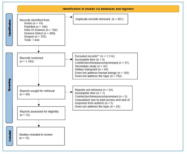
In the first phase (title and abstract screening), two researchers independently evaluated each reference and classified them as ‘included,’ ‘excluded,’ or ‘uncertain.’ In the second phase (full-text assessment), the pre-selected articles were reviewed in full by independent reviewers to confirm their eligibility. The selection process is documented in the PRISMA-ScR flowchart12 shown in Figure 1. Disagreements were resolved by discussion or consultation with a third senior reviewer when necessary.
Preferred Reporting Items for Systematic Reviews and Meta-Analyses (PRISMA) 2020 flowchart showing the process of article identification, screening, eligibility assessment, and inclusion. The flowchart details the number of records identified, excluded, and included at each stage of the scoping review. Adapted from PRISMA flowchart (Page, 2021)38.
The levels of evidence (LE) was classified for descriptive purposes only. The LE hierarchy was used as described by Wallace et al. (2022)13, as per the list below:
-
Level 1: Systematic reviews or meta-analyses of randomized controlled clinical trials.
-
Level 2: At least one well-designed randomized controlled trial.
-
Level 3: Well-designed clinical trials without randomization.
-
Level 4: Well-designed cohort and case-control studies.
-
Level 5: Systematic reviews of descriptive and qualitative studies.
-
Level 6: A single descriptive or qualitative study.
-
Level 7: Opinions of authorities or reports of expert committees.
Ethics
This review used only previously published research; therefore, ethical approval was not required.
Extraction
In total, 1,444 studies were identified, 281 of which were excluded as duplicates. After title and abstract screening, 1,114 studies were excluded; among these, 792 did not address the central topic, and three had incomplete data, 37 included individuals with coinfection or immunocompromised status, 169 were not performed in humans, and 69 addressed kidney transplantation. This resulted in the assessment of 49 full-text articles for eligibility. After this stage, five studies were excluded due to lack of access, one had incomplete information, 26 did not address the research question, and two included individuals with coinfection or immunocompromised status. The final analysis included 15 studies, as detailed in the PRISMA-ScR flowchart (Figure 1).
The extraction was designed to capture information regarding study characteristics, such as authors, study design, year of publication, country of publication, objectives, sample size, and main findings. Considering renal outcomes, evidence regarding clinical and laboratory manifestations, biomarkers, tubulointerstitial and glomerular compartment modifications, AKI, treatment toxicity, and management were collected.
RESULTS
Characteristics of the studies
Among the 15 included studies, publications ranged from 197214 to 202515, with seven published in the last decade. Nine studies were conducted in Brazil, with others from China, Iran, Spain, Somalia, and Sudan. A total of 1,209 individuals with VL were analyzed. The studies ranged from case reports to cohort studies, and described demographic data, urinary abnormalities, kidney function parameters, histological findings, AKI occurrence, treatment exposure, and outcomes (Table 1).
Clinical manifestations (signs and symptoms)
The main signs and symptoms included fever (the most common, with temperatures reaching up to 40 °C), anemia, hepatosplenomegaly, adenomegaly, jaundice, asthenia, anorexia, and vomiting. Additional symptoms included pancytopenia, hematuria, purpuric lesions, abdominal pain, diarrhea, and dyspnea16-20.
VL diagnosis
Bone marrow aspiration was the primary method for diagnosing VL14-27. One study employed Leishmania DNA detection in urine28. Additionally, Enzyme-Linked Immunosorbent Assay (ELISA) was used. Although this method has limitations, the association between clinical symptoms and response to treatment with liposomal amphotericin B supported the diagnosis21,22.
Main Findings
Kidney manifestations associated with VL ranged from asymptomatic urinary abnormalities to significant glomerular and tubulointerstitial changes with varying degrees of glomerular filtration rate (GFR) impairment, including severe cases that required dialysis or resulted in death15,17,22-24. The studies were heterogeneous and provided data on clinical manifestations, diagnostic investigations, kidney injuries, therapeutic approaches, and outcomes associated with VL (Table 2 and Table 3). The results are presented according to the renal alterations associated with VL (laboratory changes, affected renal compartments, and AKI), followed by treatment-related nephrotoxicity.
Renal Alteration
Regarding the key findings, VL can affect both glomerular and tubulointerstitial compartments15-20,22, demonstrating structural damage. AKI was reported in several studies, which also described elevated serum creatinine (sCr) levels and reduced urine output17,18,21-23.
Among urinary modifications, proteinuria was a frequent finding in VL16,19,20,28. Initial necropsy data revealed that over 80% of individuals (n = 15, 83.3%) had morphological evidence of proteinuria14. Furthermore, a study performed in Sudan indicated that approximately 40% of individuals with VL had mild kidney damage, with microalbuminuria detected using ELISA (n = 35, 39.7%) and immunoturbidimetry (n = 37, 42%)28.
Indeed, proteinuria is common in VL and can be associated with alterations in urinary sediments. One study reported the presence of proteinuria (n = 10, 90.9%), leukocyturia (n = 6, 54.5%), and hematuria (n = 7, 63.6%). Hypoalbuminemia and hypergammaglobulinemia were observed in all the individuals16. Furthermore, higher levels of proteinuria have been observed in some glomerular diseases related to VL23. In a case of membranoproliferative glomerulonephritis (MPGN) with cryoglobulinemia, the patient presented with proteinuria (1.7 g/24 hours), hematuria, and sCr elevated (2.51 mg/dL)19.
Concerning glomerular manifestations, the most common finding was MPGN in some cases associated with cryoglobulinemia15,19,23. This manifestation may be due to hypergammaglobulinemia, ruling out other possible causes of renal alterations such as autoimmune diseases19,23. Some cases were associated with severe complications such as the need for hemodialysis or death15,22. Mesangial involvement was reported in some articles14,20.
Tubulointerstitial manifestations were also observed in patients with VL. One study demonstrated that patients with AKI and VL exhibit increased levels of urinary kidney injury molecule-1 and serum NGAL (sNGAL)24. Furthermore, one report showed tubulointerstitial nephritis on anatomopathological analysis19. One study observed proximal tubule damage, with 45.4% of the patients showing increased excretion of retinol-binding protein16.
Another study reported that all patients with VL exhibited urinary concentration deficits before treatment initiation. Following treatment reassessment, most patients showed persistently reduced urinary osmolality (below 700 mOsm/L), even after water deprivation and desmopressin acetate administration. Before treatment, urinary acidification was assessed, which revealed that more than half of the patients showed acidification deficits after calcium chloride (CaCl₂) overload. Additionally, urinary oxidative stress biomarker levels were higher in the VL group before treatment than in the control group25.
Among the studies that described AKI, the incidence ranged from 26.3% to 46%18,21,22,24,26,27. A study conducted in Brazil demonstrated that sNGAL could serve as a promising early biomarker of AKI24. Furthermore, AKI was associated with disease severity, longer hospitalization time, dialysis requirement and mortality17,18,21,24. Another Brazilian study showed that individuals with higher sCr levels (classified as those with sCr > 1.3 mg/dL) required a second-line drug for treatment and had a higher mortality rate14.
Similarly, a cohort study of individuals with VL analyzed those with and without AKI and demonstrated that older age, male sex, and jaundice were associated with AKI (n = 76, 33.9%). Additionally, only 6.5% of the patients exhibited oliguria. Mortality was significantly higher in patients with AKI (n = 23, 30.2%) than in those without AKI (3.1%; p < 0.0001)20. Another study compared adults aged > 21 years with children aged ≤ 21 years and found a similar incidence of AKI (35.1% vs 39.4%, respectively)27. In the adults with hypokalemia, the use of amphotericin B, chills, and leukopenia were associated with AKI15.
In addition, a case series (n = 4) found that among patients with VL and AKI, the median urea level was 105.5 mg/dL (86-136 mg/dL), the median sCr was 2.675 mg/dL (1.8-5.0 mg/dL), all patients were men, the median age was 36.5 years (17-51 years), and the median hospitalization time was 36 days (10-48 days)18.
Treatment
Among the selected studies (n = 15), 60% (n = 9/15) reported that patients were treated with amphotericin B as a part of VL management. Of these, 26,7% of all included studies (n = 4/15) explicitly specified the liposomal formulation as amphotericin B15,19,21,27. When the treatment details were reported, the regimens included pentavalent antimonials and amphotericin B deoxycholate, with substantial variability in dosing schemes and treatment durations across studies (Table 2).
AKI after treatment
Regarding AKI following treatment, a few studies evaluated kidney function before and after amphotericin B. One study reported that patients with VL were initially treated with intravenous pentavalent antimonials (20 mg/kg/day for 20-40 consecutive days). In severe cases, amphotericin B deoxycholate was administered at a total cumulative dose of 7-20 mg/kg over 20 days. In this cohort, 39 (51.3%) and seven (9.2%) patients developed a new episode of AKI within a mean of 10 days after initiating amphotericin B treatment and pentavalent antimonial therapy, respectively. In addition, among patients with severe VL who were unresponsive to sodium antimony gluconate, treatment with amphotericin B was significantly associated with AKI (p < 0.0001). Notably, lipid-based amphotericin B formulations were not used22.
In another study, sodium antimony gluconate was used as the primary antileishmanial agent. Patients with sCr < 1.3 mg/dL were exclusively treated with pentavalent antimonial therapy. In contrast, patients with sCr > 1.3 mg/dL required adjunctive therapy, primarily amphotericin B; in a total of 15 individuals with AKI, eight developed AKI before amphotericin B treatment. All three recorded deaths occurred in the group with elevated sCr17.
Despite the nephrotoxicity associated with VL treatment, disease improvement is a key factor in reducing inflammation and facilitating AKI recovery20. One study reported a decrease in albuminuria after treatment26. A few case reports showed improvement following liposomal amphotericin B treatment19-21. Another study demonstrated a peak in sCr levels during treatment with improvements observed in the post-treatment period. However, no changes in sCr levels were recorded during amphotericin B therapy, suggesting that other factors may have contributed to the worsening of kidney function28.
Regarding glomerulonephritis treatment, two cases of MPGN associated with cryoglobulinemia were treated with liposomal amphotericin B. One of these cases also received immunosuppressive therapy; however, the outcome was fatal, while the other case showed improvement in the clinical manifestations of VL but without recovery of kidney function23. Similarly, in a case of MPGN, the patient was treated with prednisone and hemodialysis and subsequently received liposomal amphotericin B after VL diagnosis. Despite treatment, the patient remained on HD and died five months after initial presentation15. In contrast, another case of MPGN associated with cryoglobulinemia showed an improvement in kidney function after treatment with liposomal amphotericin B19.
In another case in which only mesangial alterations were observed without immune complex deposits, treatment with sodium antimony gluconate improved VL; however, proteinuria remained high. The addition of valsartan reduced the proteinuria20. Therefore, the treatment of VL is a key component of management, whereas the role of immunosuppressive therapy requires further investigation to define its benefits and risks.
DISCUSSION
The present study offers a summarized review of the associations between VL and kidney alterations, emphasizing the complexity of the disease and its potential impact on various kidney compartments, including the glomeruli and tubulointerstitial regions. This review highlights the high incidence of AKI, including severe cases that may necessitate dialysis or lead to death. The treatment of VL-related kidney complications is challenging owing to its association with impaired kidney function; however, intervention is crucial for alleviating the manifestations of VL and preventing further deterioration of the GFR.
First, the incidence of AKI was notable, although it was comparable to that observed in other neglected infectious diseases29. Despite the high incidence and severity of AKI, data on AKI in VL remain limited. Research on early diagnosis and clinical management of AKI is scarce, highlighting the urgent need to allocate resources to address this important complication of VL. Notably, some studies were conducted in hospital settings, and, in some cases, in intensive care units, where individuals tend to present with more severe cases18.
Although the criterion proposed by Kidney Disease: Improving Global Outcomes (KDIGO) is currently the most used and widely accepted in clinical practice, as well as in research, its application only became widespread over the last decade30. Many of the included studies were conducted before KDIGO implementation, limiting data standardization. Nevertheless, these studies have provided clinically relevant insights. For instance, NGAL has shown potential as an early AKI biomarker25. Several studies have identified this biomarker as an effective indicator of kidney damage31,32. Furthermore, consistent with numerous previous studies, AKI was associated with disease severity, as reflected by longer hospital stays, need for additional medications, and increased mortality33,34.
Regarding glomerular manifestations, all the included studies were case reports or case series, and both MPGN and mesangial alterations were observed. Other studies on parasitic diseases have reported comparable findings, which may be pathophysiologically related to immune complex deposition35. Some case reports have described other glomerular manifestations such as amyloidosis36. However, such data may be absent from this review owing to the decision to exclude articles describing coinfections and immunosuppressive conditions to focus specifically on the role of VL in kidney manifestations. Presentations such as amyloidosis and other glomerular diseases may occur more frequently in these excluded conditions. Therefore, further studies are required to explore this issue.
Treatment of VL is primarily based on amphotericin B, although some studies have also considered pentavalent antimonials. The toxicity of these medications differs in the context of a reduced GFR. Pentavalent antimonials are predominantly eliminated by the kidneys; consequently, toxicity, particularly cardiotoxicity and pancreatitis, increases in individuals with reduced GFR37. In contrast, amphotericin B is potentially nephrotoxic, mainly due to tubular damage and reduced renal blood flow38. As most studies were conducted in Brazil, this finding is consistent with the guidelines for the treatment of VL in the Americas, which strongly recommend the use of liposomal amphotericin B. In contrast, pentavalent antimonials or deoxycholate amphotericin B have conditional recommendations39. The management of glomerulonephritis associated with VL remains uncertain and may involve either the exclusive treatment of VL or a combination of immunosuppressive therapy and supportive treatment with antiproteinuric agents. However, further studies are required to address this issue.
This study had several limitation. First, most studies were observational or case reports. Finally, despite the limited data, this review synthesized renal manifestations and treatment. Therefore, this underscores the need to focus efforts on increasing and improving research in this area and achieving the identification of evidence regarding the potential of VL to directly cause kidney manifestations by itself.
CONCLUSION
Based on the available evidence, VL has been reported to be potentially associated with renal alterations, including histological kidney changes, laboratory abnormalities, and AKI, either associated with infection or secondary to treatment. This review mapped the reported risk groups, renal clinical manifestations, and informed early management strategies, underscoring the importance of monitoring renal alterations and searching for validated biomarkers that improve early detection in this population.
However, several gaps were identified that may help guide future research in the field of AKI. Because both VL and its treatment are potential contributors to AKI, prospective studies with better differentiation between AKI occurring before treatment, after treatment initiation, and at long-term follow-up are essential. In the field of glomerular involvement, additional studies including a larger number of individuals with glomerulonephritis are needed to better characterize the main histological alterations and define management strategies, including VL treatment alone or in combination with immunosuppressive therapy. Such efforts would strengthen the evidence base for optimal management strategies by identifying serum and renal parameters associated with worse outcomes and potentially reduce morbidity and mortality.
ACKNOWLEDGMENTS
None.
REFERENCES
- 1 Nascimento LFJ, Cirilo TM, Gomes DS, Gomes ACA, Lima VFS, Scher R, et al. Epidemiological and diagnostic aspects of feline leishmaniasis with emphasis on Brazil: a narrative review. Parasitol Res. 2022;121(1):21-34.
- 2 Erber AC, Sandler PJ, de Avelar DM, Swoboda I, Cota G, Walochnik J. Diagnosis of visceral and cutaneous leishmaniasis using loop-mediated isothermal amplification (LAMP) protocols: a systematic review and meta-analysis. Parasit Vectors. 2022;15(1):34.
- 3 Murray RP, Rosenthal KS, Pfaller MA. Microbiologia Médica. 8ª ed. Rio de Janeiro: Elsevier 2017. Cap Protozoários do sangue e dos tecidos.
-
4 World Health Organization. Leishmaniasis [Internet]. Switzerland: WHO 2023. Accessed in April, 2025. Available from: Available from: https://www.who.int/news-room/fact-sheets/detail/leishmaniasis
» https://www.who.int/news-room/fact-sheets/detail/leishmaniasis -
5 Ministério da Saúde [BR]. DATASUS [Internet]. 2023 [Accessed in August 2024]. Available from: Available from: http://tabnet.datasus.gov.br/cgi/tabcgi.exe?sinannet/cnv/leishvbr.def
» http://tabnet.datasus.gov.br/cgi/tabcgi.exe?sinannet/cnv/leishvbr.def - 6 Scarpini S, Dondi A, Totaro C, Biagi C, Melchionda F, Zama D, et al. Visceral Leishmaniasis: Epidemiology, Diagnosis, and Treatment Regimens in Different Geographical Areas with a Focus on Pediatrics. Microorganisms. 2022;10(10).
- 7 Machado CAL, Valle D, Horta MC, Meiga AYY, Sevá ADP. Patterns and drivers of Human Visceral Leishmaniasis in Pernambuco (Brazil) from 2007 to 2018. PLoS Negl Trop Dis. 2023;17(2):e0011108.
- 8 Costa CHN, Chang KP, Costa DL, Cunha FVM. From Infection to Death: An Overview of the Pathogenesis of Visceral Leishmaniasis. Pathogens. 2023;12(7).
-
9 Peters MDJ, Godfrey CM, McInerney P, Soares CB, Khalil H, Parker D. The Joanna Briggs Institute reviewers' manual 2015: methodology for JBI scoping reviews. Adelaide: The Joanna Briggs Institute. Available from:: http://joannabriggs.org/assets/docs/sumari/Reviewers-Manual_Methodology-for-JBI-Scoping-Reviews_2015_v2.pdf Cited 08 dez. 2025.
» http://joannabriggs.org/assets/docs/sumari/Reviewers-Manual_Methodology-for-JBI-Scoping-Reviews_2015_v2.pdf - 10 Lucas A, Wyatt CM. HIV at 40: kidney disease in HIV treatment, prevention, and cure. Kidney Int. 2022;102(4):740-9.
- 11 Monge-Maillo B, López-Vélez R. Leishmaniasis in transplant patients: what do we know so far? Curr Opin Infect Dis. 2024;37(5):342-8.
-
12 Fabbri S, Silva C, Hernandes E, Octaviano F, Di Thommazo A, Belgamo A. Improvements in the stArt tool to better support the systematic review process.; Limerlick, Ireland: Proceedings of the 20th International Conference on Evaluation and Assessment in Software Engineering 2016. [Internet] Available from: https://dl.acm.org/doi/10.1145/2915970.2916013.
» https://doi.org/10.1145/2915970.2916013 - 13 Wallace SS, Barak G, Truong G, Parker MW. Hierarchy of Evidence Within the Medical Literature. Hosp Pediatr. 2022;12(8):745-50.
- 14 Andrade ZA, Iabuki K. The nephropathy of kala-azar. Rev Inst Med Trop Sao Paulo. 1972;14(1):51-4.
- 15 Mansoursamaei A, Valikhani M, Rafiee H. Fatal Outcome of Membranoproliferative Glomerulonephritis in a Patient With Hidden Visceral Leishmaniasis. Case Reports in Infectious Diseases, v. 2025, n. 1, jan. 2025.
- 16 Salgado Filho N, Ferreira TM, Costa JM. Involvement of the renal function in patients with visceral leishmaniasis (kala-azar). Rev Soc Bras Med Trop. 2003;36(2):217-21.
- 17 Daher EF, Evangelista LF, Silva Júnior GB, Lima RS, Aragão EB, Arruda GA, et al. Clinical presentation and renal evaluation of human visceral leishmaniasis (kala-azar): a retrospective study of 57 patients in Brazil. Braz J Infect Dis. 2008;12(4):329-32.
- 18 Daher EF, Sampaio AM, Martiniano LVM, Vieira APF, Silva Junior GB. Acute kidney injury in visceral leishmaniasis: a cohort of 10 patients admitted to a specialized care unit in northeast of Brazil. Asian Pac J Trop Dis. 2013;3(1):41-6.
- 19 Padrón Romero M, Acevedo Ribó MM, Ahijado Hormigos FJ, Díaz Crespo F, Cueto Bravo L, Herraiz Corredor C, et al. Membranoproliferative Glomerulonephritis and Mixed Cryoglobulinemia as a Form of Presentation of Visceral Leishmaniasis. Am J Case Rep. 2020;21:e921445.
- 20 Zou L, Chen G, Zhou Y, Ye W, Wen Y, Chen L, et al. Continuous hypergammaglobulinemia and proteinuria after the recovery of the visceral Leishmaniasis: a case report. BMC Infect Dis. 2021;21(1):124.
- 21 Mohamed AH, Bashir AM. Acute kidney injury as initial presentation of visceral leishmaniasis in a young patient- A case report. Ann Med Surg (Lond). 2022;78:103821.
- 22 Oliveira MJ, Silva Júnior GB, Abreu KL, Rocha NA, Garcia AV, Franco LF, et al. Risk factors for acute kidney injury in visceral leishmaniasis (Kala-Azar). Am J Trop Med Hyg. 2010;82(3):449-53.
- 23 Ortiz M, Mon C, Herrero JC, Oliet A, Rodríguez I, Ortega O, et al. Glomerulonephritis and cryoglobulinemia: first manifestation of visceral leishmaniasis. Clin Nephrol. 2015;83(6):370-7.
- 24 Meneses GC, De Francesco Daher E, da Silva Junior GB, Bezerra GF, da Rocha TP, de Azevedo IEP, et al. Visceral leishmaniasis-associated nephropathy in hospitalised Brazilian patients: new insights based on kidney injury biomarkers. Trop Med Int Health. 2018;23(10):1046-57.
- 25 Oliveira MJ, Silva GB, Sampaio AM, Montenegro BL, Alves MP, Henn GA, et al. Preliminary study on tubuloglomerular dysfunction and evidence of renal inflammation in patients with visceral leishmaniasis. Am J Trop Med Hyg. 2014;91(5):908-11.
- 26 Rocha NA, Oliveira MJ, Franco LF, Júnior GB, Alves MP, Sampaio AM et al. Comparative Analysis of Pediatric and Adult Visceral Leishmaniasis in Brazil. Pediatr Infect Dis J. 2013;32(5):e182-5.
- 27 Corrêa-Castro G, Silva-Freitas ML, de Paula L, Soares Pereira L, Dutra MRT, Albuquerque HG, et al. A link between circulating immune complexes and acute kidney injury in human visceral leishmaniasis. Sci Rep. 2024;14(1):9870.
- 28 Elnojomi NA, Musa AM, Younis BM, Elfaki ME, El-Hassan AM, Khalil EA. Surrogate markers of subtle renal injury in patients with visceral leishmaniasis. Saudi J Kidney Dis Transpl. 2010;21(5):872-5.
- 29 Duarte DB, Lacerda MCSR, Ribeiro YJP, Ribeiro MZD, Frederico MA, Oliveira MJC. Kidney biomarkers in tropical infections: an update. Pathog Glob Health. 2020;114(6):302-8.
- 30 Khwaja A. KDIGO clinical practice guidelines for acute kidney injury. Nephron Clin Pract. 2012;120(4):c179-84.
- 31 Gupta B, Tiwari P, Subramanian A, Mahajan S, Kalaivani M, Bindra A, et al. Evaluation of plasma and urine neutrophil gelatinase-associated lipocalin (NGAL) as an early diagnostic marker of acute kidney injury (AKI) in critically ill trauma patients. J Anaesthesiol Clin Pharmacol. 2023;39(2):292-301.
- 32 Yousef Almulhim M. The efficacy of novel biomarkers for the early detection and management of acute kidney injury: A systematic review. PLoS One. 2025;20(1):e0311755.
- 33 Cheruku SR, Raphael J, Neyra JA, Fox AA. Acute Kidney Injury after Cardiac Surgery: Prediction, Prevention, and Management. Anesthesiology. 2023;139(6):880-98.
- 34 Birkelo BC, Pannu N, Siew ED. Overview of Diagnostic Criteria and Epidemiology of Acute Kidney Injury and Acute Kidney Disease in the Critically Ill Patient. Clin J Am Soc Nephrol. 2022;17(5):717-35.
- 35 Daher EF, da Silva Junior GB, Trivedi M, Fayad T, Srisawat N, Nair S, et al. Kidney complications of parasitic diseases. Nat Rev Nephrol. 2022;18(6):396-406.
- 36 Navarro M, Bonet J, Bonal J, Romero R. Secondary amyloidosis with irreversible acute renal failure caused by visceral leishmaniasis in a patient with AIDS. Nefrologia. 2006;26(6):745-6.
-
37 Marques SA, Merlotto MR, Ramos PM, Marques MEA. American tegumentary leishmaniasis: severe side effects of pentavalent antimonial in a patient with chronic renal failure. An Bras Dermatol. 2019;94(3):355-7. doi: 10.1590/abd1806-4841.20198388. PMID: 31365669; PMCID: PMC6668951.
» https://doi.org/10.1590/abd1806-4841.20198388 - 38 Dobrek L. A Synopsis of Current Theories on Drug-Induced Nephrotoxicity. Life (Basel). 2023;13(2):325.
- 39 Organização Pan-Americana de Saúde. Diretrizes para o tratamento das leishmanioses na Região das Américas. 2ª ed. Washington, DC: OPAS. 2022.
-
Data Availability Statement:
The research data are available in the Open Science Framework (OSF) repository at the following link: https://archive.org/details/osf-registrations-tgp79-v1
-
Financial Support:
None
Edited by
-
Editor-in-Chief:
Prof. Dalmo Correia Filho. Orcid: https://orcid.org/0000-0002-2174-5058
The research data are available in the Open Science Framework (OSF) repository at the following link: https://archive.org/details/osf-registrations-tgp79-v1
Data citations
Ministério da Saúde [BR]. DATASUS [Internet]. 2023 [Accessed in August 2024]. Available from: Available from: http://tabnet.datasus.gov.br/cgi/tabcgi.exe?sinannet/cnv/leishvbr.def