 Molecular and biological characterization of tobacco mild green mosaic virus from Petunia (Petunia × hybrida) in Brazil
ABSTRACT
An isolate of the tobamovirus tobacco mild green mosaic virus (TMGMV), Tobamovirus mititessellati, was found naturally infecting petunia (Petunia × hybrida) cv. Pinstripe, causing reduced leaf size, mottling, and color breaking of flowers, in an experimental field at Piracicaba, São Paulo state, Brazil. TMGMV was identified by virion morphology, cytopathology, transmission experiments, and molecular assays. The entire nucleotide sequence of this petunia isolate of TMGMV (TMGMV-Pet) was obtained and demonstrated, in essence, to be similar to previously analyzed isolates. One hundred and twenty plant species were assayed by mechanical transmission, and 35 of them from nine families, both previously reported and unreported species, were shown to be experimentally susceptible to TMGMV-Pet. On the other hand, certain species, among 85 unsusceptible hosts for TMGMV-Pet, known to be susceptible to TMGMV, failed to be infected. Cytopathological studies on naturally and experimentally infected host plants revealed that virions of TMGMV-Pet produced para-crystalline aggregates, but not angular arrays, which are considered characteristic of TMGMV. As previously described, thin sections revealed the presence of pseudo-virions in the stroma of chloroplasts in TMGMV-Pet-infected tobacco (Nicotiana tabacum), and, additionally, in other hosts such as N. clevelandii, N. megalosyphon, and petunia indicating that this process is not restricted to tobacco.
Keywords
cytopathology
; experimental host range; genome stability; pseudo-virions in chloroplasts; tobamovirus
Introduction
The solanaceous genus Petunia Juss, comprises more than 20 species and originates in South America’s tropical and subtropical regions. Most of cultivated types are hybrid (Petunia × hybrida Regel), with a wide variety in flower size and color, being a very popular ornamental worldwide, used either potted or in garden beds (Lorenzi and Souza, 2001). Similar to many other cultivated solanaceous plants, petunias are susceptible to natural infection by many viruses. Sastry et al. (2019) listed 28 viruses and four viroids infecting petunia plants in their comprehensive review of plant viruses and viroids. In Brazil, there are records of four viruses infecting petunia plants: tobacco mosaic virus (TMV), cauliflower mosaic virus, petunia vein banding virus, and an uncharacterized orthotospovirus (Kitajima, 2020).
Tobacco mild green mosaic virus (TMGMV- Tobamovirus mititessellati) belongs to the genus Tobamovirus (family Virgaviridae) and has elongated and rigid particles measuring ca. 18 nm in diameter and 300 nm long. It has positive-sense single-stranded RNA (+ssRNA) genome of 6.2 to 6.4 kb encoding four open reading frames (ORF). ORF1 and ORF2 encode two nonstructural proteins required for replication, while ORF3 and ORF4 encode the nonstructural movement protein and coat protein, respectively (Solis and Garcia-Arenal, 1990). The transmission of TMGMV occurs by contact between plants, workers’ hands, tools, and sometimes by seeds and contaminated soil (Wetter, 1986). TMGMV, also known as green-tomato atypical mosaic virus, mild strain of TMV, paratobacco mosaic virus, South Carolina mild mottling strain, TMV strain U2 and U5 and tomato atypical virus green mottling strain (Wetter, 1986; Skelton and Sansford, 2012), has been reported globally. This virus was initially described in 1929 (McKinney,1929) in the Canary Islands infecting Nicotiana glauca L. Initially many TMGMV isolates were considered strains of TMV, but detailed studies on its biophysical, biochemical, molecular, and biological properties have suggested that TMGMV represented a species distinct from TMV (Wetter, 1986). TMGMV is considered important in the tobacco industry (Wetter, 1986) and has been found worldwide infecting vegetable crops, medicinal plants, and several ornamentals, including petunia (Skelton and Sansford, 2012; Sastry et al., 2019).
In this paper, we describe the biological, molecular, and ultrastructural characterization of an isolate of TMGMV found infecting petunia in Brazil, detailing its broad experimental host range and some cytopathological peculiarities.
Materials and Methods
Mechanical transmission assays
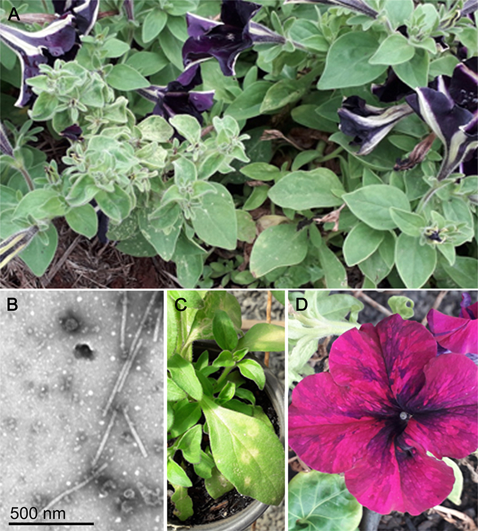
In February 2022, Petunia × hybrida cv. Pinstripe plants showing symptoms of chlorosis, sometimes mottled leaves, flower breaking, and general stunting (Figure 1A) were noticed in an experimental plot in the municipality of Piracicaba, São Paulo state, Brazil (22°42’28.5” S, 47°37’46.5” W, altitude 580 m). Twelve plants out of a total of 120 were symptomatic, and six were collected and established under greenhouse conditions for further work. Transmission electron microscopy (TEM) of leaf extracts from these symptomatic petunias revealed the presence of short, rigid, rod-shaped particles, ca. 300 nm long, similar to those of tobamoviruses (Figure 1B), suggesting a viral nature inherent in this disease. Subsequent attempts to detect and identify the putative virus responsible for the symptoms observed in Pinstripe petunia plants were made by mechanical transmission assays. Extracts of symptomatic leaves from six randomly selected field Pinstripe petunia plants were mechanically inoculated into petunia (cvs. Pinstripe, Debonaire blackcherry, and Anã compacta) and indicator assay plants (Chenopodium quinoa Willd., C. amaranticolor Coste & Reyn., Gomphrena globosa L., Datura stramonium L., N. tabacum L. cvs. TNN, and Turkish). Symptomatic leaves of field petunia cv. Pinstripe were macerated in a mortar, in the presence of 0.01 M phosphate buffer +0.1 % sodium sulfite, rubbed onto leaves of potted indicator plants, previously dusted with carborundum powder, and immediately washed with water. The plants were kept in a greenhouse and observed daily for symptom expression.

– A) Field petunia (Petunia × hybrida) cv. Pinstripe showing smaller leaves and mottling; B) Transmission electron micrograph of negatively stained leaf extract from the field, symptomatic petunia plants, showing tobamovirus-like, rod shaped particles; C) Chlorotic spots on mechanically inoculated leaf of petunia cv. Anã compacta; D) color breaking in the flower.
To evaluate the susceptibility of other plant species belonging to a large number of families, some of them previously described naturally (Skelton and Sansford, 2012; Sastry et al., 2019; Kitajima, 2020), or experimentally (Skelton and Sansford, 2012) infected by isolates of TMGMV (Tables 1 and 2), were similarly inoculated mechanically, and followed for eventual symptom expression. Leaf samples of all inoculated plants were mechanically retro-inoculated into C. quinoa to confirm infection. Samples of plants that tested positive (local chlorotic lesions in C. quinoa leaves) were also processed for TEM and tested by reverse transcription polymerase chain reaction (RT-PCR) using specific primers for TMGMV (see below).
Transmission electron microscopy
Attempts to detect virus-like particles in leaf extracts of the petunia plants from the field-infected petunia plants and experimentally inoculated plants were made by TEM of uranyl acetate, negatively stained leaf extracts. Ultrastructural analysis of symptomatic leaves was carried out on thin sections of paraformaldehyde/glutaraldehyde preserved tissues, post-fixed with osmium tetroxide and embedded in Spurr’s low viscosity epoxy resin (Kitajima and Nome, 1999). TEM examinations were completed in a JEOL JEM 1011, and images digitally recorded.
Molecular assays
For molecular detection and identification of the putative virus, total RNA was extracted from mechanically inoculated C. quinoa plants using RNeasy Plant Mini Kit (Qiagen). Next, rRNA was removed using Ribo-Zero Kit for plants (Illumina), and a cDNA library was prepared and sequenced using an Illumina NovaSeq6000 system with 100-bp paired-ends reads (Macrogen Inc.). The high-throughput sequencing (HTS) reads were trimmed by BBDuk (https://github.com/BioInfoTools/BBMap), and contigs were obtained by de novo assembly using MEGAHIT v. 1.3.1. The resulting contigs were analyzed with BlastX against the virus protein database using Geneious Prime 2023.1.2 (Dotmatics).
RT-PCR was carried out to confirm the HTS-detected virus in the symptomatic leaves of six field Pinstripe petunia plants, as well as in field plant sources of inoculum and some used in the experimental transmission assays. Total RNA was separately extracted from all samples using the PureLink Viral RNA/DNA Kit (Thermo Fisher Scientific) following the manufacturer’s instructions. A single-step RT-PCR was performed using 100-300 ng of total RNA and a specific pair of primers TMGMV_5641F (AGGAAATCTCACAACAATAG) and TMGMV_6165R (CGTCATCGAGTACGTTTTAA) which amplifies a fragment of 524 bp of TMGMV coat protein gene (Cohen et al., 2001). The fragment was amplified under the following conditions: 94 °C for 4 min; 30 cycles of 94 °C for 1 min, 48 °C for 45 s, 72 °C for 1 min; and 72 °C for 10 min. The amplicons of two samples were Sanger sequenced (Macrogen, Inc.) using forward and reverse primers, and the sequences were compared with those deposited in GenBank using the BlastN algorithm (http://blast.ncbi.nlm.nih.gov/Blast.cgi).
Available full tobamovirus genome sequences were obtained from GenBank on 23 Oct 2023. Alignments based on nucleotide sequences of the full genomes were made using ClustalW in Geneious Prime. Maximum-likelihood trees were constructed with TMGMV-Pet and 34 full-genome sequences using MrBayes v. 3.2.7 in the CIPRES Science Gateway (https://www.phylo.org/). Two independent runs were conducted simultaneously using 10 million generations which excluded 25 % of the resulting trees as burn-in (Miller et al., 2010).
All plants inoculated during the mechanical transmission experiments that showed symptoms of TMGMV infection were collected for RT-PCR tests using the primers described by Cohen et al. (2001), as described previously. The amplicons obtained were separated on a 1 % agarose gel and visualized with SYBR™ Safe DNA Gel Stain (Invitrogen).
Results
Transmission electron microscopy of leaf extracts
Preliminary evidence for the presence of a tobamovirus associated with symptomatic plants was obtained by examining negatively stained leaf extracts of field Pinstripe petunia plants by TEM, which indicated the presence of a large number of rod-shaped, tobamovirus-like particles, ca. 18 nm × 300 nm (Figure 1B). This observation triggered further biological, morphological, and molecular assays.
Mechanical transmission assays
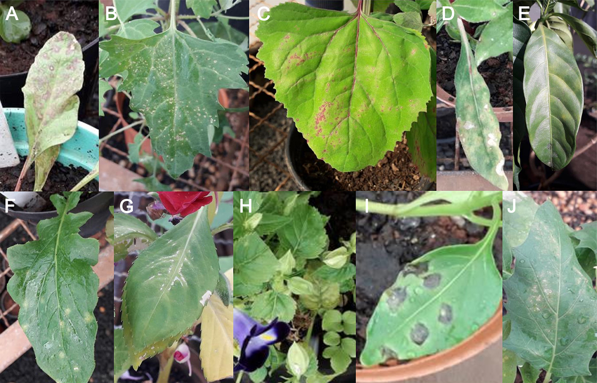
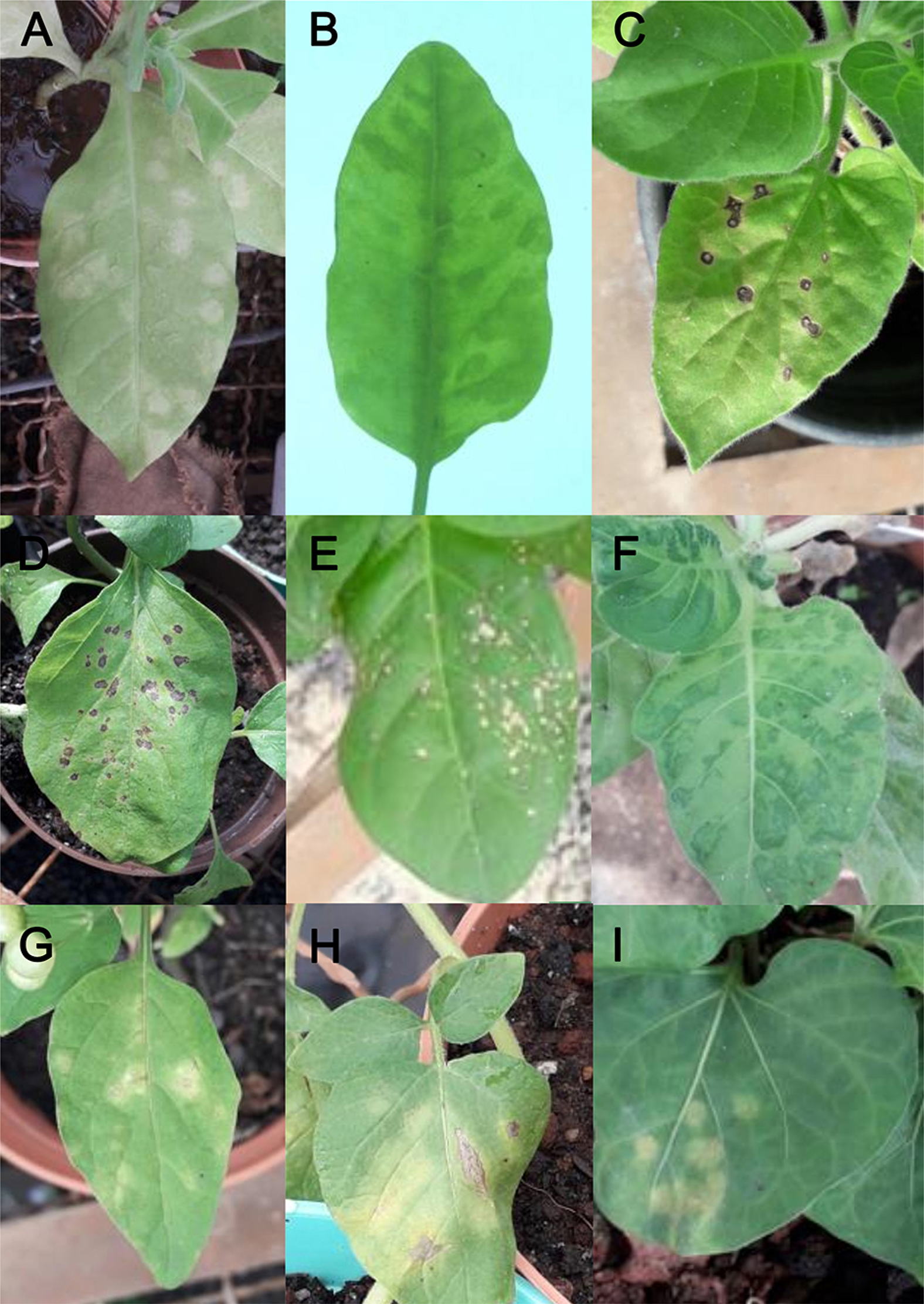
Mechanical transmission assays using leaf extracts from symptomatic field Pinstripe petunia to petunia (cvs. Pinstripe, Debonaire blackcherry, and Anã compacta) showed chlorotic spots, chlorosis, mosaic in their leaves, as well as flower breaking (Figure 1C and D). Inoculation of indicator plants such as C. quinoa (Figure 2B), C. amaranticolor (Figure 2C), tobacco (N. tabacum) cv. TNN (Figure 3E), D. stramonium (Figure 2J) and G. globosa (Figure 2D) resulted in chlorotic or necrotic local lesions on inoculated leaves. Inoculation of tobacco cv. Turkish (Figure 3F) and N. glauca (Figure 3B) resulted in systemic mild mosaic symptoms; extracts of these experimentally infected plants contained tobamovirus-like particles and tested positive in the RT-PCR assay.

– Leaf symptoms on plants experimentally infected by tobacco mild green mosaic virus isolate-Petunia. A) Local necrotic ringspots on Beta vulgaris leaf; B) local lesions in Chenopodium quinoa; C) Chenopodium amaranticolor; D) Gomphrena globosa; E) chlorotic blotches on leaves of Tabernamontana divaricata; F) local chlorotic spots in inoculated leaf of Hypochaeris brasiliensis; G) systemic vein banding in inoculated Impatiens hawkeri; H) reduced leaves and chlorosis in Torenia fournieri; I) large necrotic lesions in inoculated leaf of Capsicum annuum; J) local lesions in inoculated leaf of Datura stramonium.

– A) Systemic chlorotic patches in a leaf of Nicotiana clevelandii; B) mild mosaic on leaves of Nicotiana glauca; C) local necrotic lesions in Nicotiana glutinosa; D) Solanum melongena; E) Nicotiana tabacum cv. TNN; F) mosaic symptoms in N. tabacum cv. Turkish; G) local chlorotic blotches in Solanum Americanum; H) Solanum tuberosum; I) Solanum violifolium.
Biological tests for the detection of TMGMV in all mechanically inoculated plants (Table 1) through inoculation into C. quinoa plants, as well as examinations of leaf extracts by TEM from those plants, revealed that out of 123 plant species, mechanically inoculated with TMGMV-Pet, plants from 35 species (ten families) resulted infected (Figure 2A, E-I; Figure 3A-I) (Table 1), while plants of 87 species (38 families) were not infected (Table 2). Positive infection cases were further analyzed by examining thin sections of the leaf tissues and RT-PCR using specific primers for TMGMV. In all cases, infection by TMGMV-Pet was confirmed (Table 1).
Transmission electron microscopy of tissue sections
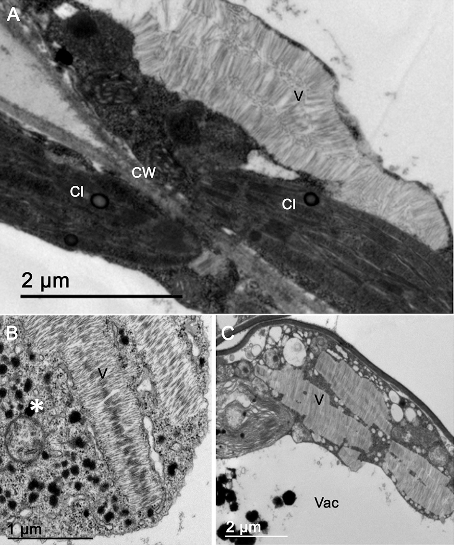
After the initial detection of rod-like particles in extracts of symptomatic field petunia plants, such particles were consistently found in assayed plants extracts to which TMGMV-Pet was experimentally transmitted. Examination of thin sections of leaf tissues from symptomatic, field Pinstripe petunia plants and from all the experimentally infected plants demonstrated the presence of rigid, rod-like particles, either randomly scattered or aggregated side-by-side, in the cytoplasm (Figure 4A-C). Viral particles in angle-layered aggregates, as mentioned by Wetter (1986), were not found. In all infected host plants examined, rod-like particles were seen forming clusters similar to those ascribed to other tobamoviruses, in para-crystalline or long arrays (Francki et al., 1985; Edwardson and Christie, 1986) (Figure 4A-C). In a few susceptible hosts such as N. clevelandii A. Grey, N. megalosyphon Van Heurck & Müll.Agr., N. tabacum cv. Turkish and petunia, virus-like particles were found in the stroma of chloroplasts (Figure 5A-C). In all susceptible hosts, in addition to the presence of putative viral particle aggregates, a structure that was interpreted as the viroplasm (X-body) (Francki et al., 1985), formed by a mixture of dense granules, elements of the endoplasmic reticulum and ribosomes, was observed in the cytoplasm (Figure 4C).

– A) Thin section from leaf palisade parenchyma cell, of field infected petunia cv. Pinstripe, showing a para-crystalline array of presumed tobacco mild green mosaic virus isolate-Petunia particles (V). B) Similar to (A), in a leaf spongy parenchyma cell from a chlorotic local lesion of experimentally infected Chenopodium quinoa. Rod-like particle aggregates are next to a cytoplasmic area rich in dense globules and ribosomes, interpreted as the so-called X body (*). C) Para-crystalline array of presumed tobacco mild green mosaic virus virions in the cytoplasm of a leaf parenchyma cell of pepper (Capsicum annuum). Cl = chloroplast; CW = cell wall; Vac = vacuole.

– A) Detail of tobamovirus-like particles (arrows) in the chloroplast’s stroma in a leaf palisade parenchyma cell of field infected, petunia cv. Pinstripe; B) similar situation in experimentally infected Nicotiana tabacum cv. Turkish (systemic mosaic); C) Nicotiana megalosyphon (local lesion); D) Nicotiana clevelandii (local lesion). C = chloroplast; CW = cell wall; V = aggregate of presumed tobacco mild green mosaic virus virions in the cytoplasm; Vac = vacuole.
Molecular assays
Longer contigs were assembled by read mapping in Geneious Prime based on the contigs related to tobamovirus sequences. The longest contig assembled contained 7,931 nucleotides (nt) corresponding to TMGMV aligned with 9,469,691 reads (out of 61,547,236) with a mean coverage of 468,809.5. The near complete genome of TMGMV-Pet contains 6,340 nt, including 59 nt in the 5’ untranslated region (UTR) and 206 nt in the 3’ UTR. This isolate of the virus has four ORF, which ORF1 (nt 60-4,889, 1,112 aa = amino acids) and ORF2 (nt 60-4,889, 1,610 aa) are separated by a leaky stop codon. ORF3 (nt 4,889-5,649, 257 aa) has a short overlap region with ORF4 (nt 5,655-6,134, 160 aa) (GenBank accession number PP623115). These data are like those described by Solis and Garcia-Arenal (1990).
The 524 bp amplicon obtained by RT-PCR with specific primers for TMGMV confirmed the presence of the virus in the six samples of petunia from the field. Comparison of the two obtained nucleotide sequences (OR756300, OR756301) with those available from GenBank showed 99.17 and 96.88 % identity with the corresponding nucleotide sequence of an isolate of TMGMV identified in infected Impatiens spp. plants from Kenya (ON013908).
The phylogenetic tree generated with the complete genome sequences of TMGMV and other tobamovirus sequences grouped the TMGMV-Pet in a clade with other TMGMV sequences (Figure 6). Furthermore, the full genome of TMGMV-Pet shares nucleotide sequence identity of at least 96.37 % with other TMGMV isolates (data not shown).

– Phylogenetic tree generated with full-genome sequences of tobacco mild green mosaic virus isolate Petunia (TMGMV_Pet, GeneBank accession number PP623115) and other tobamoviruses. The sequence of TMGMV-Pet, highlighted in red, is sub-grouped together with the recently detected Brazilian TMGMV isolate from Nicotiana glauca, in the branch corresponding to TMGMV isolates collected around the world. The bootstrap support values (1,000 replications) of branches are shown next to the corresponding nodes. The scale bar specifies the average number of substitutions per site. ToMV = tomato mosaic virus; TMV = tobacco mosaic virus; PMMoV = pepper mild mottle virus; ORSV = odontoglossum ringspot virus; CGMMV = cucumber green mottle mosaic virus. The GenBank accession numbers of viral sequences used in this analysis are shown before the viral acronym in each leaf description.
Discussion
The tobamovirus infecting petunia plants cv. Pinstripe described in this work was unequivocally identified as an isolate of TMGMV after biological, morphological, and molecular assays. Preliminary evidence obtained by examining the leaf extract, revealed the presence of tobamovirus-like particles in symptomatic petunia, mechanical transmission to indicator plants, and nucleotide sequence identity of amplicons produced by RT-PCR assays using specific primers for TMGMV, confirmed infection with this tobamovirus. Further HTS analysis produced details of the genome of this TMGMV isolated from petunia.
TMGMV has a large host range, with at least 16 plant species reported infected naturally, several of which have economic implications. After original detection in N. glauca in the Canary Islands (McKinney, 1929), TMGMV was also reported in this plant in Australia (Randles et al., 1981), USA (Bald and Goodchild, 1960; Bodaghi et al., 2004), and Brazil (Favara et al., 2019), and in tobacco (N. tabacum) plantations in Germany (Köhler and Panjan, 1943) and Turkey (Karanfil et al., 2020). There are reports on infection by TMGMV of several vegetable crops such as tomato (Solanum lycopersicon L.) in Iran (Alishiri et al., 2011), Capsicum spp. in Italy and USA (Wetter, 1986), Turkey (Balsak et al., 2022), Korea (Choi et al., 2002), Venezuela (Córdoba et al., 2006), Tunisia (Font et al., 2009), Panama (Herrera-Vásquez et al., 2009), and Taiwan (Li and Chang, 2005), and cucurbits in China (Tang et al., 2017). TMGMV has been described infecting ornamentals such as petunia in France (Parrella et al., 2006), and Israel (Gera et al., 2011), Tabernaemontana divaricata (L.) R.Br. ex. Roem. & Schult (Cohen et al., 2001) and Torenia sp. (Gera et al., 2011), in Israel, and Calibrachoa sp. (Gera et al., 2011) in Israel and the USA (Sabanadzovic et al., 2009). In China, there is a report of TMGMV infecting the medicinal plant Rhemannia glutinosa Libosch (Qin et al., 2022). TMGMV isolates have been shown to have high genetic stability (Rodrígues-Cerezo et al., 1991; Fraile et al., 1996; Karanfil et al., 2023; Andrés-Torán et al., 2023; Zamfir et al., 2023). In China, an infectious clone of TMGMV was constructed based on an isolate from pepper (Gao et al., 2023). This study indicated that this isolate of TMGMV from petunia could experimentally infect many plant species, though, in Brazil, to date only N. glauca is naturally infected by TMGMV.
The near-complete genome of TMGMV-Pet showed the highest nucleotide sequence identity values (97-99 %), with the complete genome of TMGMV isolates DSMZ PV-1112 (GenBank OP525296) and Ng92/73 (GenBank MH730970) and BR (MK005155). Since these viruses were collected from Europe and South America, these extremely high percentages reveal conserved viral sequences across continents and likely from different hosts. The low genetic diversity among TMGMV isolates has been previously reported after comparison involving full-genome sequences of isolates from Australia, Egypt, Greece, Spain, and the USA (Rodríguez-Cerezo et al., 1991; Fraile et al., 1996). The same evidence was also obtained in a study comparing the full genome sequence of several TMGMV isolates of plants collected in Spain (period 2015-2017) and Turkey (2019-2020) (Andrés-Torán et al., 2023; Karanfil et al., 2023; Zamfir et al., 2023).
Thirty-five plant species from nine botanical families were infected in mechanical transmission assays with the isolate TMGMV-Pet to assess their susceptibility. Infection was confirmed by TEM of leaf extracts and leaf tissue sections and by RT-PCR using specific primers. Plants from nine of these species have already been previously reported to have been naturally infected with TMGMV. Seven new species may be included in the list of experimental hosts of this virus (Skelton and Sansford, 2012) (Table 1). Out of the total of 130 plant species tested, 85 species (39 families) were not infected with TMGMV in present experiments (Table 2). Among them, Osteospermum sp. has been described as being naturally infected with TMGMV (Skelton et al., 2010), as well as tomato (S. lycopersicon) (Alishiri et al., 2011), and Ocimum basilicum plants were previously reported as being experimentally susceptible to TMGMV (Skelton and Sansford, 2012), but were unsusceptible to TMGMV-Pet. Such variation in host susceptibility is likely related to the virus and host variability.
The cytopathology of TMGMV-infected tissues in all susceptible host plants is consistent with the pattern previously described for most tobamoviruses (Francki et al., 1985; Wetter, 1986; Edwardson and Christie, 1986). Viral particles were found in para-crystalline or elongated arrays in the cytoplasm and occasionally randomly scattered (Figure 4A-C). Although Wetter (1986) has emphasized that TMGMV characteristically produces particles organized in angle-layered arrays, we could not find such arrays in any of the samples examined. It should be mentioned that in at least four of the susceptible hosts analyzed (N. clevelandii, N. tabacum, N. megalosyphon, and petunia) tobamovirus-like particles were also seen in the stroma of chloroplasts (Figure 5A-D). Esau and Cronshaw (1967) described tobamovirus-like particles within chloroplast in TMV-infected tobacco leaf cells, followed by Shalla (1968), who gave a similar description in tobacco leaf cells infected by TMGMV [at the time considered an isolate U of the tobacco mosaic virus – TMV-U (Wetter, 1986)]. Further studies have shown that these particles inside chloroplast stroma were not infectious and were pseudo-virions, i.e., tobamovirus coat protein associated with plastid RNA (Shalla et al., 1975; Rochon and Siegel, 1984). Siegel (1971) first reported the presence of non-infective, pseudo-virions of TMV in extracts of TMV-infected tobacco tissues, and that some of these particles contained RNA complementary to the chloroplast genome. Reinero and Beachy (1986) detected TMV coat protein associated with chloroplast’s membrane. The entrance of TMV-RNA into chloroplast in vivo was demonstrated by Schoelz and Zaitlin (1989). Accumulation of tobamovirus coat protein in chloroplasts is considered related to symptom expression (Zhao et al., 2016). Viral coat protein has been reported to be associated with plastid’s membrane in other viruses, as well as tobamovirus, such as potato virus X genus Potexivurs, potato virus Y genus Potyvirus, and cucumber mosaic virus genus Cucumovirus, apparently without forming pseudo-virions (Zhao et al., 2016). Tobamovirus pseudo-virions have been reported within chloroplasts only in infected N. tabacum (Shalla, 1968). In this work, they were also observed in other host plant species such as petunia, N. megalosyphon, and N. clevelandii infected by TMGMV-Pet. However, in none of the remaining 30 species infected with TMGMV-Pet were pseudo-virions found in chloroplasts. Thus, the formation and accumulation of pseudo-virions in chloroplasts may depend on the combination of virus isolate and plant species.
A survey of plant viruses in the sewage waters of Brasília, Federal District, Brazil, demonstrated the presence of TMGMV among several other plant viruses by metagenomic analysis (Duarte et al., 2023). Brasília, the capital of Brazil, hosts embassies from numerous countries worldwide, and its population is made up of people from across the nation. This diversity continually introduces edible, medicinal or ornamental plants, some potentially carrying viruses, which may eventually find their way into the city’s sewage system. Therefore, the presence of various plant viruses, including several still unreported in Brazil, in the sewage waters was not entirely surprising. It has been reported that among other plant viruses, TMGMV was detected in 2016 (Duarte et al., 2023), before the first formal report in the state of São Paulo state (Brazil), infecting N. glauca in 2019 (Favara et al., 2019). This suggests that TMGMV isolates have been circulating for a long time in many Brazilian regions and that field petunias found infected by TMGMV in the present study could have been infected locally during manipulation, from a still unknown source.
Acknowledgments
This research study received financial support from Fundação de Amparo à Pesquisa do Estado de São Paulo (FAPESP – 2021/02179-4). The first author is the recipient of a postdoctoral scholarship from FAPESP (2020/05563-7). The second and third authors are doctoral students with a Fundação Coordenação de Aperfeiçoamento de Pessoal de Nível Superior (CAPES) doctoral scholarship (88887.702325/2022-00 and 88887.701957/2022-00, respectively). The fourth author had a FAPESP scientific initiation scholarship (2022/05043-9). The fifth author has a postdoctoral scholarship from CAPES (PNPD20132154-33141010001P4-PNPDd-IBSP, 88882.157041/2017-01). The seventh author receives a Conselho Nacional de Desenvolvimento Científico e Tecnológico (CNPq) research dedication scholarship 1A (30.3715/2021-9).
References
-
Alishiri A, Rakhshandehroo F, Zamanizadeh HR. 2011. First report of Tobacco mild green mosaic virus infecting tomato in Iran. New Disease Reports 23: 30. https://doi.org/10.5197/j.2044-0588.2011.023.030
» https://doi.org/10.5197/j.2044-0588.2011.023.030 -
Andrés-Torán R, Guidoum L, Zamfir AD, Mora MA, Moreno-Vásquez S, Garcia-Arenal F. 2023. Tobacco mild green mosaic virus (TMGMV) isolates from different plant families show no evidence of differential adaptation to their host of origin. Viruses 15: 2384. https://doi.org/10.3390/v15122384
» https://doi.org/10.3390/v15122384 - Bald J, Goodchild DJ. 1960. Tobacco mosaic virus in Nicotiana glauca. Phytopathology 50: 497-499.
-
Balsak SC, Avcu SG, Buzkan N. 2022. Detection and molecular characterization of tobacco mild green mosaic virus isolates infecting peppers in Turkey. Journal of Agriculture and Nature 25: 490-496. https://doi.org/10.18016/ksutarimdoga.vi.1048580
» https://doi.org/10.18016/ksutarimdoga.vi.1048580 -
Bodaghi S, Mathews DM, Dodds JA. 2004. Natural incidence of mixed infections and experimental cross protection between two genotypes of Tobacco mild green mosaic virus Phytopathology 94: 1337 1341. https://doi.org/10.1094/PHYTO.2004.94.12.1337
» https://doi.org/10.1094/PHYTO.2004.94.12.1337 -
Choi S, Kim JH, Ryu KH, Choi JK, Chae SY, Kim JS, et al. 2002. First report of Tobacco mild green mosaic virus infecting pepper in Korea. The Plant Pathology Journal 18: 323-327. https://doi.org/10.5423/PPJ.2002.18.6.323
» https://doi.org/10.5423/PPJ.2002.18.6.323 -
Cohen J, Rosner A, Kagan S, Lampel M, Maslenin L, Zeidan M, et al. 2001. A new disease in Tabernaemontana associated with Tobacco mild green mosaic virus. Annals of Applied Biology 138: 153-159. https://doi.org/10.1111/j.17447348.2001.tb00097.x
» https://doi.org/10.1111/j.17447348.2001.tb00097.x -
Córdoba C, García-Rández A, Montaño N, Jordá C. 2006. First report of Tobacco mild green mosaic virus in Capsicum chinense in Venezuela. Plant Disease 90: 1108. https://doi.org/10.1094/PD-90-1108A
» https://doi.org/10.1094/PD-90-1108A -
Duarte MF, Andrade IA, Silva JMF, Melo FL, Machado AM, Inoue-Nagata AK, et al. 2023. Metagenomic analyses of plant virus sequences in sewage water for plant virus monitoring. Tropical Plant Pathology 48: 408-416. https://doi.org/10.1007/s40858-023-00575-8
» https://doi.org/10.1007/s40858-023-00575-8 -
Edwardson JR, Christie RG. 1986. Tobacco mosaic virus cytopathological effects. p.153-166. In: Van Regenmortel MHV, Fraenkel-Conrat H. eds. The plant viruses: the rod-shaped plant viruses. Springer, Boston, MA, USA. https://doi.org/10.1007/978-1-4684-7026-0_7
» https://doi.org/10.1007/978-1-4684-7026-0_7 -
Esau K, Cronshaw J. 1967. Relation of tobacco mosaic virus to the host cells. Journal of Cell Biology 33: 665-678. https://doi.org/10.1083/jcb.33.3.665
» https://doi.org/10.1083/jcb.33.3.665 -
Favara GM, Camelo-Garcia VM, Nagata T, Silva JMF, Saito M, Rezende JAM, et al. 2019. Tobacco mild green mosaic virus found naturally infecting Nicotiana glauca in Brazil. Australasian Plant Disease Notes 14: 13. https://doi.org/10.1007/s13314-019-0345-6
» https://doi.org/10.1007/s13314-019-0345-6 -
Font MI, Córdoba-Sellés MC, Cebrián MC, Herrera-Vásquez JA, Alfaro-Fernández A, Boubaker A, et al. 2009. First report of tobacco mild green mosaic virus infecting Capsicum annuum in Tunisia. Plant Disease 93: 761. https://doi.org/10.1094/PDIS-93-7-0761B
» https://doi.org/10.1094/PDIS-93-7-0761B -
Fraile A, Malpica JM, Aranda MA, Rodríguez-Cerezo E, García-Arenal F. 1996. Genetic diversity in tobacco mild green mosaic tobamovirus infecting the wild plant Nicotiana glauca. Virology 223: 148-155. https://doi.org/10.1006/viro.1996.0463
» https://doi.org/10.1006/viro.1996.0463 -
Francki RIB, Milne RG, Hatta T. 1985. Atlas of Plant Viruses. CRC Press. Boca Raton, FL, USA. http://dx.doi.org/10.1201/9781351069946-1
» http://dx.doi.org/10.1201/9781351069946-1 -
Herrera-Vásquez JA, Córdoba-Sellés MC, Cebrián MC, Alfaro-Fernández A, Jordá C. 2009. First report of Pepper mild mottle virus and Tobacco mild green mosaic virus infecting pepper in Panama. Plant Pathology 58: 786. https://doi.org/10.1111/j.1365-3059.2009.02030.x
» https://doi.org/10.1111/j.1365-3059.2009.02030.x -
Gao X, Tu L, Yang L, Liu Y, Gao D, Sun F, et al. 2023. Construction of an infectious clone of tobacco mild green mosaic virus isolate infecting pepper from Jiangsu based on genomic clone. Scientia Agricultura Sinica 56: 1494-1502 (in Chinese, with abstract in English). https://doi.org/10.3864/j.issn.0578-1752.2023.08.006
» https://doi.org/10.3864/j.issn.0578-1752.2023.08.006 -
Gera A, Beckelman E, Katzir P, Tam Y, Spiegel S, Zeidan M. 2011. New and emerging viruses in ornamental crops. Acta Horticulturae 901: 105-112. https://doi.org/10.17660/ActaHortic.2011.901.13
» https://doi.org/10.17660/ActaHortic.2011.901.13 -
Karanfil A, Sari M, Korkmaz S. 2020. First report of tobacco mild green mosaic virus in Turkey. Journal of Plant Pathology 102: 547. https://doi.org/10.1007/s42161-019-00436-x
» https://doi.org/10.1007/s42161-019-00436-x -
Karanfil A, Randa-Zelyüt F, Korkmaz S. 2023. Population structure and genetic diversity of tobacco mild green mosaic virus variants in western Anatolia of Turkey. Physiological and Molecular Plant Pathology 125: 102008. https://doi.org/10.1016/j.pmpp.2023.102008
» https://doi.org/10.1016/j.pmpp.2023.102008 - Kitajima EW, Nome CF. 1999. Microscopia electrónica en virologia vegetal. p. 59-87. In: Do Campo D, Lenardón S. eds. Métodos para detectar patógenos sistémicos. IFFIVE/INTA, Córdoba, Argentina (in Spanish).
-
Kitajima EW. 2020. An annotated list of plant viruses and viroids described in Brazil (1926-2018). Biota Neotropica 20: e20290932. https://doi.org/10.1590/1676-0611-BN-2019-0932
» https://doi.org/10.1590/1676-0611-BN-2019-0932 -
Köhler E, Panjan M. 1943. Das Paramosaikvirus der Tabakpflanze. Berichte der Deutschen Botanischen Gesellschaft 61: 175-180. https://doi.org/10.1111/j.1438-8677.1943.tb01748.x (in German).
» https://doi.org/10.1111/j.1438-8677.1943.tb01748.x -
Li C-Y, Chang Y-C. 2005. First identification of Tobacco mild green mosaic virus on Capsicum annuum in Taiwan. Plant Pathology 54: 258. https://doi.org/10.1111/j.1365-3059.2005.01171.x
» https://doi.org/10.1111/j.1365-3059.2005.01171.x - Lorenzi H, Souza HM. 2001. Plantas Ornamentais no Brasil. 3ed. Instituto Plantarum, Nova Odessa, SP, Brazil (in Portuguese).
- McKinney HH. 1929. Mosaic diseases in the Canary Islands, West Africa and Gibraltar. Journal of Agricultural Research 39: 557-578.
-
Miller MA, Pfeiffer W, Schwartz T. 2010. Creating the CIPRES science gateway for inference of large phylogenetic trees. p. 1-8. Proceedings of the Gateway Computing Environments Workshop. New Orleans, LA, USA. https://doi.org/10.1109/GCE.2010.5676129
» https://doi.org/10.1109/GCE.2010.5676129 - Parrella G, Verdin E, Gognalons P, Marchoux G. 2006. Detection and characterization of tobacco mild green mosaic virus (TMGMV) large type isolate from trailing petunia in France. Communications in Agricultural and Applied Biological Sciences 71: 1237-1244.
-
Qin YH, Wang F, Lu CT, Wang FL, Wen Y, Liu YX, et al. 2022. First report of Tobacco Mild Green Mosaic Virus infecting Rehmannia glutinosa in China. Plant Disease 106: 3004. https://doi.org/10.1094/PDIS-10-21-2283-PDN
» https://doi.org/10.1094/PDIS-10-21-2283-PDN -
Randles JW, Palukaitis P, Davies C. 1981. Natural distribution, spread, and variation in the tobacco mosaic virus infecting Nicotiana glauca in Australia. Annals of Applied Biology 98: 109-119. https://doi.org/10.1111/j.1744-7348.1981.tb00428.x
» https://doi.org/10.1111/j.1744-7348.1981.tb00428.x -
Reinero A, Beachy RN. 1986. Association of TMV coat protein with chloroplast membranes in virus-infected leaves. Plant Molecular Biology 6: 291-301. https://doi.org/10.1007/BF00034936
» https://doi.org/10.1007/BF00034936 -
Rochon DA, Siegel A. 1984. Chloroplast DNA transcripts are encapsidated by tobacco mosaic virus coat protein. Proceedings of the National Academy of Sciences U.S.A. 81: 1719-1723. https://doi.org/10.1073/pnas.81.6.1719
» https://doi.org/10.1073/pnas.81.6.1719 -
Rodríguez-Cerezo E, Elena SF, Moya A, García-Arenal F. 1991. High genetic stability in natural populations of the plant RNA virus tobacco mild green mosaic virus. Journal of Molecular Evolution 32: 328-332. https://doi.org/10.1007/BF02102191
» https://doi.org/10.1007/BF02102191 -
Sabanadzovic S, Henn A, Ghanem-Sabanadzovic NA, Lawrence A. 2009. First report on Tobacco mild green mosaic virus in Calibrachoa plants (Calibrachoa × hybrida) in Mississippi. Plant Disease 93: 1354. https://doi.org/10.1094/PDIS-93-12-1354A
» https://doi.org/10.1094/PDIS-93-12-1354A -
Sastry KS, Mandal B, Hammond J, Scott SW, Briddon RW. 2019. Encyclopedia of Plant Virus and Viroids. Springer Nature, New Delhi, India. https://doi.org/10.1007/978-81-322-3912-3
» https://doi.org/10.1007/978-81-322-3912-3 -
Schoelz JE, Zaitlin M. 1989. Tobacco mosaic virus RNA enters chloroplasts in vivo. Proceedings of the National Academy of Sciences USA 86: 4496-4500. https://doi.org/10.1073/pnas.86.12.4496
» https://doi.org/10.1073/pnas.86.12.4496 -
Shalla TA. 1968. Virus particles in chloroplasts of plants infected with the U5 strain of tobacco mosaic virus. Virology 35: 194-203. https://doi.org/10.1016/0042-6822 (68)90260-2
» https://doi.org/10.1016/0042-6822 (68)90260-2 -
Shalla TA, Petersen LJ, Giunched L. 1975. Partial characterization of virus-like particles in chloroplasts of plants infected with the U5 strain of TMV. Virology 66: 94-105. https://doi.org/10.1016/0042-6822 (75)90181-6
» https://doi.org/10.1016/0042-6822 (75)90181-6 -
Siegel A. 1971. Pseudovirions of tobacco mosaic virus. Virology 46: 50-59. https://doi.org/10.1016/0042-6822 (71)90005-5
» https://doi.org/10.1016/0042-6822 (71)90005-5 -
Skelton A, Nixon T, Monger W, Bennett S, Daly M, Hobden E, et al. 2010. Tobacco mild green mosaic virus in Impatiens and Osteospermun: new hosts and first report in the UK. Plant Pathology 59: 1160. https://doi.org/10.1111/j.1365-3059.2010.02338.x
» https://doi.org/10.1111/j.1365-3059.2010.02338.x -
Skelton A, Sansford C. 2012. Pest risk analysis for tobacco mild green mosaic virus. The Food and Environmental Research Agency. Available at: https://pra.eppo.int/pra/f5a54917-9a1d-4c32-bc85-aa325fcddac5 [Accessed Dec 15, 2023]
» https://pra.eppo.int/pra/f5a54917-9a1d-4c32-bc85-aa325fcddac5 -
Solis I, Garcia-Arenal F. 1990. The complete nucleotide sequence of the genomic RNA of the tobamovirus tobacco mild green mosaic virus. Virology 177: 553-558 https://doi.org/ 10.1016/0042-6822(90)90520-2
» https://doi.org/ -
Tang Q-J, Liu M-Y, Li X, Zhao S-Q, Dai L-Y. 2017. Pumpkin: a new natural host of Tobacco mild green mosaic virus in China. Plant Disease 101: 1063. https://doi.org/10.1094/PDIS-03-16-0317-PDN
» https://doi.org/10.1094/PDIS-03-16-0317-PDN -
Wetter C. 1986. Tobacco mild green mosaic virus. p. 205-219. In: Van Regenmortel MHV, Fraenkel-Conrat H. eds. The plant viruses. Springer, Boston, MA, USA. https://doi.org/10.1007/978-1-4684-7026-0_10
» https://doi.org/10.1007/978-1-4684-7026-0_10 -
Zamfir AD, Babalola BM, Fraile A, McLeish MJ, García-Arenal F. 2023. Tobamoviruses show broad host range and little genetic diversity among four habitat types of a heterogeneous ecosystem. Phytopathology 113: 1697-1707. https://doi.org/10.1094/PHYTO-11-22-0439-V
» https://doi.org/10.1094/PHYTO-11-22-0439-V -
Zhao J, Zhang X, Hong Y, Liu Y. 2016. Chloroplast in plant-virus interaction. Frontiers in Microbiology 7: 1565. https://doi.org/10.3389/fmicb.2016.01565
» https://doi.org/10.3389/fmicb.2016.01565
Edited by
-
Edited by:
Jorge Alberto Marques Rezende