Efficacy of in vitro antimicrobial intracanal medicaments against C. albicans and E. faecalis in primary teeth
Abstract
To exclusively evaluate, in vitro, the efficacy of five intracanal medicaments against Candida albicans and Enterococcus faecalis in infected single-rooted primary teeth. Forty-three teeth were selected, out of which 42 were simultaneously contaminated with C. albicans and E. faecalis, verified by measuring the initial quantification (IQ), whereas one tooth was used as sterile control. Forty-two teeth were then randomly assigned to six groups (n = 7 teeth in each); one was the control group, while the others received calcium hydroxide (CH), camphorated paramonochlorophenol (CMCP), CH/CMCP, chlorhexidine (CHX), or iodoform paste (IP). Two samples from each group were then randomly selected and evaluated under a scanning electron microscope along with the sterile control (n = 1) for qualitative analyses (n = 13). The remaining samples (n = 30) were collected after 7 days of intracanal medicaments (final quantification - FQ) and 7 days after medicament removal (residual quantification – RQ). ANOVA and Tukey’s and c2 tests (p £ 0.05) were performed for the statistical analysis. All the medicaments, except CMCP, significantly reduced the levels of C. albicans from IQ to FQ (p = 0.046). Only CHX (p = 0.000) and IP (p = 0.007) showed a significant reduction from IQ to FQ against E. faecalis. The 2% CHX and IP demonstrated a favorable performance as an intracanal medicament, with a good antimicrobial activity against C. albicans and E. faecalis.
Tooth, Deciduous; Candida albicans; Enterococcus faecalis; Pulpectomy
Introduction
An endodontic infection in primary teeth with pulp necrosis or periapical lesions has a polymicrobial nature with a predominance of anaerobic microorganisms.1,2 Candida albicans and Enterococcus faecalis are commonly found in the oral environment, accounting for 15% and 35% of this type of infection, respectively.3 These microorganisms are seen as the main reason for persistent or secondary root canal infection in primary and permanent teeth and as a potential cause of endodontic therapy failure.
The difficulty in completely eliminating C. albicans and E. faecalis from the root canal system, even after biomechanical preparation, has been discussed in the literature. This difficulty arises from the limitations of instrumentation techniques and from the complex internal morphology of primary teeth.5Retrospective studies have reported a limited survival rate of pulpectomies in primary teeth, with failure rates ranging from 62.9 to 65.7% after a 12- month follow-up.6,7 Reduction of root canal infection is certainly one of the main contributing factors for successful endodontic treatments.4Intracanal medicaments may act against resistant pathogens and maximize the disinfection of root canal systems.8 Furthermore, these medicaments can be placed into root canals of primary teeth when abscesses, swellings, or periapical/inter-radicular involvement are present9 or in specific situations deemed necessary by the dentists,10 when, for instance, a two-visit approach is necessary to treat uncooperative very young children.
There are several substances used as intracanal medicaments in multiple-appointment scenarios. Calcium hydroxide (CH) is most commonly employed in primary teeth with necrotic pulp because of its antimicrobial activity, induction of repair processes, and capacity to inactivate bacterial endotoxin (lipopolysaccharide - LPS).11 Camphorated paramonochlorophenol (CMCP), chlorhexidine (CHX), and iodoform pastes (IP) are also employed as intracanal medicaments, either associated or not with CH. Despite reports on the cytotoxicity of CMCP, it is still widely used as an antiseptic with sedative properties9for treating the pulp in primary teeth and for increasing the bacterial spectrum of CH.12On the other hand, CHX has garnered attention as an intracanal medicament because of its broad antimicrobial spectrum and high substantivity.13 Another alternative is IP,14 especially in cases of severe infections.15
No studies in the reviewed literature have shown the success rate of all these intracanal drugs for primary teeth in a single experiment. In addition, there is no study investigating those intracanal medicaments, regardless of the influence of cleaning methods. Thus, an in vitro comparison of these intracanal medicaments is relevant for elucidating their mechanisms of action and assisting in clinical decision. The aim of this study was to exclusively evaluate, in vitro, the efficacy of five intracanal medicaments against C. albicans and E. faecalis in infected single-rooted primary teeth, regardless of the irrigants used.
Methods
This study was approved by the Research Ethics Committee of the Institute of Public Health Studies, Universidade Federal do Rio de Janeiro, Brazil. A pilot study was undertaken to establish the amount of BHI agar necessary for immersion of the apical portion of the tooth, determine the amount of medication and the volume of irrigants, and verify the dilutions for the quantification of colony-forming units(CFUs). Sample size was calculated by the G*Power statistical software, version 3.1.9.6 (Düsseldorf, Germany), as proposed by Gomes et al.34 The software was used to perform a priori power analysis for the study, utilizing the Wilcoxon-Mann-Whitney test for validation of the measurement model. Based on the data obtained from the study, the effect size was set at 2.59. The required power of the test was kept at 0.80, with an alpha error of 0.05, suggesting four specimens per group. To account for possible losses, seven specimens per group were used. One tooth served as the sterile, and the sample then consisted of 43 avulsed or extracted primary anterior teeth, donated by the parents who had signed an informed consent.
Inclusion criteria
The teeth should be single-rooted and have adequate coronal structure to retain a temporary restoration and at least two-thirds of the root portion intact. In addition, the teeth were measured with a caliper and radiographed in the mesiodistal and buccolingual planes to confirm their integrity and the presence of a single canal in order to be included in the study.
Exclusion criteria
Teeth with resorption of the external surfaces of the roots and with another root canal evidenced by the radiographs were excluded. Teeth previously subjected to any type of root canal manipulation were also excluded from the study.
Specimen preparation
All teeth were disinfected with a 4% thymol solution. The selected teeth (n = 42) were then kept in a 0.9% sterile saline solution until use. The pulp chambers were accessed using a #4 KG Sorensen diamond bur (Barueri, São Paulo, Brazil) at high speed (Kavo Instruments; Berlin, Germany) under water cooling. The root canals were enlarged using Kerr files up to size #45 (Mani Inc., Utsunomiya, Japan) and their final apical diameters were standardized. Between each use of an endodontic file, the root canals were irrigated with 5 mL of 0.9% sterile saline solution. For the final irrigation, 3 mL of 6% citric acid (Fórmula & Ação Farmácia Magistral Ltda; São Paulo, Brazil) was used for 1 min to remove the smear layer, followed by 5 mL of 0.9% sterile saline solution and 3 mL of 0.06% sodium thiosulfate (Fórmula & Ação Farmácia Magistral Ltda; São Paulo, Brazil) in order to neutralize the substances used, thus preparing the canals for the microbiological step.16
Following the preparation procedure, the apices of the teeth were sealed with thread-seal tape (Tigre, Joinville, Brazil) and individually sterilized with ethylene oxide (BIOXXI – Serviços de Esterilização Ltda.; Rio de Janeiro, Brazil). The teeth were then randomly distributed and fixed in 24-well microplates with brain heart infusion agar (BHI – Difco; Rio de Janeiro, Brazil). The entire root length was immersed in the BHI agar, and the crown was gripped with the aid of sterilized clinical forceps to keep the tooth straight, until the agar solidified, thus mimicking the shape of a socket. After agar solidification, the teeth were gently removed from the BHI agar by grasping the crown with sterilized clinical forceps, the thread-seal tape was removed from the apex, and the teeth were then repositioned in the agar to induce microbial contamination.
Microbial contamination with C. albicans and E. faecalis
The mixed contamination consisted of C. albicans ATCC 10231 (American Type Culture Collection) and E. faecalis ATCC 29212, previously grown in BHI broth at 37 ± 1 oC for 24 h. E. faecalis was incubated in an anaerobic chamber with 5% CO2. Each tooth was inoculated with 20 mL of cellular suspensions containing 1.5 x 10⁸ cells of C. albicans and E. faecalis. The plates with these specimens were incubated in an anaerobic chamber with 5% CO2 at 37oC for 72 h .17 Twenty microliters of fresh BHI broth was added to the root canals every 24 h to feed the microorganisms.
Measurement of contamination
Microbial contamination was accessed after 72 h. The initial quantification (IQ) was confirmed by five sterile paper points size #40 (Cell Packs; Rio de Janeiro, Brazil), each inserted into the root canals for 1 min . They were then transferred to a microtube (Eppendorf; Alfa-lab., São Paulo, Brazil) with sterile phosphate-buffered saline (PBS) and vigorously vortexed for 1 min. The samples were sequentially diluted up to 10-5 and 10-6 for C. albicans and E. faecalis, respectively. Thereafter, 100 µL of the microbial suspension was transferred to cell culture plates to quantify the growth of C. albicans with Sabouraud dextrose agar (SDA-– Difco; Rio de Janeiro, Brazil) and E. faecalis with Mitis salivarius (MS – Difco; Rio de Janeiro, Brazil). The SDA plates were incubated at 37 oC for 48 h and the MS plates were incubated at 37 oC for 24 h in an anaerobic chamber with 5% CO2. After these procedures, CFUs were counted.18
Effectiveness of intracanal medicaments
After IQ, the sample was randomly distributed into six groups (n = 7 teeth in each). In each group, one tooth was used as control (no intracanal medication) whereas the others received the intracanal medicaments (Table 1).
CH and CH/CMCP were mechanically inserted into the canal with the aid of a sterile syringe and a needle. In the CMCP group, a sterile cotton ball weighing 1.2 to 1.5 µg was moistened with 0.5 µL of CMCP and then inserted into the cervical third of the canal.
Both CHX and IP were inserted into the canal using disposable sterile syringes. All teeth were then sealed with gutta-percha (Dentsply; Rio de Janeiro, Brazil) and incubated in an anaerobic chamber for 7 days. No medication was used in the control group; therefore, the sample was only sealed with gutta-percha.
After 7 days, the temporary seal was removed and the teeth were irrigated with 10 mL of 0.9% sterile saline solution under constant aspiration to remove the intracanal medicaments. A second microbial sample (final quantification – FQ) was then taken, following the same procedures as for the IQ.
Following the FQ, the teeth were temporary resealed with gutta-percha without any medication to monitor the residual effect. After 1 week, the gutta-percha was removed, and a third microbial sample collection and quantification was performed (residual quantification – RQ).
Scanning electron microscopy
After sample preparation and before microbial contamination, one sample (n = 1) was randomly selected to confirm the effectiveness of the sterilization process, through qualitative analysis conducted under a scanning electron microscope (SEM). All teeth were then contaminated, and two samples from each group (n = 12) were selected for qualitative analysis using SEM (JEOL JSM-5800, Japan) after FQ (Figure 1).

In order to prepare the samples for SEM, the 12 selected teeth were irrigated with 10 mL of sterile 0.9% saline under constant aspiration to remove the medicaments fixed in a solution of glutaraldehyde buffered with 2.5% phosphatase for 4 days.19
In order to observe any effects inside the teeth, the samples were cleaved. Initially, two grooves were made with a stainless steel disc (American Burrs, Palhoça, Brazil) on the buccal and palatal surfaces of the root, following pre-marked lines. With a retractable utility knife, the teeth were then split into two parts. Finally, the samples were prepared for observation using SEM.
Statistical analysis
The CFU data were subjected to analysis of variance (ANOVA) and to a parametric test (Tukey’s test) at a significance level of 5%. The data were also subjected to paired analyses (c2 test) using the SPSS statistical package v. 21.0 (SPSS Inc, Chicago, USA).
Results
The CFU counts for C. albicans and E. faecalis in each group are presented in Tables 2 and 3, respectively. The tables also show the p-value of the paired analyses for determination of the reduction in quantification from IQ to FQ and from FQ to RQ. The c2 test showed that all medicaments, except for CMCP, significantly decreased the levels of C. albicans from IQ to FQ (Table 2). However, CHX and IP were the only groups to show a significant reduction of E. faecalis from IQ to FQ (Table 3). None of the intracanal medicaments were able to maintain the reduced levels of both microorganisms from FQ to RQ. A significant increase in the count of C. albicans and of E. faecalis was observed from FQ to RQ in the CHX and CH-CMCP groups, respectively. In addition, a significant decrease of C. albicans was observed in the control group. Tukey’s test showed the variation between the results of the microbial sample collections. A significant difference was observed from IQ to FQ for both microorganisms in all groups (p < 0.000), but no statistically significant difference was observed from FQ to RQ (C. albicans. p = 0.029 / E. faecalis. p = 0.043).
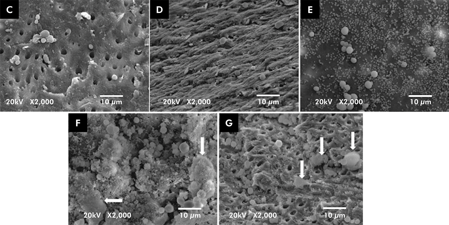
The SEM images confirmed a positive correlation between the quantitative (CFU) and qualitative (SEM) analyses. Figure 2A shows microorganisms organized with coaggregation characteristics, as can also be seen in Figure 2B and in the control group. Figure 3 shows a few microorganisms in all groups; however, there is some residual paste in F and G. Both Figures (2 and 3) clearly show the difference in size between the fungus (C. albicans) and the bacterium (E. faecalis).

Representative SEM photomicrograph of the teeth showing the presence of the two microorganisms after use of the five experimental medicaments: iodoform paste (c); chlorhexidine (d); camphorated paramonochlorophenol (e); calcium hydroxide (f); and calcium hydroxide-camphorated paramonochlorophenol (g). The white arrow indicates irregular shapes and represents the residual paste observed in f and g.
Discussion
The present study exclusively evaluated the antimicrobial efficacy of five different intracanal medicaments against C. albicans and E. faecalis infection in the root canal system of deciduous teeth. The results demonstrated that CMCP was the least efficient medicament against C. albicans, whereas CHX and IP were the only ones to show a significant reduction against E. faecalis. None of the intracanal medicaments tested in this study had any residual effect to protect the teeth against C. albicans and E. faecalis.
The elimination of microorganisms from the root canal system is important for a favorable prognosis in endodontic treatment. Considering the unique anatomy of primary teeth, some areas are inaccessible to irrigants and instruments, such as accessory canals, lateral canals, isthmi, dentinal tubules, and apical ramifications. Consequently, biomechanical preparation alone cannot effectively eliminate microorganisms from an infected root canal system, and the use of an interappointment medicament may be useful to maximize the reduction of any bacteria.5,8 Therefore, a two-visit protocol could be beneficial for necrotic pulp conditions with periapical radiolucency in primary teeth,9 but clinician’s expertise and individual circumstances should be taken into account as well.10
C. albicans and E. faecalis were chosen as test specimens because of their therapy-resistant pulp/apical infection and association with failed pulpectomies in primary teeth.20,21 The current protocol did not include chemomechanical preparation to remove the influence of cleaning methods and exclusively evaluate the efficiency of intracanal medicament against C. albicans and E. faecalis and the benefits in improving the cleaning of primary teeth. Therefore, the teeth were placed in a nutritional medium that mimicked necrosis, a condition in which microorganisms obtain nutrients through the foramen.
Ferguson et al.22 showed the antifungal effectiveness of several intracanal medicaments against C. albicans and they observed that CMCP was the most effective medicament, followed by CH-CMCP paste.22 However, the present study showed that CMCP was the least effective medicament against C. albicans and the second least effective against E. faecalis. This might have occurred because of the low concentration used and also the fact that the medicament did not have direct contact with the microorganisms.23 CMCP activity is based on the evaporation of camphor and on the precipitation of paramonochlorophenol. It is not possible to increase its concentration by injecting it directly into the canal because of its cytotoxicity to human periodontal ligament cells.24 Nowadays, CMCP is used mainly as an additive at low concentrations in mixed pastes where it has been shown to be more effective in eliminating E. faecalis than when used alone.25
Although CH has excellent properties such as antibacterial activity, high alkalinity (pH = 12.8), induction of mineralized tissue formation, biocompatibility, and inactivation of bacterial endotoxin,8,11 it did not perform well in this study. A few studies have reported that CH is a weak bactericide against E. faecalis.25-27 In attempt to enhance its bacterial action, the combination of other agents such as CMCP or CHX with CH has been proposed in order to increase the chances of successful pulp therapy,4,11,13 in line with Siqueira et al.28 These authors investigated the antifungal ability of various medicaments against several fungi such as C. albicans, whereas the CH-CMCP paste showed the most noticeable antifungal effect.28
However, in our study, CH-CMCP did not show a favorable result, as no antimicrobial effects of CH were significantly increased by adding CMCP, in addition to its low efficiency against E. faecalis. Some authors have reported that E. faecalis and C. albicans can survive in dentinal tubules after long periods of CH therapy.20,29,30 Safavi and Nakayama31 reported that higher concentration of viscous vehicles may decrease the effectiveness of CH as intracanal medicament,31 which might not have occurred in the present study, in which a commercially available prefabricated calcium hydroxide paste was used. CH therapy should maintain pH above 12.6 to be effective; however, it usually drops to 9.5 upon setting, resulting in decreased antimicrobial activity.20 In this study, we tested CH-CMCP and found no efficiency against E. faecalis, as shown by SEM (Figure 3G). Figure 2 shows that the small size of E. faecalis would have allowed it to penetrate into the dentinal tubules, preventing the medicaments from making physical contact. The reduced size of the bacterium, as reported by a few studies, is probably due to its ability to adapt to a changing environment.19,22 Unlike E. faecalis, C. albicans may have been affected by irrigants/medicaments through the fluid dynamics in the root canal system due to its larger size. This was observed in our study, as C. albicans showed a significant reduction in the control group, which might have been due to the irrigation steps used. On the other hand, the formation and structure of C. albicans biofilm in vivo helps to retain this fungus in the root canal system.32 However, in our in vitro study, we may not have provided the appropriate conditions for biofilm formation within the canals and, consequently, the fungus was unable to adhere and was therefore easy to remove.

Representative SEM photomicograph of Candida albicans, the largest microorganism (blue arrow), and Enterococcus faecalis, the smaller ones (red arrow), after 72 h, in initial quantification (a) and control group after 7 days without medication (b).
In our study, 2% CHX gel showed a statistically significant decrease in the growth of C. albicans and E. faecalis. CHX is an antimicrobial agent with optimal activity at pH 5.5 to 7.0.11Many studies have shown maximum inhibition of C. albicans and E. faecalis.21,23,27,33,34The gel formulation may provide longer contact of the microorganisms with chlorhexidine gluconate, inhibiting their growth.35 An in vitro study by Mathew et al. (2022) investigated the antimicrobial efficacy of six intracanal medicaments against C. albicans and E. faecalis. As a result, CHX exhibited higher antibacterial and antifungal activity against those species. Its effectiveness may be due to its high concentration (2%), turning it into a broad-spectrum antibacterial agent with positively charged molecules that interact with the negatively charged phosphate groups on the bacterial cell wall, allowing the CHX molecule to penetrate into the bacteria and produce toxic effects. In addition, 2% CHX gel is able to diffuse into dentinal tubules and reach the outer root surface.11,35 However, Gomes et al.33 reported that CHX had no substantivity. In our study, there was an increase in the residual quantification results (CFU count), which corroborates the findings of other studies.21,36 Other authors have suggested that the potential substantivity of CHX could decrease over time.21
The IP showed a favorable performance as well as a statistically significant decrease in the growth of both microorganisms. This medication is a mixture of antimicrobial agents: Rifocort®, a corticosteroid, iodoform, a topical germicidal agent,9,14and CMCP, a bacteriostatic agent.12. Silva et al.37 investigated the bactericidal and bacteriostatic effects of a few iodoform and CH pastes against S. mutans, S. aureus, E. faecalis, and C. albicans. They concluded that only IP (iodoform 0.08 g, CMCP 0.13 g, and Rifocort, 0.08 g) had a bactericidal and bacteriostatic effect against all the tested microorganisms, which is consistent with our findings.37 However, Srikumar et al.38 showed poor antifungal effect of IP (CH and iodoform) as intracanal medicament on C. albicans.38 This discrepancy with our study could be explained by the components associated with iodoform, considering that they have different mechanisms of action.
Bacteriostatic activity was verified in an in vitro experiment in which IP was used as the filling material.14 IP showed bactericidal action against most microorganisms, except for E. faecalis and Bacillus subtilis, which is at odds with the findings of our study. Our experiment evaluated IP as an intracanal medicament, and its superiority may be attributed to the antimicrobial associated with the synergistic action of its components. In addition, a systematic review by Silva Junior et al.39 found better performance of iodoform-based than noniodoform-based filling materials in primary teeth pulpectomies. Their explanation for that relies on the high antimicrobial property of iodoform,39 in agreement with the favorable performance of IP we found in the present study.
None of the tested drugs had a residual effect to protect the teeth against C. albicans and E. faecalis and, in some cases, such as CHX, they even allowed for a significant increase in the growth of C. albicans. This is possible because microorganisms resume their physiological development when in a nutritional medium, mimicking necrosis or periapical lesion for which intracanal medicaments are inefficient or absent.40 This may occur in vivo, where the fungi are more usually found in periapical lesions and obtain nutrients through the foramen and accessory canal. Therefore, filling the root canal with broad-spectrum antimicrobial material is pivotal for effective reduction of microorganisms in case of infection. In addition, the use of resorbable biocompatible materials and well-sealed crown restorations play an important role for a favorable prognosis.9
Any direct correlation of antimicrobial evaluation of primary teeth between in vitro and in vivo studies should be viewed with caution. In vitro tests may not show the full clinical potential of a material being tested, but the results presented here can be used to select materials that should be tested in vivo.
Conclusion
It is possible to conclude that all the intracanal medicaments, except for CMCP, reduced the levels of C. albicans. However, only IP and CHX showed a significant reduction against E. faecalis. The 2% CHX gel and IP demonstrated a favorable performance as intracanal medicaments, which had a good antimicrobial activity against C. albicans and E. faecalis. Nevertheless, none of the intracanal medicaments exhibited any residual effect to protect the teeth against the investigated microorganisms.
Acknowledgments
Emily Feitosa Rêgo acknowledges the support from Coordenação de Aperfeiçoamento de Pessoal de Nível Superior (CAPES) – Finance code 001, and Dr Laura Primo Guimarães acknowledges the support from the Fundação de Amparo à Pesquisa do Estado do Rio de Janeiro – FAPERJ (E-26/210.352/2019 - APQ1).
References
-
1 Nóbrega LM, Montagner F, Ribeiro AC, Mayer MA, Gomes BP. Bacterial diversity of symptomatic primary endodontic infection by clonal analysis. Braz Oral Res. 2016 Oct;30(1):e103. https://doi.org/10.1590/1807-3107BOR-2016.vol30.0103
» https://doi.org/10.1590/1807-3107BOR-2016.vol30.0103 -
2 Ito IY, Matoba Junior F, Paula-Silva FW, Silva LA, Leonardo MR, Nelson-Filho P. Microbial culture and checkerboard DNA-DNA hybridization assessment of bacteria in root canals of primary teeth pre- and post-endodontic therapy with a calcium hydroxide/chlorhexidine paste. Int J Paediatr Dent. 2011 Sep;21(5):353-60. https://doi.org/10.1111/j.1365-263X.2011.01131.x
» https://doi.org/10.1111/j.1365-263X.2011.01131.x -
3 Hedge AM, Pallavi KL. Prevalence pf selected microorganisms in the pulp space of human deciduous teeth with irreversible pulpitis. Endodontology. 2013;25(1):107-11. https://doi.org/10.4103/0970-7212.352299
» https://doi.org/10.4103/0970-7212.352299 -
4 Dutta B, S Dhull K, Das D, Samir PV, K Verma R, Singh N. Evaluation of antimicrobial efficacy of various intracanal medicaments in primary teeth: an in vivo study. Int J Clin Pediatr Dent. 2017;10(3):267-71. https://doi.org/10.5005/jp-journals-10005-1448
» https://doi.org/10.5005/jp-journals-10005-1448 -
5 Hidalgo LR, Silva LA, Leoni GB, Mazzi-Chaves JF, Carvalho EE, Consolaro A, et al. Mechanical preparation showed superior shaping ability than manual technique in primary molars: a micro-computed tomography study. Braz Dent J. 2017;28(4):453-60. https://doi.org/10.1590/0103-6440201601552
» https://doi.org/10.1590/0103-6440201601552 -
6 Silva BS, Nicoloso GF, Ruiz LF, Melo TA, Casagrande L. Does endodontic re-treatment in primary teeth increase the functional tooth retention? A clinical, retrospective, university-based study. Pesqui Bras Odontopediatria Clin Integr. 2019;19(1):e3902. https://doi.org/10.4034/PBOCI.2019.191.10
» https://doi.org/10.4034/PBOCI.2019.191.10 -
7 Brustolin JP, Mariath AA, Ardenghi TM, Casagrande L. Survival and Factors associated with failure of pulpectomies performed in primary teeth by dental students. Braz Dent J. 2017;28(1):121-8. https://doi.org/10.1590/0103-6440201601009
» https://doi.org/10.1590/0103-6440201601009 -
8 Gondim JO, Neto JJ, Gomes DA, Azevedo ER, Jeremias F, Giro EM. In vivo study of an intracanal dressing of calcium hydroxide/chlorhexidine in necrotic primary teeth. Braz J Oral Sci. 2014;13(1):70-5. https://doi.org/10.1590/1677-3225v13n1a14
» https://doi.org/10.1590/1677-3225v13n1a14 -
9 Cassol DV, Duarte ML, Pintor AV, Barcelos R, Primo LG. Iodoform vs calcium hydroxide/zinc oxide based pastes: 12-month findings of a randomized controlled trial. Braz Oral Res. 2019 Feb;33:e002. https://doi.org/10.1590/1807-3107bor-2019.vol33.0002
» https://doi.org/10.1590/1807-3107bor-2019.vol33.0002 - 10 Coll JA, Dhar V, Vargas K, Chen CY, Crystal YO, AlShamali S, et al. Use of non-vital pulp therapies in primary teeth. Pediatr Dent. 2020 Sep;42(5):337-49.
-
11 Bedran NR, Nadelman P, Magno MB, Neves AA, Ferreira DM, Pintor AVB, et al. Does calcium hydroxide reduce endotoxins in infected root canals? Systematic review and meta-analysis. J Endod. 2020 Nov;46(11):1545-58. https://doi.org/10.1016/j.joen.2020.08.002
» https://doi.org/10.1016/j.joen.2020.08.002 -
12 Jara M, Salcedo-Moncada D, Ayala G, Watanabe R, Alvítez-Temoche D, Mayta-Tovalino F. Antibacterial efficacy of calcium hydroxide with iodoform versus calcium hydroxide with camphorated paramonochlorophenol as intrachannel pastes on an Enterococcus faecalis biofilm: a comparative in vitro study. J Int Soc Prev Community Dent. 2020 Sep;10(5):555-60. https://doi.org/10.4103/jispcd.JISPCD_177_20
» https://doi.org/10.4103/jispcd.JISPCD_177_20 -
13 Gomes BP, Vianna ME, Zaia AA, Almeida JF, Souza-Filho FJ, Ferraz CC. Chlorhexidine in endodontics. Braz Dent J. 2013;24(2):89-102. https://doi.org/10.1590/0103-6440201302188
» https://doi.org/10.1590/0103-6440201302188 -
14 Praetzel JR, Ferreira FV, Weiss RN, Friedrich RS, Guedes-Pinto AC. Antimicrobial action of a filling paste used in pulp therapy in primary teeth under different storage conditions. J Clin Pediatr Dent. 2008;33(2):113-6. https://doi.org/10.17796/jcpd.33.2.d5jh650g20g686g5
» https://doi.org/10.17796/jcpd.33.2.d5jh650g20g686g5 - 15 Garcia-Godoy F. Evaluation of an iodoform paste in root canal therapy for infected primary teeth. ASDC J Dent Child. 1987;54(1):30-4.
-
16 Siqueira JF Jr, Antunes HS, Rôças IN, Rachid CT, Alves FR. Microbiome in the apical root canal system of teeth with post-treatment apical periodontitis. PLoS One. 2016 Sep;11(9):e0162887. https://doi.org/10.1371/journal.pone.0162887
» https://doi.org/10.1371/journal.pone.0162887 -
17 Saber SD, El-Hady SA. Development of an intracanal mature Enterococcus faecalis biofilm and its susceptibility to some antimicrobial intracanal medications; an in vitro study. Eur J Dent. 2012 Jan;6(1):43-50. https://doi.org/10.1055/s-0039-1698929 PMID:22229006
» https://doi.org/10.1055/s-0039-1698929 - 18 Valera MC, Salvia AC, Maekawa LE, Camargo SE, Carvalho CA, Camargo CH, et al. Antimicrobial analysis of chlorhexidine gel and intracanal medicaments against microorganisms inoculated in root canals. Minerva Stomatol. 2010;59(7-8):415-21.
-
19 Tannure PN, Barcelos R, Portela MB, Gleiser R, Primo LG. Histopathologic and SEM analysis of primary teeth with pulpectomy failure. Oral Surg Oral Med Oral Pathol Oral Radiol Endod. 2009 Jul;108(1):e29-33. https://doi.org/10.1016/j.tripleo.2009.03.014
» https://doi.org/10.1016/j.tripleo.2009.03.014 -
20 Singh S, Srivastava B, Gupta K, Gupta N, Singh R, Singh S. Comparative evaluation of antifungal efficacy of five root canal sealers against clinical isolates of Candida albicans: a microbiological study. Int J Clin Pediatr Dent. 2020;13(2):119-23. https://doi.org/10.5005/jp-journals-10005-1718
» https://doi.org/10.5005/jp-journals-10005-1718 -
21 Vaghela DJ, Kandaswamy D, Venkateshbabu N, Jamini N, Ganesh A. Disinfection of dentinal tubules with two different formulations of calcium hydroxide as compared to 2% chlorhexidine: as intracanal medicaments against Enterococcus faecalis and Candida albicans: an in vitro study. J Conserv Dent. 2011 Apr;14(2):182-6. https://doi.org/10.4103/0972-0707.82625
» https://doi.org/10.4103/0972-0707.82625 -
22 Ferguson JW, Hatton JF, Gillespie MJ. Effectiveness of intracanal irrigants and medications against the yeast Candida albicans. J Endod. 2002 Feb;28(2):68-71. https://doi.org/10.1097/00004770-200202000-00004
» https://doi.org/10.1097/00004770-200202000-00004 - 23 Mohammadi Z, Shalavi S. Is chlorhexidine an ideal vehicle for calcium hydroxide? A microbiologic review. Iran Endod J. 2012;7(3):115-22.
-
24 Chang YC, Tai KW, Chou LS, Chou MY. Effects of camphorated parachlorophenol on human periodontal ligament cells in vitro. J Endod. 1999 Dec;25(12):779-81. https://doi.org/10.1016/S0099-2399 (99)80295-7
» https://doi.org/10.1016/S0099-2399 (99)80295-7 -
25 Lima RA, Carvalho CB, Ribeiro TR, Fonteles CS. Antimicrobial efficacy of chlorhexidine and calcium hydroxide/camphorated paramonochlorophenol on infected primary molars: a split-mouth randomized clinical trial. Quintessence Int. 2013 Feb;44(2):113-22. https://doi.org/10.3290/j.qi.a28932
» https://doi.org/10.3290/j.qi.a28932 - 26 Mozayeni MA, Haeri A, Dianat O, Jafari AR. Antimicrobial effects of four intracanal medicaments on enterococcus faecalis: an in vitro study. Iran Endod J. 2014;9(3):195-8.
-
27 Onçag O, Cogulu D, Uzel A. Efficacy of various intracanal medicaments against Enterococcus faecalis in primary teeth: an in vivo study. J Clin Pediatr Dent. 2006;30(3):233-7. https://doi.org/10.17796/jcpd.30.3.r773j6l674lw0268
» https://doi.org/10.17796/jcpd.30.3.r773j6l674lw0268 -
28 Siqueira JF Jr, Rôças IN, Magalhães FA, Uzeda M. Antifungal effects of endodontic medicaments. Aust Endod J. 2001 Dec;27(3):112-4. https://doi.org/10.1111/j.1747-4477.2001.tb00471.x
» https://doi.org/10.1111/j.1747-4477.2001.tb00471.x -
29 Siqueira JF Jr, Sen BH. Fungi in endodontic infections. Oral Surg Oral Med Oral Pathol Oral Radiol Endod. 2004 May;97(5):632-41. https://doi.org/10.1016/j.tripleo.2003.12.022
» https://doi.org/10.1016/j.tripleo.2003.12.022 -
30 Verma R, Sharma DS, Pathak AK. Antibacterial efficacy of pastes against e faecalis in primary root dentin: a confocal microscope study. J Clin Pediatr Dent. 2015;39(3):247-54. https://doi.org/10.17796/1053-4628-39.3.247
» https://doi.org/10.17796/1053-4628-39.3.247 -
31 Safavi K, Nakayama TA. Influence of mixing vehicle on dissociation of calcium hydroxide in solution. J Endod. 2000 Nov;26(11):649-51. https://doi.org/10.1097/00004770-200011000-00004
» https://doi.org/10.1097/00004770-200011000-00004 -
32 Ponde NO, Lortal L, Ramage G, Naglik JR, Richardson JP. Candida albicans biofilms and polymicrobial interactions. Crit Rev Microbiol. 2021 Feb;47(1):91-111. https://doi.org/10.1080/1040841X.2020.1843400
» https://doi.org/10.1080/1040841X.2020.1843400 -
33 Gomes BP, Vianna ME, Sena NT, Zaia AA, Ferraz CC, Souza Filho FJ. In vitro evaluation of the antimicrobial activity of calcium hydroxide combined with chlorhexidine gel used as intracanal medicament. Oral Surg Oral Med Oral Pathol Oral Radiol Endod. 2006 Oct;102(4):544-50. https://doi.org/10.1016/j.tripleo.2006.04.010
» https://doi.org/10.1016/j.tripleo.2006.04.010 -
34 Gomes BP, Montagner F, Berber VB, Zaia AA, Ferraz CC, Almeida JF, et al. Antimicrobial action of intracanal medicaments on the external root surface. J Dent. 2009 Jan;37(1):76-81. https://doi.org/10.1016/j.jdent.2008.09.009
» https://doi.org/10.1016/j.jdent.2008.09.009 -
35 Mathew R, Sukumaran AS, Singh P, Varughese AV. Evaluation of the efficacy of different intracanal medicaments against Candida albicans and Enterococcus faecalis: an in-vitro study. Indian J Dent Res. 2022;33(4):440-4. https://doi.org/10.4103/ijdr.ijdr_10_22
» https://doi.org/10.4103/ijdr.ijdr_10_22 -
36 Paquette L, Legner M, Fillery ED, Friedman S. Antibacterial efficacy of chlorhexidine gluconate intracanal medication in vivo. J Endod. 2007 Jul;33(7):788-95. https://doi.org/10.1016/j.joen.2005.12.017
» https://doi.org/10.1016/j.joen.2005.12.017 - 37 Silva CM, Candelária LF, Bombana AC. Comparative study of the antimicrobial effect of five root canal filling materials for deciduous teeth. J Bras Odontoped Odontol Bebê. 2002;5(28):502-10.
-
38 Srikumar GP, Kumar RS, Bardia S, Geojan NE, Nishad G, Bhagat P. Antifungal effectiveness of various intracanal medicaments against Candida albicans: an in vitro study. J Contemp Dent Pract. 2020 Sep;21(9):1042-7. https://doi.org/10.5005/jp-journals-10024-2923
» https://doi.org/10.5005/jp-journals-10024-2923 -
39 Silva Junior MF, Wambier LM, Gevert MV, Chibinski AC. Effectiveness of iodoform-based filling materials in root canal treatment of deciduous teeth: a systematic review and meta-analysis. Biomater Investig Dent. 2022 May;9(1):52-74. https://doi.org/10.1080/26415275.2022.2060232
» https://doi.org/10.1080/26415275.2022.2060232 -
40 Rath S, Bal SC, Dubey D. Oral biofilm: development mechanism, multidrug resistance, and their effective management with novel techniques. Rambam Maimonides Med J. 2021 Jan;12(1):e0004. https://doi.org/10.5041/RMMJ.10428
» https://doi.org/10.5041/RMMJ.10428