
1. General Aspects of Old Age, Risk Factors, and Prevention
1.1. Demographic and Epidemiological Aspects
Since the second half of the 20th Century, survival has been democratized in numerous countries around the world. This means that more people are reaching more advanced ages. In Brazil, in 1980, of every 100 female live births, 30 could be expected to reach their 80th birthday; in 2013, this number increased to 55. The average lifespan of the Brazilian population, consequently, increased nearly 12 years during this period. One of the factors responsible for this phenomenon was the decrease in advanced age mortality, which was the result of the control of theretofore lethal diseases. However, many of these diseases that ceased to be lethal are still not curable. As a consequence, the aged population, which continues aging and becomes more heterogeneous, has grown. This heterogeneity is due to differentiated gender, age, and epidemiological profile, among other factors. For example, of the approximately 26 million people age 60 or over, 56.4% were women, and 13.8% were age 80 or over. It is worth highlighting that not only is the aging process expected to increase, but the aged populated itself is also expected to age further. Or be it, the population age 80 or over is the one that grows the most, given the reduced mortality in this age group11. Instituto Brasileiro de Geografia e Estatística. (IBGE). Pesquisa Nacional de Saúde (PNS). Rio de Janeiro; 2013. (Table 1). It is known that advanced age leads to the need to live with chronic, incapacitating diseases, which may compromise individual autonomy. In 2013, only 22.3% of elderly Brazilians declared that they had no chronic diseases. Approximately half, 48.6%, declared that they had 1 or 2 diseases, and 29.1% declared 3 or more. Women have a higher likelihood of contracting a disease than men, 81.2%, compared to 73.1%. This higher proportion of women in the elderly age group means a higher proportion of people with chronic morbidity11. Instituto Brasileiro de Geografia e Estatística. (IBGE). Pesquisa Nacional de Saúde (PNS). Rio de Janeiro; 2013. (Table 2). Within diseases reported, cardiovascular diseases (CVD) are predominant. For example, 62.0% of men and 67.4% of women declare that they have hypertension, and 23.2% and 36.9% of men and women, respectively, declare high cholesterol. These diseases also constitute the main cause of death in the elderly population, accounting for 34.2% and 35.2% of deaths in men and women, respectively. Within CVD, acute myocardial infarction (AMI) and stroke stand out22. Brasil. Ministério da Saúde. Sistema de informação sobre a mortalidade. Brasília; 2013. (Tables 3 and 4).
Percentage distribution of the elderly population by sex and age11. Instituto Brasileiro de Geografia e Estatística. (IBGE). Pesquisa Nacional de Saúde (PNS). Rio de Janeiro; 2013.
Proportion of elderly people with chronic diseases by number of pathological conditions11. Instituto Brasileiro de Geografia e Estatística. (IBGE). Pesquisa Nacional de Saúde (PNS). Rio de Janeiro; 2013.
Main causes of death in the elderly population by age22. Brasil. Ministério da Saúde. Sistema de informação sobre a mortalidade. Brasília; 2013.
Main causes of death due to circulatory system disease by sex22. Brasil. Ministério da Saúde. Sistema de informação sobre a mortalidade. Brasília; 2013.
This indicates a greater need for prevention, with lifestyle changes, alcohol and tobacco control, better diet, and physical exercise being able to contribute to a reduction in CVD. In summary, it is possible to affirm that humanity seems to be making the dream of long life come true, but it is necessary to avoid the Tithonus trap. Tithonus was a mythical Trojan hero who was granted eternal life; he forgot, however, to ask for eternal youth. Eventually he was transformed into a cricket. Ulysses, on the other hand, declined the gift of immortality, Ulysses, on the other hand, declined the gift of immortality, preferred remain owner of his destiny and his soul (Homero). Or be it, living a long life, with autonomy, should be humanity’s dream.
1.2. Interpretation of Frailty
Frailty is a biological syndrome characterized by decreased homeostatic reserve and resistance to various stressors. It results in cumulative decreases in multiple physiological systems and leads to increased vulnerability and unfavorable clinical outcomes, such as falls, impaired mobility and functional decline, hospitalization, institutionalization, and a higher risk of death.33. Fried LP, Tangen CM, Walston J, Newman AB, Hirsch C, Gottdiener J et al. Frailty in older adults evidence for a phenotype. J Gerontol A Biol Sci Med Sci. 2001;56(3):M146-56. This state of vulnerability causes an apparently minor injury (e.g., infection, introduction of a new medication, or even a small surgery) to lead to an evident, disproportional change in the patient’s state of health; these changes may by exemplified as alterations from independent to dependent status, from able to move to immobile, from balance and stable gait speed to risk of falling, or from lucid to delirious.44. Fried LP, Ferrucci L, Darer J, Williamson JD, Anderson G. Untangling the concepts of disability, frailty, and comorbidity: implications for improved targeting and care. J Gerontol A Biol Sci Med Sci. 2004;59(3):255-63.,55. Morley JE, Vellas B, van Kan A, Anker SD, Bauer JM, Bernabei R et al. Frailty consensus: a call to action. J Am Med Dir Assoc. 2013;14(6):392-7.
There is an overlap, but not a concurrence in the incidence of frailty, incapacity, and multimorbidity (coexistence of two or more chronic diseases). Although they are less frequent, there are frail individuals who have neither incapacity nor multimorbidity.44. Fried LP, Ferrucci L, Darer J, Williamson JD, Anderson G. Untangling the concepts of disability, frailty, and comorbidity: implications for improved targeting and care. J Gerontol A Biol Sci Med Sci. 2004;59(3):255-63. Sarcopenia (decreased muscle mass and function) is a component of the syndrome of frailty, which is more multifaceted and complex than sarcopenia alone.55. Morley JE, Vellas B, van Kan A, Anker SD, Bauer JM, Bernabei R et al. Frailty consensus: a call to action. J Am Med Dir Assoc. 2013;14(6):392-7.
Clinical presentation results not only from a single well defined disease, but rather from the accumulation of impairments in multiple organic systems, and it occurs when the accumulated effects of these impairments compromises the organism’s compensatory capacity. A systematic review demonstrated that the prevalence of frailty among community-dwelling elderly people was 10.7% (varying from 4.0% to 59.1%).66. Collard RM, Boter H, Schoevers RA, Oude Voshaar RC. Prevalence of frailty in community-dwelling older persons: a systematic review. J Am Geriatr Soc. 2012;60(8):1487-92.
In CVD patients, frailty confers a 2-fold risk of death, and this effect continues after adjusting for comorbidities and age. Numerous studies have also demonstrated an increase in the prevalence of frailty among patients with CVD, such as coronary artery disease (CAD), heart failure (HF), heart valve disease, etc. A higher risk of complications and mortality has also been identified in frail elderly patients who undergo cardiovascular interventions such as surgery and angioplasty.77. Afilalo J, Alexander KP, Mack MJ, Maurer MS, Green P, Allen LA et al. Frailty assessment in the cardiovascular care of older adults. J Am Coll Cardiol. 2014;63(8):747-62.
Frailty may potentially be prevented or treated, and many studies have demonstrated that exercise, protein/caloric supplementation, vitamin D supplementation, and reduction and optimization of polypharmacy may decrease levels of frailty, thus minimizing adverse outcomes and risks of interventions.55. Morley JE, Vellas B, van Kan A, Anker SD, Bauer JM, Bernabei R et al. Frailty consensus: a call to action. J Am Med Dir Assoc. 2013;14(6):392-7.,88. Puts MT, Toubasi S, Andrew MK, Ashe MC, Ploeg J, Atkinson E et al. Interventions to prevent or reduce the level of frailty in community-dwelling older adults: a scoping review of the literature and international policies. Age Ageing. 2017;46(3):383-92.
The identification of frail elderly patients is advocated so that multidimensional interventions may be implemented, mainly physical and nutritional rehabilitation, which reduces or postpones adverse outcomes and provides risk prognosis. It is necessary to emphasize that the identification of frailty does not need to be seen as a reason to exclude or suspend treatment, but rather as a means of programming individualized, patient-centered interventions.55. Morley JE, Vellas B, van Kan A, Anker SD, Bauer JM, Bernabei R et al. Frailty consensus: a call to action. J Am Med Dir Assoc. 2013;14(6):392-7.,77. Afilalo J, Alexander KP, Mack MJ, Maurer MS, Green P, Allen LA et al. Frailty assessment in the cardiovascular care of older adults. J Am Coll Cardiol. 2014;63(8):747-62.
Fried et al. (2001), in a longitudinal cardiovascular cohort study, identified the following manifestations for this syndrome: unintentional weight loss, muscular weakness, exhaustion (fatigue), decreased gait speed, and decreased degree of physical activity. Based on this, they proposed diagnostic criteria known as the “Fried et al. Frailty Phenotype”,33. Fried LP, Tangen CM, Walston J, Newman AB, Hirsch C, Gottdiener J et al. Frailty in older adults evidence for a phenotype. J Gerontol A Biol Sci Med Sci. 2001;56(3):M146-56. or “Cardiovascular Health Study Frailty Screening Scale”.33. Fried LP, Tangen CM, Walston J, Newman AB, Hirsch C, Gottdiener J et al. Frailty in older adults evidence for a phenotype. J Gerontol A Biol Sci Med Sci. 2001;56(3):M146-56.,55. Morley JE, Vellas B, van Kan A, Anker SD, Bauer JM, Bernabei R et al. Frailty consensus: a call to action. J Am Med Dir Assoc. 2013;14(6):392-7. These criteria have been criticized, insofar as those referring to exhaustion and decreased physical activity are not objective and are difficult to evaluate in daily practice with elderly patients. Other indexes and scales for diagnosis have been proposed, such as Rockwood Clinical Frailty Scale,99. Rockwood K, Mitnitski A. Frailty in relation to the accumulation of deficits. J Gerontol A Biol Sci Med Sci. 2007;62(7):722-7. the Gérontopôle Frailty Screening Tool,1010. Demougeot L, van Kan GA, Vellas B, de Souto Barreto P. Frailty detection with the Gérontopôle Frailty Screening Tool (GFST). J Frailty Aging. 2013;2(3):150-2. the FRAIL scale proposed by Van Kan and Morley,1111. Abellan van Kan G, Rolland YM, Morley JE, Vellas B. Frailty: toward a clinical definition. J Am Med Dir Assoc. 2008;9(2):71-2. the Groningen Frailty Indicator,1212. Steverink N, Slaets JPJ, Schuurmans H, van Lis M. Measuring frailty. development and testing of the Groningen Frailty Indicator (GFI). Gerontologist. 2001;41:236-7. the Tilburg Frailty Indicator,1313. Santiago LM, Luz LL, Mattos IE, Gobbens RJ. Adaptação transcultural do instrumento Tilburg Frailty Indicator (TFI) para a população brasileira. Cad Saúde Pública. 2012;28(9):1795-801. the PRISMA-7 questionnaire,1414. Raîche M, Hébert R, Dubois MF. PRISMA-7: a case-finding tool to identify older adults with moderate to severe disabilities. Arch Gerontol Geriatr. 2008;47(1):9-18. the VES-13 Scale,1515. Saliba D, Elliott M, Rubenstein LZ, Solomon DH, Young RT, Kamberg CJ et al. The Vulnerable Elders Survey: a tool for identifying vulnerable older people in the community. J Am Geriatr Soc. 2001;49(12):1691-9. and the Edmonton Frailty Scale.1616. Fabrício-Wehbe SC, Cruz IR, Haas VJ, Diniz MA, Dantas RA, Rodrigues RA. Adaptação cultural e validade da Edmonton Frail Scale - EFS em uma amostra de idosos brasileiros. Rev Lat Am Enferm. 2009;17(6):1330-6. The latter five instruments have been transculturally adapted and/or validated in Brazil. Studies have demonstrated that the 5-meter gait speed test is a useful tool for evaluating frailty in elderly patients referred for percutaneous aortic valve implantation.1717. Chen MA. Frailty and cardiovascular disease: potential role of gait speed in surgical risk stratification in older adults. J Geriatr Cardiol. 2015;12(1):44-56.,1818. Alfredsson J, Stebbins A, Brennan JM, Matsouaka R, Afilalo J, Peterson ED et al. Gait speed predicts 30-day mortality after transcatheter aortic valve replacement: results from the Society of Thoracic Surgeons / American College of Cardiology Transcatheter Valve Therapy Registry. Circulation. 2016;133(14):1351-9. The incorporation of this tool into the Society of Thoracic Surgeons (STS) score improved its ability to predict adverse events. For a given STS score, the risk of mortality or morbidity was 2–3 times greater in patients with slow gait speed.1717. Chen MA. Frailty and cardiovascular disease: potential role of gait speed in surgical risk stratification in older adults. J Geriatr Cardiol. 2015;12(1):44-56.,1818. Alfredsson J, Stebbins A, Brennan JM, Matsouaka R, Afilalo J, Peterson ED et al. Gait speed predicts 30-day mortality after transcatheter aortic valve replacement: results from the Society of Thoracic Surgeons / American College of Cardiology Transcatheter Valve Therapy Registry. Circulation. 2016;133(14):1351-9. Regardless of the instrument used to screen and identify, the syndrome of frailty should be investigated in all individuals over age 70 and in elderly patients with CVD, even if they are below this age group, and prevention and treatment measures should be put into practice.55. Morley JE, Vellas B, van Kan A, Anker SD, Bauer JM, Bernabei R et al. Frailty consensus: a call to action. J Am Med Dir Assoc. 2013;14(6):392-7.,77. Afilalo J, Alexander KP, Mack MJ, Maurer MS, Green P, Allen LA et al. Frailty assessment in the cardiovascular care of older adults. J Am Coll Cardiol. 2014;63(8):747-62.,88. Puts MT, Toubasi S, Andrew MK, Ashe MC, Ploeg J, Atkinson E et al. Interventions to prevent or reduce the level of frailty in community-dwelling older adults: a scoping review of the literature and international policies. Age Ageing. 2017;46(3):383-92.
1.3. Particularities in the Evaluation of Elderly Patients
Aging is a risk factor for most CVD, as well as numerous comorbidities, making the elderly the most heterogeneous and most complex adult age group.1919. Ramos LR, Toniolo J, Cendoroglo MS, Garcia JT, Najas MS, Perracini M et al. Two-year follow-up study of elderly residents in S. Paulo, Brazil: methodology and preliminary results. Rev Saúde Pública. 1998;32(5):397-407. Generally speaking, the healthcare system is poorly prepared to attend patients with multimorbidities, given that they require greater individualization, as well as assistance from a multiprofessional team that works integratedly.2020. Smith SM, Soubhi H, Fortin M, Hudon C, O'Dowd T. Managing patients with multimorbidity: systematic review of interventions in primary care and community settings. BMJ. 2012 Sep 3;345:e5205.,2121. Studenski S, Perera S, Patel K, Rosano C, Faulkner K, Inzitari M et al. Gait speed and survival in older adults. JAMA. 2011;305(1):50-8.
Interventions which are clearly beneficial in adults are, generally, also beneficial for elderly patients. However, the peculiarities which exist regarding evaluation of elderly patients are fundamental for their individual treatment. The evaluation of elderly patients should be performed using the Broad Geriatric Assessment (Avaliação Geriátrica Ampla, AGA).2222. Freitas EV, Costa EF, Galera CS. Avaliação geriátrica ampla. In: In: Freitas EV, Py L. Tratado de geriatria e gerontologia. 4. ed. Rio de Janeiro: Guanabara Koogan; 2016. p. 152-66. This is a multidimensional, generally interdisciplinary, diagnostic process for determining impairments, inabilities, and disadvantages in elderly patients and, thus, planning their medium- to long-term care and assistance. The AGA prioritizes functional status and quality of life, facilitating communication between interdisciplinary team members. It should be applied to frail elderly patients and patients with multimorbidities. The AGA is also an important predictor of unfavorable outcomes, i.e., it has prognostic value for surgery, oncology, and orthopedic patients.2222. Freitas EV, Costa EF, Galera CS. Avaliação geriátrica ampla. In: In: Freitas EV, Py L. Tratado de geriatria e gerontologia. 4. ed. Rio de Janeiro: Guanabara Koogan; 2016. p. 152-66. The AGA is fundamental in the context of evaluating elderly patients. It includes, at least, 4 principal dimensions, which are functional capacity, medical conditions, social functionality, and mental health.2222. Freitas EV, Costa EF, Galera CS. Avaliação geriátrica ampla. In: In: Freitas EV, Py L. Tratado de geriatria e gerontologia. 4. ed. Rio de Janeiro: Guanabara Koogan; 2016. p. 152-66.
Independent elderly patients with a long life expectancy should be treated comprehensively in a manner that combines prevention and intervention. On the other hand, pre-frail and frail patients require more attention regarding their individual needs and priorities, as well as risk-benefit assessment for individualized therapeutic decisions.1919. Ramos LR, Toniolo J, Cendoroglo MS, Garcia JT, Najas MS, Perracini M et al. Two-year follow-up study of elderly residents in S. Paulo, Brazil: methodology and preliminary results. Rev Saúde Pública. 1998;32(5):397-407.,2020. Smith SM, Soubhi H, Fortin M, Hudon C, O'Dowd T. Managing patients with multimorbidity: systematic review of interventions in primary care and community settings. BMJ. 2012 Sep 3;345:e5205. Goals to be reached should, equally, depend on functional status, without contraindicating any treatment whatsoever exclusively on account of age.
Considering the high prevalence of multimorbidities and the high evolution of therapeutic options, polypharmacy has become very frequent in elderly patients, posing further challenges to case management.1919. Ramos LR, Toniolo J, Cendoroglo MS, Garcia JT, Najas MS, Perracini M et al. Two-year follow-up study of elderly residents in S. Paulo, Brazil: methodology and preliminary results. Rev Saúde Pública. 1998;32(5):397-407.,2020. Smith SM, Soubhi H, Fortin M, Hudon C, O'Dowd T. Managing patients with multimorbidity: systematic review of interventions in primary care and community settings. BMJ. 2012 Sep 3;345:e5205. Understanding the advantages and disadvantages of every treatment is fundamental to adequate elderly treatment. This may only be scaled through the AGA. Familiarity with the AGA is, thus, essential to the evaluation and introduction of a determined treatment in an elderly patient.2121. Studenski S, Perera S, Patel K, Rosano C, Faulkner K, Inzitari M et al. Gait speed and survival in older adults. JAMA. 2011;305(1):50-8.,2222. Freitas EV, Costa EF, Galera CS. Avaliação geriátrica ampla. In: In: Freitas EV, Py L. Tratado de geriatria e gerontologia. 4. ed. Rio de Janeiro: Guanabara Koogan; 2016. p. 152-66.
1.4. Particularities in the Treatment of Elderly Patients
In treating elderly patients, priority is given to the patient who is ill, rather than to the illness, and to controlling the disease, rather than curing it. It is essential to know the disease, the patient who has the disease, and the treatment. CVD is frequent, and, even when there are few manifestations, it brings increased risks; elderly patients with diseases present comorbidities and high biopsychosocial vulnerability; treatments are more susceptible to undesired effects. Thus, evaluation of multiple clinical and psychosocial domains is fundamental. Owing to the fact that evidence is often lacking, conduct should be individualized. Decisions should be shared, and it is necessary to consider risk-benefit ratio and life expectancy. In elderly patients, treatment indication requires more caution. Although therapeutic goals are less precise, excluding them solely on the basis of age implies omission.2323. Wajngarten M. Geriatric cardiology: a subspecialty or a need? Arq Bras Cardiol. 2006;87(3):e 8-9.–2626. Bansilal S, Castellano JM, Garrido E, Wei HG, Freeman A, Spettell C et al. Assessing the impact of medication adherence on long-term cardiovascular outcomes. J Am Coll Cardiol. 2016;68(8):789-801.
Orientations regarding lifestyle changes are recommended, as in younger age groups. This may, however, cause undesired effects, especially if the changes are misunderstood or misapplied. Changing old habits requires attention.
Pharmacological treatments should: prioritize conditions and restrict number of medications, simplify posology, evaluate and stimulate satisfactory adherence even in secondary prevention following AMI,2626. Bansilal S, Castellano JM, Garrido E, Wei HG, Freeman A, Spettell C et al. Assessing the impact of medication adherence on long-term cardiovascular outcomes. J Am Coll Cardiol. 2016;68(8):789-801. provide orientation regarding problems related to self-medication, consider modifications in pharmacology related to age which, generally, recommend reducing doses, and evaluate possible drug interactions, given that “polypharmacy” is common. Beers Criteria, informally known as the “Beers List,” are a reference on safety in prescribing medications to the elderly. They were created in 1991, by the geriatrician Mark H. Beers, and they are periodically revised, the 2015 version being the most recent.2727. American Geriatrics Society 2015 Beers Criteria Update Expert Panel. American Geriatrics Society 2015 updated Beers criteria for potentially inappropriate medication use in older adults. J Am Geriatr Soc. 2015;63(11):2227-46.
In the United States, more than one third of emergency room visits due to adverse effects of substances occur in individuals over age 65. They imply hospitalization of more than 40% of cases, and this frequency is increasing over time. Of these visits, nearly 60% were related to the use of anticoagulants, antidiabetic agents, and opioid analgesics, and nearly 2% were related to restricted use medications, in accordance with the Beers Criteria.2828. American Geriatrics Society 2015 Updated Beers Criteria for potentially inappropriate medication use in older adults. [Internet]. [Cited in 2016 Sep 10]. Available from: https://www.guidelinescentral.com/summaries/American-geriatrics-society-2015.
https://www.guidelinescentral.com/summar...
Recently, the Food and Drug Administration Adverse Event Reporting System (FAERS) drew attention to evaluating the eventual need for regulatory action for the following: the anticoagulants apixaban, edoxaban, rivaroxaban, and dabigatran, due to reports of vasculitis; ivabradine, due to potential signs of ventricular arrhythmias; and midodrine, due to reports of interactions with monoamine oxidase inhibitors (MAOI) which could trigger a stroke.2929. Shehab N, Lovegrove MC, Geller AI, Rose KO, Weidle NJ, Budnitz DS. US Emergency Department Visits for Outpatient Adverse Drug Events, 2013-2014. JAMA. 2016;316(20):2115-25.
Interventional treatments should be carefully based on criteria, with the participation of heart teams, and performed by experienced and qualified teams, given that they present more frequent and severe complications.
A noteworthy example of this scenario is the need for hospitalization and admission to skilled nursing facilities in 4 of every 5 elderly patients who received an implantable cardioverter-defibrillator for secondary prevention of sudden cardiac death, even though they survived at least 2 years.3030. Betz JK, Katz DF, Peterson PN, Borne RT, Al-Khatib SM, Wang Y et al. Outcomes among older patients receiving implantable cardioverter-defibrillators for secondary prevention: from the NCDR ICD Registry. J Am Coll Cardiol. 2017;69(3):265-74.
1.5. Diabetes Mellitus in Elderly Patients
The National Health Survey conducted by the Brazilian Institute of Geography and Statistics (IBGE, 2013) showed a 19.9% prevalence of diabetes mellitus in individuals in the 65–74 age group.11. Instituto Brasileiro de Geografia e Estatística. (IBGE). Pesquisa Nacional de Saúde (PNS). Rio de Janeiro; 2013. In diabetic adults, there is an increase in mortality and a decrease in functional capacity with consequent increase in the risk of institutionalization.3131. Kirkman MS, Briscoe VJ, Clark N, Florez H, Haas LB, Halter JB et al. Diabetes in older adults. Diabetes Care. 2012;35(12):2650-64. The presence of multimorbidities and comorbidities associated with this group’s high heterogeneity means that the elderly are often excluded from randomized clinical trials, making disease management more difficult in this population.3131. Kirkman MS, Briscoe VJ, Clark N, Florez H, Haas LB, Halter JB et al. Diabetes in older adults. Diabetes Care. 2012;35(12):2650-64.,3232. American Diabetes Association. 11. Older adults. Diabetes Care. 2017;40(Suppl 1):S99-104.
Diagnostic criteria for diabetes mellitus in the elderly are similar to those in younger populations: (1) fasting blood glucose ≥ 126 mg/dL; or (2) random blood glucose ≥ 200 mg/dL, associated with disease symptoms; or (3) blood glucose 2 hours after a 75-g glucose load ≥ 200 mg/dL; or (4) glycated hemoglobin (HbA1C) ≥ 6.5% (provided that the laboratory is standardized). The American Diabetes Association (ADA) recommends that individuals who are overweight as a risk factor and all adults ≥ age 45 be screened for diabetes every 1 to 3 years, with fasting blood glucose, glycated hemoglobin dosage, or oral glucose tolerance test, for the benefit of early diagnosis, early treatment, and prevention of complications.3131. Kirkman MS, Briscoe VJ, Clark N, Florez H, Haas LB, Halter JB et al. Diabetes in older adults. Diabetes Care. 2012;35(12):2650-64.
Elderly individuals with diabetes are at a higher risk of developing geriatric syndromes, such as polypharmacy, cognitive impairment, urinary incontinence, falls, and chronic pain. When individuals with these syndromes develop diabetes, their clinical condition worsens. Thus, in addition to screening for complications, multidimensional evaluation of elderly diabetic individuals is also fundamental. It becomes imperative to perform AGA with mental, functional, nutritional, and social evaluations for these individuals in order to define goals to be met for each patient.3232. American Diabetes Association. 11. Older adults. Diabetes Care. 2017;40(Suppl 1):S99-104. The objective should be defined between two options: intensive blood glucose control, with less progression of chronic complications; or standard blood glucose control, in order to avoid only symptoms of hyperglycemia and acute complications.
The United Kingdom Prospective Diabetes Study (UKPDS), although it excluded elderly patients, showed the benefits of intensive blood glucose control in individuals as they age, with posterior follow-up.3333. Turner RC, Holman RR, Cull CA, Stratton IM, Matthews, DR, Frighi V et al.; UK Prospective Diabetes Study Group. Intensive blood-glucose control with sulphonylureas or insulin compared with conventional treatment and risk of complications in patients with type 2 diabetes (UKPDS33). Lancet. 1998;353(9131):837-53.,3434. Holman RR, Paul SK, Bethel MA, Matthews DR, Neil HA. 10-year follow-up of intensive glucose control in type 2 diabetes. N Engl J Med. 2008;359(15):1577-89. There are 3 main randomized clinical trials with the participation of elderly patients and intensive blood glucose control. The Action to Control Cardiovascular Risk in Diabetes (ACCORD) study was interrupted due to mortality in the youngest group; however, hypoglycemia and other adverse effects of treatment were more common in elderly patients;3535. Gerstein HC, Miller ME, Byimgton RP, Goff DC Jr, Bigger JT, Buse JB et al.; Action to control cardiovascular risk in diabetes study group: effects of intensive glucose lowering in type 2 diabetes. N Engl J Med. 2008;358(24):2545-59.,3636. Milleer ME, Bonds DE, Gerstein HC, Seaquist ER, Bergenstal RM, Calles-Escandon J et al.; ACCORD Investigators. The effects of baseline characteristics, glycaemia treatment approach, and glycated haemoglobin concentration on the risk of severe hypoglycaemia: post hoc epidemiological analysis of the ACCORD study: BMJ. 2010 Jan 8;340:b5444. in the Action in Diabetes and Vascular Disease: Preteraax and Diamicron MR Controlled Evaluation (ADVANCE) study, the risk of hypoglycemia and hospitalization increased significantly;3737. Patel A, Mac Mahon S, Chalmers J, Neal B, Billot L, Woodward M et al.; ADVANCE Collaborative Group. Intensive blood glucose control and vascular outcomes in patients with type 2 diabetes. N Engl J Med. 2008;358(24):2560-72. and in the Veterans Affairs Diabetes Trial (VADT) study, there were no benefits, with the exception of decreased progression of microalbuminuria.3838. Duckworth W, Abraira C, Moritz TE, Reda D, Emanuele N, Reaven PD et al; VADT Investigators. Glucose control and vascular complications in veterans with type 2 diabetes. N Engl J Med. 2009;360(2):129-39. Erratum in: N Engl J Med. 2009;361(10):1028. Two retrospective studies (U.K. General Practice Research Database, 20093939. Currie CJ, Peters JR, Tynan A, Evans M, Heine RJ, Bracco OL et al. Survival as a function of HbA(1c) in people with type 2 diabetes: a retrospective cohort study. Lancet. 2010;375(9713):481-9. and The Diabetes and Aging Study, 2011) show a U-shaped curve relating mortality and blood glucose levels.4040. Huang ES, Liu JY, Moffer HH, John PM, Karter AJ. Glycemic control, complications, and death in older diabetic patients: the Diabetes and Aging Study. Diabetes Care. 2011;34(6):1329-36. Individualization of treatment is, thus, imperative in elderly patients in accordance with their clinical, functional, and life expectancy profile, as demonstrated in Table 5, with treatment goals for arterial hypertension and dyslipidemia in elderly patients with diabetes.
1.6. Tobacco Use
The influence of tobacco use in elderly individuals occurs due to anatomical and physiological alterations in a cumulative process which leads to endothelial dysfunction, increased platelet adhesion, decreased high-density lipoprotein cholesterol (HDL-c), and increased low-density lipoprotein cholesterol (LDL-c), among other alterations.4141. Sociedade Brasileira de Diabetes. Atualização Brasileira sobre Diabetes. Síndrome metabólica. Rio de Janeiro: Diagraphic; 2005. Tobacco use is common in the elderly population, and it is an important cause of morbidity and mortality, including CVD, peripheral vascular disease, cerebrovascular disease, cancer, and obstructive pulmonary disease. On the other hand, the tobacco cessation has benefits, even in elderly patients, with respect to the prevention of these diseases or, at least, to slowing the decline of pulmonary function.4242. Little D’A. A review of smoking in the elderly. Geriatrics & Aging. 2002;5(9):11-4. The Systolic Hypertension in the Elderly Program Study4343. Siegel D, Kuller L, Lazarus NB, Black D, Feigal D, Hughes G et al. Predictors of cardiovascular events and mortality in the Systolic Hypertension in the Elderly Program Pilot Project. Am J Epidemiol. 1987;126(3):385-99. observed patients with an average age of 72 and showed a significant increase in AMI, sudden death, and stroke in smokers, compared with non-smokers. Exposure to long periods of passive tobacco use increases the risk of developing CAD. Kawachi et al. (1997)4444. Kawachi I, Colditz GA, Speltzer FE, Manson JE, Stampfer MJ, Willett WC et al. A prospective study of passive smoking and coronary artery disease. Circulation. 1997;95(10):2374-9. followed 32,000 non-smoking women, between the ages of 36 and 71, for 10 years, and found that the relative risk of developing coronary heart disease increased in women exposed to smoking. Occasional exposure to cigarettes increased their relative risk by 1.58, and regular exposure increased the relative risk by 1.91.4545. Jajich CL, Ostfeld AM, Freeman DH Jr. Smoking and CHD: mortality in the elderly. JAMA. 1984;252(20):2831-4. Tobacco use constitutes a risk factor for dementia, and cessation may reduce the burden of dementia. Passive exposure to smoking may also increase the risk of dementia.4646. World Health Organization (WHO). Tobacco knowledge summaries: tobacco and dementia. Geneva; 2014. Studies show that elderly smokers have a lower intention of quitting in comparison with younger smokers; they have, on the other hand, a higher likelihood of success when they do try to stop smoking.4747. Dwyer J. Exposure to environmental tobacco smoke and coronary risk. Circulation. 1997;96(5):1367-9.,4848. Hall SM, Humfleet GL, Gorecki JA, Muñoz RF, Reus VI, Prochaska JJ. Older versus younger treatment-seeking smokers: differences in smoking behavior, drug and alcohol use, and psychosocial and physical functioning. Nicotine Tob Res. 2008;10(3):463-70. Success in stopping is frequently achieved after an acute coronary event, aggravation of chronic obstructive pulmonary disease (COPD), or symptomatic and limiting peripheral arterial disease. Medical advice to cease smoking should be firm, with emphasis placed on the short- and medium-term benefits. Aggressive practices related to tobacco cessation should be adopted.4949. 2008 PHS Guideline Update Panel, Liaisons, and Staff. Treating tobacco use and dependence: 2008 Update U.S. Public Health Service Clinical Practice Guideline executive summary. Respir Care. 2008;53(9):1217-22.,5050. Bratzler DW, Oehlert WH, Austelle A. Smoking in the elderly – it´s never too late to quit. J Okla State Med Assoc. 2002;95(3):185-91. Evidence shows the efficiency of using the 4 As method in elderly patients, namely: ask, advise, assist, and arrange follow up.5151. Buckland A, Connolly MJ. Age-related differences in smoking cessation advice and support given to patients hospitalized with smoking-related illness. Age Ageing. 2005;34(6):639-42.,5252. Andrews JO, Heath J, Graham-Garcia J. Management of tobacco dependence in older adults: using evidence-based strategies J Gerontol Nurs. 2004;30(12):13-24. Different approaches, such as interventions through individual counseling performed by healthcare professionals, age-appropriate self-help material, use of nicotine (transdermal patches or chewing gum), or use of specific medications, e.g. bupropion), have also been shown to be efficient in treating tobacco use.5353. Tait RJ, Hulse GK, Waterreus A, Flicker L, Lautenschlager NT, Jamrozik K et al. Effectiveness of a smoking cessation intervention in older adults. Addiction. 2007;102(1):148-55.–5555. Morgan GD, Noll EL, Orleans CT, Rimer BK, Amfoh K, Bonney G. Reaching midlife and older smokers: tailored interventions for routine medical care. Prev Med. 1996;25(3):346-54.
1.7. Obesity
The prevalence of overweight status and obesity has increased over the past decades in all age groups, including the elderly.5656. World Health Organization (WHO). The challenge of obesity in the WHO European region and the strategies for response. Denmark: World Health Organization; 2007. p. 1-59.,5757. Eiben G, Dey DK, Rothenberg E, Steen B, Björkelund C, Bengtsson C et al. Obesity in 70-year-old Swedes: secular changes over 30 years. Int J Obes (Lond). 2005,29(7):810-7.
Both obesity and overweight status have been associated with the risk of all cause and CVD mortality, in the general population.5858. McTigue K, Larson JC, Valoski A, Burke G, Kotchen J, Lewis CE et al. Mortality and cardiac and vascular outcomes in extremely obese women. JAMA. 2006;296(1):79-86.–6060. Pischon T, Boeing H, Hoffmann K, Bergmann M, Schulze MB, Overvad K et al. General and abdominal adiposity and risk of death in Europe. N Engl J Med. 2008;359(20):2105-20. Erratum in: N Engl J Med. 2010;362(25):2433.
The majority of these studies mainly involved young adult patients, making this relationship less evident in the elderly.6161. Janssen I, Katzmarzyk PT, Ross R. Body mass index is inversely related to mortality in older people after adjustment for waist circumference. J Am Geriatr Soc. 2005;53(12):2112-8.–6464. Zamboni M, Mazzali G, Zoico E, Harris TB, Meigs JB, Di Francesco V et al. Health consequences of obesity in the elderly: a review of four unresolved questions. Int J Obes (Lond). 2005;29(9):1011-29.
Some meta-analysis studies have reported that overweight and obese elderly individuals, when compared with elderly individuals within the normal weight range, had lower mortality rates and lower or no risk of CVD. This effect has been called the “obesity paradox”.6565. Flegal KM, Kit BK, Orpana H, Graubard BI. Association of all-cause mortality with overweight and obesity using standard body mass index categories: a systematic review and meta-analysis. JAMA. 2013;309(1):71-82.–6767. Janssen I, Mark AE. Elevated body mass index and mortality risk in the elderly. Obes Rev. 2007;8(1):41-59.
In addition to possible confounding factors in these studies, other reasons may be involved. The index used to measure and classify body mass was the body mass index (BMI). Degrees of obesity adopted by the World Health Organization (WHO), with respect to BMI, are: overweight (25.0 to 29.9 kg/m2) and obese (over 30.0 kg/m2).6868. World Health Organization (WHO). Consultation on obesity. Geneva; 1999. (WHO technical report series; 894). Variables such as age, sex, and race may affect BMI. With aging, changes in body composition occur, such as increased visceral fat and decreased muscle mass. Loss of height may also occur, owing to compression of vertebral bodies or kyphosis. In this manner, BMI becomes less precise in measuring fat mass. When used alone, it is not able to be an accurate predictor of CVD risk in elderly patients. For instance, some elderly individuals may be considered overweight by body fat patterns without having a BMI over 25 kg/m2.
Using BMI alone, we may be underestimating the degree of adiposity in individuals who lost muscle mass. Central obesity and nutrition are factors which seem more important in relation to mortality and CVD risk in this population. Some authors suggest that waist circumference (WC) could be a particularly important measure for elderly patients, which would be better than BMI at evaluating risk, given its association with visceral adiposity.6969. Martínez-González MA, García-Arellano A, Toledo E, Bes-Rastrollo M, Bulló M, Corella D et al. Obesity indexes and total mortality among elderly subjects at high cardiovascular risk: The PREDIMED Study. PLoS One. 2014;9(7):e103246.,7070. de Hollander EL, Bemelmans WJ, Boshuizen HC, Friedrich N, Wallaschofski H, Guallar-Castillón P et al; WC elderly collaborators. The association between waist circumference and risk of mortality considering body mass index in 65- to 74-year-olds: a meta-analysis of 29 cohorts involving more than 58 000 elderly persons. Int J Epidemiol. 2012;41(3):805-17.
Another study indicates that the presence or absence of metabolic syndrome is more important than BMI in obese elderly patients, thus dividing this population into “healthy obese” (without metabolic syndrome) and obese with metabolic syndrome. The latter group has been strongly associated with increased risk of CVD regardless of BMI.7171. Dhana K, Koolhaas CM, van Rossum EF, Ikram MA, Hofman A, Kavousi M et al. Metabolically healthy obesity and the risk of cardiovascular disease in the elderly population. PLoS One. 2016;11(4):e0154273.
More studies are necessary to clarify the interrelation between aging, obesity, and cardiovascular risk and what the best measures parameter(s) would be. Weight management in the elderly and efforts to promote healthy aging should be based on an individual approach, taking into consideration the maintenance of muscle mass and strength, comorbidities, functional and social status, physical activity, and quality of life. Intentional weight loss in obese elderly patients improves their cardiovascular risk profile, reduces chronic inflammation, and is correlated with improved quality of life. Unintentional weight loss requires careful clinical assessment of the underlying cause. Furthermore, the identification of elderly patients with sarcopenic obesity is relevant to prognosis. Sarcopenia and sarcopenic obesity have been associated with a higher risk of CVD, especially in elderly men with this type of obesity.
1.8. Sedentarism
Regular physical activity is essential to healthy aging. Considering that aging in inevitable, the rhythm and magnitude of decline in physiological function may be influenced by an intervention comprising exercise/physical activity (Table 6).7272. Thompson PD, Arena R, Riebe D, Pescatello LS; American College of Sports Medicine. ACSM new preparticipation health screening recommendations from ACSMs Guidelines for Exercise Testing and Prescription. Ninth edition. Curr Sports Med Rep. 2013;12(4):215-7.,7373. Whaley MH, Brubaker PH, Otto RM, Armstrong LE. ACSM’S guidelines for exercise testing & prescription. Philadelphia: Lippincott Williams & Wilkins; 2006.
Aging is associated with skeletal muscle mass loss; reduced muscle strength, flexibility, cardiac output, and pulmonary function; changes in hormonal and immune system regulation; reduced bone density, and higher prevalence and incidence of sedentarism.7474. Canada Public Health Agency of Canada. Physical activity tips for older adults (65 years and older). [Internet]. [Cited in 2017 Dec 10]. Available from: https://www.canada/ca/content/dam/phac-aspx/migration/
https://www.canada/ca/content/dam/phac-a...
In sedentary elderly patients, walking may be a practical solution, evaluating heart rate (HR) before and after exercise. It is necessary to recognize that elderly people do not represent a uniform group of patients and chronological age in itself does not identify this special group.7575. US Department of Health and Physical Activity Guidelines Advisory Committee. Report 2008. [Internet]. [Cited in 2017 Dec 10]. Available from: https://health.gov/paguidelines/report/pdf/committeeReport.pdf.
https://health.gov/paguidelines/report/p...
Sedentarism is an important risk factor for CAD in elderly individuals. Some studies demonstrate that the relative risk of CAD attributable to sedentarism is comparable to that of hypertension, hyperlipidemia, and tobacco use. Sedentarism, as an important risk factor, is, in most cases, directly or indirectly associated with the causes or aggravation of various diseases, such as obesity, diabetes, arterial hypertension, anxiety, depression, dyslipidemia, atherosclerosis, respiratory disease, osteoporosis, and cancer.7676. Gravina, CF, Crespan SM, Araújo N. Envelhecimento e risco cardiovascular. In: Sociedade de Cardiologia do Estado de São Paulo (SOCESP). Tratando de Cardiologia da Socesp. São Paulo: Manole; 2009.,7777. Janssen I, Jolliffe CJ. Influence of physical activity on mortality in elderly with coronary artery disease. Med Sci Sports Exerc. 2006;38(3):418-7. Systematic physical exercise helps control systemic arterial hypertension (SAH), by reducing peripheral arterial resistance, increasing HDL-c, reducing obesity and triglycerides, improving blood glucose control, preventing coronary disease, and decreasing mortality.7777. Janssen I, Jolliffe CJ. Influence of physical activity on mortality in elderly with coronary artery disease. Med Sci Sports Exerc. 2006;38(3):418-7.,7878. World Health Organization (WHO). Physical activity and older adults. [Cited in 2017 Feb 16]. Available from: http://www.who.int/dietpshysicalactivity/factsheet-olderadults/en.
http://www.who.int/dietpshysicalactivity...
Furthermore, it improves sleep quality, cognitive function, and short-term memory; decreases degree of depression; reduces or slows the onset of dementia; reduces the risk of colon, breast, prostate, and rectal cancer; increases bone density; and decreases the incidence of femur and vertebrae fractures.7777. Janssen I, Jolliffe CJ. Influence of physical activity on mortality in elderly with coronary artery disease. Med Sci Sports Exerc. 2006;38(3):418-7.
In elderly patients, pre-exercise clinical evaluation is very important. The goal of exercise and cardiovascular rehabilitation in elderly patients is to improve their functional capacity as much as possible. These objectives are reached through programs that aim to increase aerobic capacity, muscle strength, and flexibility.7272. Thompson PD, Arena R, Riebe D, Pescatello LS; American College of Sports Medicine. ACSM new preparticipation health screening recommendations from ACSMs Guidelines for Exercise Testing and Prescription. Ninth edition. Curr Sports Med Rep. 2013;12(4):215-7.,7979. Meneghello RS, Araújo CG, Stein R, Mastrocolla LE, Albuquerque PF, Serra SM et al; Sociedade Brasileira de Cardiologia. [III Guidelines of Sociedade Brasileira de Cardiologia on the exercise test]. Arq Bras Cardiol. 2010;95(5 Suppl 1):1-26.–8282. Costa RC, Carrera MA. Ergometria, ergoespirometria, cintilografia e ecocardiografia de esforço. São Paulo: Atheneu; 2007. p. 59-64.
The amount of physical activity should be individualized, considering each patient’s comorbidities and peculiarities.7373. Whaley MH, Brubaker PH, Otto RM, Armstrong LE. ACSM’S guidelines for exercise testing & prescription. Philadelphia: Lippincott Williams & Wilkins; 2006.,7474. Canada Public Health Agency of Canada. Physical activity tips for older adults (65 years and older). [Internet]. [Cited in 2017 Dec 10]. Available from: https://www.canada/ca/content/dam/phac-aspx/migration/
https://www.canada/ca/content/dam/phac-a...
,7979. Meneghello RS, Araújo CG, Stein R, Mastrocolla LE, Albuquerque PF, Serra SM et al; Sociedade Brasileira de Cardiologia. [III Guidelines of Sociedade Brasileira de Cardiologia on the exercise test]. Arq Bras Cardiol. 2010;95(5 Suppl 1):1-26.
Elderly individuals should spend more time warming up before and cooling down after activity. The warm-up phase includes flexibility and movement exercises, which facilitate musculoskeletal biomechanics. The post-exercise cool-down phase allows for the gradual dissipation of body heat and consequent peripheral vasodilatation. Musculoskeletal injuries may be decreased by avoiding high impact activities, such as running and jumping. Extreme care is necessary for activities using free weights, given the risk of accidents, especially in less skilled or more frail elderly patients.7272. Thompson PD, Arena R, Riebe D, Pescatello LS; American College of Sports Medicine. ACSM new preparticipation health screening recommendations from ACSMs Guidelines for Exercise Testing and Prescription. Ninth edition. Curr Sports Med Rep. 2013;12(4):215-7.,8080. Freitas EV, Py L. Tratado de geriatria e gerontologia. 4. ed. Rio de Janeiro: Guanabara Koogan; 2016. p. 667-74. Walking briskly is an excellent way to obtain physical conditioning, with gradual increases in pace and distance covered.8181. Price GM, Uauy R, Breeze E, Bulpitt CJ, Fletcher AE. Weight, shape and mortality risk in older persons: elevated waist-hip ratio, not high body mass index, is associated with a greater risk of death. Am J Clin Nutr. 2006;84(2):449-60. Elderly patients should be instructed to reduce exercise intensity on humid or hot days, given that skin blood flow decreases with age, consequently lowering the efficiency of sweating and thermal regulation.7777. Janssen I, Jolliffe CJ. Influence of physical activity on mortality in elderly with coronary artery disease. Med Sci Sports Exerc. 2006;38(3):418-7. Practicing resistance exercise twice weekly is also recommended.
Pre-participation assessment should begin with patient history and clinical exam, focusing on the particularities of this population, which often has silent atherosclerotic disease. Complementary investigation should be oriented by clinical data, avoiding high costs, which are sometimes prohibitive and may discourage physical exercise. Resting electrocardiogram (EKG) for the elderly has limited application as a pre-selection exam for physical activity.
If possible, an exercise test (ET) should be performed in all elderly patients before initiating physical activity. The prevalence of coronary disease increases with age; the rationale behind the ET in this population may, thus, be even greater than in the general adult population.7272. Thompson PD, Arena R, Riebe D, Pescatello LS; American College of Sports Medicine. ACSM new preparticipation health screening recommendations from ACSMs Guidelines for Exercise Testing and Prescription. Ninth edition. Curr Sports Med Rep. 2013;12(4):215-7.,7979. Meneghello RS, Araújo CG, Stein R, Mastrocolla LE, Albuquerque PF, Serra SM et al; Sociedade Brasileira de Cardiologia. [III Guidelines of Sociedade Brasileira de Cardiologia on the exercise test]. Arq Bras Cardiol. 2010;95(5 Suppl 1):1-26.,8080. Freitas EV, Py L. Tratado de geriatria e gerontologia. 4. ed. Rio de Janeiro: Guanabara Koogan; 2016. p. 667-74. The ET is a procedure during which the patient undergoes programmed and individualized exercise, with the aim of evaluating clinical, metabolic, hemodynamic, autonomic, electrocardiographic, and, eventually, ventilatory responses to exercise. In elderly patients, modified protocols are used to perform the ET.7979. Meneghello RS, Araújo CG, Stein R, Mastrocolla LE, Albuquerque PF, Serra SM et al; Sociedade Brasileira de Cardiologia. [III Guidelines of Sociedade Brasileira de Cardiologia on the exercise test]. Arq Bras Cardiol. 2010;95(5 Suppl 1):1-26. If there are contraindications to performing the ET, stress EKG or scintigraphy should be performed. A Holter monitor is used to stratify risk in elderly patients with arrhythmias detected during EKG or ET, as well as those with a history of syncope.7272. Thompson PD, Arena R, Riebe D, Pescatello LS; American College of Sports Medicine. ACSM new preparticipation health screening recommendations from ACSMs Guidelines for Exercise Testing and Prescription. Ninth edition. Curr Sports Med Rep. 2013;12(4):215-7.,8282. Costa RC, Carrera MA. Ergometria, ergoespirometria, cintilografia e ecocardiografia de esforço. São Paulo: Atheneu; 2007. p. 59-64.
Adherence to physical activity in this group has been increasingly positive. It is always necessary to consider that an active or latent pathological process may by present in an elderly individual and that the ET may contribute to defining it.8383. Moreira MC, Montenegro ST, Paola AA. Livro texto da Sociedade Brasileira de Cardiologia. 2. ed. Barueri (SP): Manole; 2015. p.128.65.,8484. American College of Sports Medicine. ACSM’s Guidelines for exercise testing & prescription. 7. ed. Baltimore: Lippincott Williams & Wilkins; 2006.
1.9. Dyslipidemia in Elderly Patients
Dyslipidemia is a frequent diagnosis in elderly patients, mainly in women, owing to the fact that LDL-c levels tend to rise as they advance in age, especially after menopause; in men, however, LDL-c tends to decrease after age 55. Unlike in young adults, cases of de novo dyslipidemia are rare, and cases of dyslipidemia secondary to hypothyroidism (especially in women), diabetes mellitus, glucose intolerance, nephrotic syndrome, obesity, alcoholism, or use of medications such as thiazide diuretics and non-selective beta-adrenergic receptor blockers, are more common.8585. Katzel LI, Blumenthal J, Sorkin JD, Goldberg AP. Dyslipoproteinemia. In: Halter JB, Ouslander JG, Tinetti ME et al. (editors). Hazzard’s geriatric medicine and gerontology. 6. ed. New York: McGraw-Hill; 2009. p. 1325-42.
In relation to treatment, as elderly patients are often already at high risks (owing to the factor of age), the approach to dyslipidemia, regarding therapeutic decisions, should give greater consideration to the patient’s good general and mental state, his or her socio-economic conditions, family support, comorbidities present, and the use of other drugs that may influence adherence to and maintenance of therapy. Non-pharmacological orientations should follow the same principals of indication for young adults, more carefully observing caloric, protein, and vitamin intake needs and physical conditions for practicing exercise (recommendation I, evidence B). It is necessary to reiterate the importance of ceasing habits of smoking and excessive consumption of alcoholic beverages. After 90 days, if there is no response, drug treatment may be initiated, with the following precautions: (1) always start with low doses and, if necessary, increase, progressively; (2) analyze the cost-benefit ratio; and (3) verify the existence of socioeconomic conditions for maintaining long-term treatment and performing periodical clinical and laboratory exams, due to the higher likelihood of collateral effects and drug interactions.8585. Katzel LI, Blumenthal J, Sorkin JD, Goldberg AP. Dyslipoproteinemia. In: Halter JB, Ouslander JG, Tinetti ME et al. (editors). Hazzard’s geriatric medicine and gerontology. 6. ed. New York: McGraw-Hill; 2009. p. 1325-42.
For hypercholesterolemia, statins are the first choice.8686. Long SB, Blaha MJ, Blumenthal RS, Michos ED. Clinical utility of rosuvastatin and other statins for cardiovascular risk reduction among the elderly. Clin Interv Aging. 2011;6:27-35. Tolerance is good; there is not a high incidence of undesired effects, even though muscle pain, cramps, and weakness, which are sometimes confounded with osteomuscular disease, may occur, even in low doses. Evidence from subgroup analyses in primary and secondary prevention studies and the Pravastatin in Elderly Individuals at Risk of Vascular Disease (PROSPER) study,8787. Shepherd J, Blauw GJ, Murphy MB, Bollen EL, Buckley BM, Cobbe SM et al. PROSPER study group. PROspective Study of Pravastatin in the Elderly at Risk. Pravastatin in elderly individuals at risk of vascular disease (Prosper): a randomised controlled trial. Lancet. 2002;360(9346):1623-30. specially designed for elderly patients with or without previous manifestations of atherosclerosis, demonstrated the following benefits to treatment for this age group: reduction of coronary events (grade of recommendation IIa, level of evidence B), stroke (grade of recommendation IIa, level of evidence B), and preservation of cognitive functions (grade of recommendation IIb, level of evidence B). When maximum statin dosages are not sufficient to meet recommended LDL-c goals, ezetimibe may be associated with the statins (grade of recommendation IIb, level of evidence B).8888. Reiner Z, Catapano AL, De Backer G, Graham I, Taskinen MR, Wiklund O et al.; European Association for Cardiovascular Prevention & Rehabilitation, ESC Committee for Practice Guidelines (CPG) 2008-2010 and 2010-2012 Committees. ESC/EAS Guidelines for the management of dyslipidaemias: The Task Force for the management of dyslipidaemias of the European Society of Cardiology (ESC) and the European Atherosclerosis Society (EAS). Eur Heart J. 2011;32(14):1769-818.
In cases of hypertriglyceridemia, fibrates are used (provided there are no gallstones or renal insufficiency). Fibrates and statins may be associated in cases of mixed dyslipidemia (elevated LDL-c and triglycerides), mainly with reduced HDL-c (grade of recommendation IIb, level of evidence D).8888. Reiner Z, Catapano AL, De Backer G, Graham I, Taskinen MR, Wiklund O et al.; European Association for Cardiovascular Prevention & Rehabilitation, ESC Committee for Practice Guidelines (CPG) 2008-2010 and 2010-2012 Committees. ESC/EAS Guidelines for the management of dyslipidaemias: The Task Force for the management of dyslipidaemias of the European Society of Cardiology (ESC) and the European Atherosclerosis Society (EAS). Eur Heart J. 2011;32(14):1769-818.
In secondary dyslipidemias, the fundamental concern is treating the triggering disease and substituting or removing inductor drugs. We should remember that elderly individuals, generally, use other drugs metabolized by cytochrome P450 (CYP450), which have the possibility of interacting with lipid-lowering agents, thus altering their blood concentration (grade of recommendation IIb, level of evidence D).8888. Reiner Z, Catapano AL, De Backer G, Graham I, Taskinen MR, Wiklund O et al.; European Association for Cardiovascular Prevention & Rehabilitation, ESC Committee for Practice Guidelines (CPG) 2008-2010 and 2010-2012 Committees. ESC/EAS Guidelines for the management of dyslipidaemias: The Task Force for the management of dyslipidaemias of the European Society of Cardiology (ESC) and the European Atherosclerosis Society (EAS). Eur Heart J. 2011;32(14):1769-818.
1.10. Depression and Cardiovascular Disease
Depression and anxiety are highly prevalent in individuals with CAD and other CVD. They have been also been considered independent risk factors for CAD and CVD, in addition to altering their natural history.8989. Nasser FJ, Almeida MM, Silva LS, Almeida RG, Barbirato GB, Mendlowicz MV et al. Psychiatric disorders and cardiovascular system: heart-brain interaction. Int J Cardiovasc Sci. 2016;29(1):65-75.,9090. Pająk A, Jankowski P, Kotseva K, Heidrich J, de Smedt D, De Bacquer D et al.; EUROASPIRE Study Group. Depression, anxiety, and risk factor control in patients after hospitalization for coronary heart disease: the EUROASPIRE III Study. Eur J Prev Cardiol. 2013;20(2):331-40.
Depression is disproportionally more frequent among CAD patients, with prevalence between 20% and 40%. It has also been reported that depression is prospectively associated with an increased risk of developing CAD,9191. Whooley MA. To screen or not to screen? Depression in patients with cardiovascular disease. J Am Coll Cardiol. 2009;54(10):891-3.,9292. Gale CR, Batty GD, Osborn DP, Tynelius P, Rasmussen F. Mental disorders across the adult life course and future coronary heart disease: evidence for general susceptibility. Circulation. 2014;129(2):186-93. Erratum in: Circulation. 2015;131(20):e501. including AMI,9393. Gustad LT, Laugsand LE, Janszky I, Dalen H, Bjerkeset O. Symptoms of anxiety and depression and risk of acute myocardial infarction: the HUNT 2 study. Eur Heart J. 2014;35(21):1394-403. at some point during life, as well as an increased risk of mortality.9494. Lichtman JH, Froelicher ES, Blumenthal JA, Carney RM, Doering LV, Frasure-Smith N et al.; American Heart Association Statistics Committee of the Council on Epidemiology and Prevention and the Council on Cardiovascular and Stroke Nursing. Depression as a risk factor for poor prognosis among patients with acute coronary syndrome: systematic review and recommendations: a scientific statement from the American Heart Association. Circulation. 2014;129(12):1350-69. A 60-month follow-up study of 158 patients who suffered AMI revealed that greater depression was a significant predictor of mortality and adverse cardiac events.9595. Messerli-Bürgy N, Molloy GJ, Poole L, Wikman A, Kaski JC, Steptoe A. Psychological coping and recurrent major adverse cardiac events following acute coronary syndrome. Br J Psychiatry. 2015;207(3):256-61.
Collateral effects of antidepressants on the cardiovascular system have been reported. These include bradycardia, tachycardia, hypertension, hypotension, orthostatic hypotension (OH), EKG alterations, altered electrolytes, reduced cardiac conduction, arrhythmias, and sudden cardiac death.9696. Yekehtaz H, Farokhnia M, Akhondzadeh S. Cardiovascular considerations in antidepressant therapy: an evidence-based review. J Tehran Heart Cent. 2013;8(4):169-76.
1.10.1. Treating Depression and Anxiety in Patients with Cardiovascular Disease
First generation antidepressants include MAOI and tricyclic and tetracyclic antidepressants (TCA and TeCA, respectively); second-generation antidepressants include selective serotonin reuptake inhibitors (SSRI), selective norepinephrine reuptake inhibitors (SNRI), and atypical antidepressants.8989. Nasser FJ, Almeida MM, Silva LS, Almeida RG, Barbirato GB, Mendlowicz MV et al. Psychiatric disorders and cardiovascular system: heart-brain interaction. Int J Cardiovasc Sci. 2016;29(1):65-75.,9696. Yekehtaz H, Farokhnia M, Akhondzadeh S. Cardiovascular considerations in antidepressant therapy: an evidence-based review. J Tehran Heart Cent. 2013;8(4):169-76.
Even though MAOI (phenelzine, tranylcypromine, moclobemide, selegiline, etc.) are effective, they present several unfavorable collateral effects, mainly OH, tachycardia, and hypertensive crises; the latter are also associated with stroke and acute aortic dissection and should, thus, be avoided in patients with CAD.8989. Nasser FJ, Almeida MM, Silva LS, Almeida RG, Barbirato GB, Mendlowicz MV et al. Psychiatric disorders and cardiovascular system: heart-brain interaction. Int J Cardiovasc Sci. 2016;29(1):65-75.,9696. Yekehtaz H, Farokhnia M, Akhondzadeh S. Cardiovascular considerations in antidepressant therapy: an evidence-based review. J Tehran Heart Cent. 2013;8(4):169-76.
The cardiovascular collateral effects of TCA (imipramine, amitriptyline, nortriptyline, desipramine, clomipramine, doxepin, maprotiline, etc.) are fairly well known, namely, increased HR, OH, slowed cardiac conduction, and increased QT interval variability.8989. Nasser FJ, Almeida MM, Silva LS, Almeida RG, Barbirato GB, Mendlowicz MV et al. Psychiatric disorders and cardiovascular system: heart-brain interaction. Int J Cardiovasc Sci. 2016;29(1):65-75.,9797. Noordam R, Aarts N, Leening MJ, Tiemeier H, Franco OH, Hofman A et al. Use of antidepressants and the risk of myocardial infarction in middle-aged and older adults: a matched case-control study. Eur J Clin Pharmacol. 2016;72(2):211-8. These effects, which have been reported not only in patients with CVD but also in people without previous cardiac disease, in addition to their anticholinergic action, make this class of drugs inappropriate for treating depression in elderly patients.9696. Yekehtaz H, Farokhnia M, Akhondzadeh S. Cardiovascular considerations in antidepressant therapy: an evidence-based review. J Tehran Heart Cent. 2013;8(4):169-76.
SSRI (fluoxetine, sertraline, paroxetine, citalopram, escitalopram, fluvoxamine, etc.) are considered the medications of choice for treating depression and anxiety in most cases, due to their acceptable safety profile and higher margins of non-toxic levels in comparison with other classes of antidepressants.8989. Nasser FJ, Almeida MM, Silva LS, Almeida RG, Barbirato GB, Mendlowicz MV et al. Psychiatric disorders and cardiovascular system: heart-brain interaction. Int J Cardiovasc Sci. 2016;29(1):65-75.,9696. Yekehtaz H, Farokhnia M, Akhondzadeh S. Cardiovascular considerations in antidepressant therapy: an evidence-based review. J Tehran Heart Cent. 2013;8(4):169-76.
Regarding efficacy of SSRI in decreasing symptoms of depression, all meta-analyses of selected indicators have shown that these antidepressants are more effective than placebo.9898. Pizzi C, Rutjes AW, Costa GM, Fontana F, Mezzetti A, Manzoli L. Meta-analysis of selective serotonin reuptake inhibitors in patients with depression and coronary heart disease. Am J Cardiol. 2011;107(7):972-9.
SSRI may cause prolonged QT intervals (reported mainly with fluoxetine and citalopram), but they do not generally lead to life-threatening arrhythmias in therapeutic doses. Citalopram appears to be the most cardiotoxic SSRI (conduction disturbances and arrhythmias).9696. Yekehtaz H, Farokhnia M, Akhondzadeh S. Cardiovascular considerations in antidepressant therapy: an evidence-based review. J Tehran Heart Cent. 2013;8(4):169-76.
Most causes of prolonged QT interval and subsequent torsade de pointes (TdP) induced by SSRI are observed in patients with underlying vulnerabilities, such as congenital long QT syndrome, recent AMI, hypokalemia, or hypomagnesemia, or in cases of substance overdoses.9696. Yekehtaz H, Farokhnia M, Akhondzadeh S. Cardiovascular considerations in antidepressant therapy: an evidence-based review. J Tehran Heart Cent. 2013;8(4):169-76.
Within this class, there is some evidence that escitalopram and sertraline have the best balance between effectiveness and acceptability for pharmacological treatment of depression in cardiac patients.9999. Hare DL, Toukhsati SR, Johansson P, Jaarsma T. Depression and cardiovascular disease: a clinical review. Eur Heart J. 2014;35(21):1365-72.
In summary, SSRI probably do not cause adverse effects when used according to the recommended dosages, and it has been suggested that, through complex mechanisms, they may bring some benefits to the cardiovascular system, such as lower rates of AMI in comparison with other types of antidepressants, especially TCA.9696. Yekehtaz H, Farokhnia M, Akhondzadeh S. Cardiovascular considerations in antidepressant therapy: an evidence-based review. J Tehran Heart Cent. 2013;8(4):169-76.
As there are still no robust clinical orientations, patient treatments should be individualized in relation to potential risks and benefits. Additional studies are necessary to verify the exact cardiovascular safety profile.9696. Yekehtaz H, Farokhnia M, Akhondzadeh S. Cardiovascular considerations in antidepressant therapy: an evidence-based review. J Tehran Heart Cent. 2013;8(4):169-76.
Regarding selective serotonin and norepinephrine reuptake inhibitors (SSNRI) (venlafaxine, desvenlafaxine, reboxetine, duloxetine, etc.), venlafaxine is associated with severe cardiotoxicity, only when given in high doses. Left ventricular (LV) failure, even in patients with no prior history of CVD, has also been reported in the literature.8989. Nasser FJ, Almeida MM, Silva LS, Almeida RG, Barbirato GB, Mendlowicz MV et al. Psychiatric disorders and cardiovascular system: heart-brain interaction. Int J Cardiovasc Sci. 2016;29(1):65-75.,9696. Yekehtaz H, Farokhnia M, Akhondzadeh S. Cardiovascular considerations in antidepressant therapy: an evidence-based review. J Tehran Heart Cent. 2013;8(4):169-76. It is recommended to monitor blood pressure (BP) in patients who take SSNRI (especially venlafaxine), given that it has been reported to increase in epidemiological studies.9696. Yekehtaz H, Farokhnia M, Akhondzadeh S. Cardiovascular considerations in antidepressant therapy: an evidence-based review. J Tehran Heart Cent. 2013;8(4):169-76.
Regarding atypical antidepressants (mirtazapine, agomelatine, nefazodone, trazodone, etc.), mirtazapine, in high doses, may cause hypotension and affect HR. Trazodone has minimal cholinergic activity; it may cause OH, and, in excess, prolonged QT and slowed atrioventricular conduction.9696. Yekehtaz H, Farokhnia M, Akhondzadeh S. Cardiovascular considerations in antidepressant therapy: an evidence-based review. J Tehran Heart Cent. 2013;8(4):169-76.
In addition to pharmacological treatment, psychotherapy and the prescription of non-medical treatments, such as physical activity, especially aerobic exercise and cardiac rehabilitation, are also indicated. These improve prognosis and patient quality of life and reduce risks of evolution of CAD and CVD.8989. Nasser FJ, Almeida MM, Silva LS, Almeida RG, Barbirato GB, Mendlowicz MV et al. Psychiatric disorders and cardiovascular system: heart-brain interaction. Int J Cardiovasc Sci. 2016;29(1):65-75.,9999. Hare DL, Toukhsati SR, Johansson P, Jaarsma T. Depression and cardiovascular disease: a clinical review. Eur Heart J. 2014;35(21):1365-72.
1.11. Other Cardiovascular Risk Factors
Traditional risk factors explain only half of CVD cases, which present high morbimortality rates. Several studies have been developed to look for possible new risk factors, known as emerging risk factors, as well as means of early diagnosis of CVD by investigating signs of subclinical atherosclerosis. The emerging risk factors covered in these Guidelines are hyperuricemia, C-reactive protein (CRP), vitamin D, genetic factors, coronary calcium score (CCS), and investigation of subclinical atherosclerosis.
1.11.1. Hyperuricemia
Recent epidemiological studies have demonstrated that hyperuricemia is frequently observed in patients with CVD or high risks thereof, such as arterial hypertension, CAD, peripheral vascular disease, HF, and stroke.100100. Chobanian AV, Bakris GL, Black HR, Cushman WC, Green LA, Izzo JL Jr et al.; National Heart, Lung, and Blood Institute Joint National Committee on Prevention, Detection, Evaluation, and Treatment of High Blood Pressure; National High Blood Pressure Education Program Coordinating Committee. The Seventh Report of the Joint National Committee on Prevention, Detection, Evaluation, and Treatment of High Blood Pressure: the JNC 7 report. JAMA. 2003;289(19):2560-72. Erratum in: JAMA 2003;290(2):197.
A recent meta-analysis of observational prospective studies on hyperuricemia and risk of stroke demonstrated a significant increase in the risk of stroke incidence and mortality, based on studies that adjusted traditional stroke risk factors, such as age, sex, hypertension, hypercholesterolemia, and blood glucose. Several pathophysiological mechanisms have been postulated, including endothelial dysfunction, oxidative metabolism, and platelet adhesiveness and aggregation. The role of hyperuricemia as an independent risk factor for CAD, however, remains controversial.101101. Kim SY, Guevara JP, Kim KM, Choi HK, Heitjan DF, Albert DA. Hyperuricemia and coronary heart disease: a systematic review and meta-analysis. Arthritis Care Res (Hoboken). 2010;62(2):170-80.
1.11.2. C-Reactive Protein
The role of inflammation in the propagation of atherosclerosis and susceptibility to adverse cardiovascular events is well established. Even though CRP is involved in the immunological process which triggers vascular remodeling and platelet deposition and is associated with increased CVD risk, there is no definitive evidence for its role as a causal factor of atherothrombosis. The Jupiter study analyzed 9,261 elderly patients of both sexes, using ultrasensitive CRP (US-CRP) levels to determine whether or not they would receive rosuvastatin; the results were similar to those found in younger individuals, namely, a reduced occurrence of cardiovascular events.102102. Gravina CF, Franken R, Wenger N, Freitas EV, Batlouni M, Rich M et al.; Sociedade Brasileira de Cardiologia. [II Guidelines of Brazilian Society of Cardiology in geriatric cardiology]. Arq Bras Cardiol. 2010;95(3 Suppl 2):e16-76.
Notwithstanding the publication of guidelines on the use of US-CRP for predicting CVD risk by several professional organizations, there is still a lack of consensus regarding optimal clinical use of US-CRP.103103. Shepherd J, Blauw GJ, Murphy MB, Bollen EL, Buckley BM, Cobbe SM et al. PROSPER study group. PROspective Study of Pravastatin in the Elderly at Risk. Pravastatin in elderly individuals at risk of vascular disease (Prosper): a randomised controlled trial. Lancet. 2002;360(9346):1623-30.
1.11.3. Vitamin D
Recent studies show evidence of a strong association between vitamin D deficiency and the presence of SAH, metabolic syndrome, diabetes, and atherosclerosis. It is thus considered an emerging risk factor for CVD.104104. Monteiro Júnior FC, Mandarino N, Salgado JV, Lages JS, Salgado Filho N. Vitamin D deficiency: a new cardiovascular risk fator? Rev Bras Cardiol. 2014;27(5):356-65.
The mechanisms by which vitamin D exercises its role as a cardiovascular protector are still not well established. In the Third National Health and Nutrition Examination Survey (NHANES III), which involved 3,408 elderly patients, followed up for 7 years, after adjusting for cardiovascular risk, season of the year, and demographic data, verified that vitamin D levels are negatively associated with mortality risk, with this association being even stronger for cardiovascular mortality.105105. Martins D, Wolf M, Pan D, Zadshir A, Tareen N, Thadhani R et al. Prevalence of cardiovascular risk factors and the serum levels of 25-hydroxyvitamin D in the United States: data from the Third National Health and Nutrition Examination Survey. Arch Intern Med. 2007;167(11):1159-65.
A meta-analysis of 19 prospective studies with more than 65,000 patients demonstrated that the risk of all CVD, as well as cardiovascular death and CAD, was lower in patients with higher levels of vitamin D.106106. Anderson TJ, Grégoire J, Hegele RA, Couture P, Mancini GB, McPherson R et al. 2012 update of the Canadian Cardiovascular Society guidelines for the diagnosis and treatment of dyslipidemia for the prevention of cardiovascular disease in the adult. Can J Cardiol. 2013;29(2):151-67.,107107. Wang L, Song Y, Manson JE, Pilz S, März W, Michaëlsson K et al. Circulating 25-hydroxy-vitamin D and risk of cardiovascular disease. Circ Cardiovasc Qual Outcomes. 2012;5(6):819-29.
1.11.4. Genetic Factors
Aging is characterized by the complex interaction of cellular and molecular mechanisms that lead to a series of functional problems. These problems are intimately associated with one another; they include poor vasodilatation, increased arterial stiffness, and evident extracellular matrix remodeling, diffuse carotid intimal thickening, and endothelial dysfunction.
The mechanisms by which age truly contributes to cardiovascular risk continue to be the object of speculation. Although this paradigm explains vascular aging, considering classic risk factors as causal mechanisms, a recently proposed alternative view on vascular aging has emerged, which presents new mechanistic alternatives for understanding the vascular aging process. In this new paradigm, causal mechanisms of the aging process in itself, most notably genomic instability, including telomeric wear, drive the harmful changes that increasingly occur with biological aging.108108. Bautista-Niño PK, Portilla-Fernandez E, Vaughan DE, Danser AH, Roks AJ. DNA damage: a main determinant of vascular aging. Int J Mol Sci. 2016;17(5). pii: E748.
1.11.5. Coronary Calcium Score
CCS represents an important risk marker for cardiovascular events, especially in predicting risk of AMI in subsequent years, with a score of 0 demonstrating an almost null possibility of coronary events in subsequent years. A score above 100, however, is considered an aggravating risk factor, and scores over 400 indicate a high risk of coronary events.109109. Kuller LH, Arnold AM, Psaty BM, Robbins JA, O'Leary DH, Tracy RP et al. 10-year follow-up of subclinical cardiovascular disease and risk of coronary heart disease in the Cardiovascular Health Study. Arch Intern Med. 2006;166(1):71-8.
1.11.6. Investigating Subclinical Atherosclerosis
This is indicated to better stratify cardiovascular risk in elderly patients, with the aim of better identifying cases that will require more aggressive therapy. The Cardiovascular Health Study followed up elderly patients for 10 years and demonstrated that the subclinical atherosclerosis index was a better predictor of cardiovascular events than traditional risk factors in asymptomatic elderly adults. This index is composed of the ankle-brachial index (ABI), carotid artery stenosis, carotid intima-media complex thickness, altered EKG or echocardiogram, positive response to the Rose questionnaire or the intermittent claudication questionnaire.110110. Mozaffarian D, Furberg CD, Psaty BM, Siscovick D. Physical activity and incidence of atrial fibrillation in older adults: the cardiovascular health study. Circulation. 2008;118(8):800-7. Carotid artery ultrasonography is an important resource for evaluating elderly patients. Patients with carotid blockage of 50% or more are considered at a high risk of coronary events.111111. Wang TJ, Gona P, Larson MG, Tofler GH, Levy D, Newton-Cheh C et al. Multiple biomarkers for the prediction of first major cardiovascular events and death. N Engl J Med. 2006;355(25):2631-9.
Ankle-brachial index
Peripheral arterial obstructive disease (PAOD) is strongly related to coronary events, and it may be assessed by the ABI, a low-cost, easily applicable exam. ABI < 0.9 is positively associated with a higher number of coronary events and with death of cardiovascular etiology. Its indication is always applicable when there are alterations in the clinical exam which suggest peripheral arterial disease, as well as excluding intermittent claudication (grade of recommendation IIa, level of evidence C). The recommendations of a recent American scientific statement highlight the strong, consistent association of advanced age with PAOD prevalence and incidence. Age > 70 is an independent risk factor for developing PAOD involving lower extremities, notwithstanding other risk factors, with a prevalence rate of > 20% in men and women in this age group. Given the strong effect of age on the prevalence of PAOD, the statement endorses the use of ABI as a class I recommendation (level of evidence C).112112. Rich MW, Chyun DA, Skolnick AH, Alexander KP, Forman DE, Kitzman DW et al. Knowledge gaps in cardiovascular care of older adults: a scientific statement from the American Heart Association, American College of Cardiology, and American Geriatrics Society: executive summary. J Am Geriatr Soc. 2016;64(11):2185-92.
1.12. Aorta and Carotid Artery Disease
1.12.1. Thoracic Aortic Aneurysm
Bicuspid aortic valve (BAV) is the most frequent modality of congenital heart disease (1% to 2%), and it may occur with thoracic aortic aneurysm (TAA), with a high risk of undergoing expansion. As many as 50% of patients with BAV develop ascending aorta dilation. Factors that contribute to progression of TAA in the presence of SAH include obesity and increase in age. As these 3 conditions are frequently present together in elderly adults, TAA has been underdiagnosed in this age group. It is estimated that TAA is present in at least 3% to 4% of elderly individuals.
Patients with TAA are in primary prevention. One of the complications of TAA is acute dissection, whose frequency is 2 times higher in men than in women. Rupture, however, is responsible for 60% of deaths attributed to TAA.
Current guidelines consider a cutoff point for surgery indication for ascending TAA of 5.5 cm for patients without Marfan or BAV and 5.0 cm in the presence of one of these clinical conditions (Table 7). TAA with diameters ≥ 4 cm require annual measurement, preferably by angiotomography (gold standard, but subject to radiation) or magnetic angioresonance. In non-genetic cases of TAA of ≥ 5 cm, measurements should be performed biannually. EKG tends to underestimate aorta caliber.113113. Erbel R, Aboyans V, Boileau C, Bossone E, Bartolomeo RD, Eggebrecht H et al.; ESC Committee for Practice Guidelines. 2014 ESC Guidelines on the diagnosis and treatment of aortic diseases. Document covering acute and chronic aortic diseases of the thoracic and abdominal aorta of the adult. The Task Force for the Diagnosis and Treatment of Aortic Diseases of the European Society of Cardiology (ESC). Eur Heart J. 2014;35(41):2873-926.–116116. Hiratzka LF, Bakris GL, Beckman JA, Bersin RM, Carr VF, Casey DE Jr et al.; American College of Cardiology Foundation/American Heart Association Task Force on Practice Guidelines; American Association for Thoracic Surgery; American College of Radiology; American Stroke Association; Society of Cardiovascular Anesthesiologists; Society for Cardiovascular Angiography and Interventions; Society of Interventional Radiology; Society of Thoracic Surgeons; Society for Vascular Medicine. 2010 ACCF/AHA/AATS/ACR/ASA/SCA/SCAI/SIR/STS/SVM Guidelines for the diagnosis and management of patients with thoracic aortic disease. A Report of the American College of Cardiology Foundation/American Heart Association Task Force on Practice Guidelines, American Association for Thoracic Surgery, American College of Radiology, American Stroke Association, Society of Cardiovascular Anesthesiologists, Society for Cardiovascular Angiography and Interventions, Society of Interventional Radiology, Society of Thoracic Surgeons, and Society for Vascular Medicine. J Am Coll Cardiol. 2010;55(14):e27-129. Erratum in: J Am Coll Cardiol. 2013;62(11):1039-40.
Elective TAA surgery mortality in highly specialized centers is 2.9%. The risk of stroke or paraplegia is much higher in descending aorta. In this case, the option of endovascular intervention, with stent collocation, presents lower risk of paraplegia.
1.12.2. Abdominal Aortic Aneurysm
Abdominal aortic aneurysms (AAA) tend to affect elderly individuals (≥ age 65) and are atherosclerotic in nature; in this manner, AAA places patients in secondary prevention. Tobacco use is the main etiological factor of AAA, which is 3 to 5 times more common in smokers than in non-smokers. AAA is also common in patients with peripheral arterial disease (PAD).115115. Rooke TW, Hirsch AT, Misra S, Sidawy AN, Beckman JA, Findeiss L et al.; American College of Cardiology Foundation Task Force; American Heart Association Task Force. Management of patients with peripheral artery disease (Compilation of 2005 and 2011 ACCF/AHA Guideline Recommendations). A report of the American College of Cardiology Foundation/American Heart Association Task Force on Practice Guidelines. J Am Coll Cardiol. 2013;61(14):1555-70.
AAA is found in 1.3% of men between the ages of 45 and 54, and in 12.5% of those between the ages of 75 and 84. In women, the maximum prevalence was 5.2% in the elderly age group, being found in 0% of young women. The fact that men smoke more than women likely contributes to this pronounced difference between age groups by sex. This notwithstanding, evolution and prognosis of AAA are worse in women.113113. Erbel R, Aboyans V, Boileau C, Bossone E, Bartolomeo RD, Eggebrecht H et al.; ESC Committee for Practice Guidelines. 2014 ESC Guidelines on the diagnosis and treatment of aortic diseases. Document covering acute and chronic aortic diseases of the thoracic and abdominal aorta of the adult. The Task Force for the Diagnosis and Treatment of Aortic Diseases of the European Society of Cardiology (ESC). Eur Heart J. 2014;35(41):2873-926.–115115. Rooke TW, Hirsch AT, Misra S, Sidawy AN, Beckman JA, Findeiss L et al.; American College of Cardiology Foundation Task Force; American Heart Association Task Force. Management of patients with peripheral artery disease (Compilation of 2005 and 2011 ACCF/AHA Guideline Recommendations). A report of the American College of Cardiology Foundation/American Heart Association Task Force on Practice Guidelines. J Am Coll Cardiol. 2013;61(14):1555-70.
Initial discriminatory evaluation by ultrasonography is recommended, especially in male patients who have been smokers, starting at age 65. In the event that the result is normal, there is no need for periodic reevaluation.113113. Erbel R, Aboyans V, Boileau C, Bossone E, Bartolomeo RD, Eggebrecht H et al.; ESC Committee for Practice Guidelines. 2014 ESC Guidelines on the diagnosis and treatment of aortic diseases. Document covering acute and chronic aortic diseases of the thoracic and abdominal aorta of the adult. The Task Force for the Diagnosis and Treatment of Aortic Diseases of the European Society of Cardiology (ESC). Eur Heart J. 2014;35(41):2873-926.–115115. Rooke TW, Hirsch AT, Misra S, Sidawy AN, Beckman JA, Findeiss L et al.; American College of Cardiology Foundation Task Force; American Heart Association Task Force. Management of patients with peripheral artery disease (Compilation of 2005 and 2011 ACCF/AHA Guideline Recommendations). A report of the American College of Cardiology Foundation/American Heart Association Task Force on Practice Guidelines. J Am Coll Cardiol. 2013;61(14):1555-70.
AAA with diameters of ≥ 4 cm require annual measurement, which may only be performed by abdominal ultrasonography, which, in this area, has excellent sensitivity and specificity. If it is ≥ 5 cm, screening should be performed biannually. The cutoff point for indicating intervention is 5.5 cm. Open surgery poses a higher risk, but it lasts longer and should preferably be indicated in younger individuals with longer life expectancy. Endovascular intervention has evolved considerably and should preferably be indicated in older patients or patients considered high risk for surgery.113113. Erbel R, Aboyans V, Boileau C, Bossone E, Bartolomeo RD, Eggebrecht H et al.; ESC Committee for Practice Guidelines. 2014 ESC Guidelines on the diagnosis and treatment of aortic diseases. Document covering acute and chronic aortic diseases of the thoracic and abdominal aorta of the adult. The Task Force for the Diagnosis and Treatment of Aortic Diseases of the European Society of Cardiology (ESC). Eur Heart J. 2014;35(41):2873-926.–116116. Hiratzka LF, Bakris GL, Beckman JA, Bersin RM, Carr VF, Casey DE Jr et al.; American College of Cardiology Foundation/American Heart Association Task Force on Practice Guidelines; American Association for Thoracic Surgery; American College of Radiology; American Stroke Association; Society of Cardiovascular Anesthesiologists; Society for Cardiovascular Angiography and Interventions; Society of Interventional Radiology; Society of Thoracic Surgeons; Society for Vascular Medicine. 2010 ACCF/AHA/AATS/ACR/ASA/SCA/SCAI/SIR/STS/SVM Guidelines for the diagnosis and management of patients with thoracic aortic disease. A Report of the American College of Cardiology Foundation/American Heart Association Task Force on Practice Guidelines, American Association for Thoracic Surgery, American College of Radiology, American Stroke Association, Society of Cardiovascular Anesthesiologists, Society for Cardiovascular Angiography and Interventions, Society of Interventional Radiology, Society of Thoracic Surgeons, and Society for Vascular Medicine. J Am Coll Cardiol. 2010;55(14):e27-129. Erratum in: J Am Coll Cardiol. 2013;62(11):1039-40.
1.12.2.1. Carotid Arteries
There is no solid evidence regarding the eventual advantage of interventional treatment in intensive clinical control of cardiovascular risk factors, especially if we consider the use of full dosages of latest generation of statins, although many services opt for aggressive treatment, based only on registers and specialist opinion.117117. Brott TG, Hobson RW 2nd, Howard G, Roubin GS, Clark WM, Brooks W et al.; CREST Investigators. Stenting versus endarterectomy for treatment of carotid-artery stenosis. N Engl J Med. 2010;363(1):11-23. Erratum in: N Engl J Med. 2010;363(2):198. N Engl J Med. 2010;363(5):498.,118118. Brott TG, Howard G, Roubin GS, Meschia JF, Mackey A, Brooks W et al.; CREST Investigators. Long-term results of stenting versus endarterectomy for carotid-artery stenosis. N Engl J Med. 2016;374(11):1021-31.
Routine carotid ultrasonography is only indicated for patients who have suffered stroke/transient ischemic attack (TIA), or when physical examination identifies decreased, absent, or asymmetric pulse or carotid murmur.
1.12.3. The Original and 10-Year CREST Studies
The original Carotid Revascularization Endarterectomy vs. Stenting Trial (CREST) study (n = 2,502) aimed to observe the medium- and long-term reduction in risk of ischemic stroke associated with carotid endarterectomy (CEA) and angioplasty with carotid-artery stenting (CAS) in patients with significant carotid atherosclerotic disease. The proportion of cerebrovascular asymptomatic patients and of those who had suffered stroke/TIA was very similar. The main study objective was to evaluate the risk of death, AMI, or stroke during the first 30 days after the procedure and of ipsilateral stroke during the following 4 years. They did not, however, find an optimized clinical treatment group. The risk of minor stroke was higher in the CAS group during the first 30 days, whereas the risk of AMI was higher in the CEA group. At the end of the 4-year period, the risk of stroke was low and similar in both groups analyzed (2.0% and 2.4%; p = 0.85). The main conclusion was that both CEA and CAS may be alternatively indicated as interventional carotid treatments. Additional findings suggest that CEA seems to be more beneficial in elderly patients, while CAS would be more useful in subpopulations under age 65.117117. Brott TG, Hobson RW 2nd, Howard G, Roubin GS, Clark WM, Brooks W et al.; CREST Investigators. Stenting versus endarterectomy for treatment of carotid-artery stenosis. N Engl J Med. 2010;363(1):11-23. Erratum in: N Engl J Med. 2010;363(2):198. N Engl J Med. 2010;363(5):498.
The main lesson of the 10-year CREST study was that, once the initial critical phase was over, patients who underwent interventional treatment tended to evolve very well long-term. The 10-year risk of stroke was 6.9% in the CAS group and 5.6% in the CEA group, with no significant statistical difference (p = 0.96). The primary composite endpoint (death, AMI, and stroke) occurred in 11.8% of participants in the CAS group and in 9.9% of those in the CEA group, with no statistical difference (p = 0.51). Nevertheless, the primary composite endpoint death/stroke over 10 years was worse in the CAS group (11.0% vs. 7.9%; hazard ratio [HR]: 1.37%; p = 0.04).118118. Brott TG, Howard G, Roubin GS, Meschia JF, Mackey A, Brooks W et al.; CREST Investigators. Long-term results of stenting versus endarterectomy for carotid-artery stenosis. N Engl J Med. 2016;374(11):1021-31.
The Asymptomatic Carotid Trial (ACT) 1 study (n = 1,453) included patients with significant asymptomatic carotid disease, randomized into interventional treatment by CEA (n = 364; 25%) or CAS (n = 1,089; 75%). Elderly patients > age and those who had suffered stroke/TIA during the past 180 days were excluded. The carotid anatomical pattern was required to be viable for both procedures, with a minimum degree of stenosis of 70% diagnosed by either ultrasonography or angiography.119119. Rosenfield K, Matsumura JS, Chaturvedi S, Riles T, Ansel GM, Metzger DC et al.; ACT I Investigators. Randomized trial of stent versus surgery for asymptomatic carotid stenosis. N Engl J Med. 2016;374(11):1011-20.,120120. Spence JD, Naylor AR. Endarterectomy, stenting, or neither for asymptomatic carotid-artery stenosis. N Engl J Med. 2016;374(11):1087-8.
The main objective was to demonstrate the noninferiority of CAS to CEA in relation to a composite endpoint, represented by death, AMI, and stroke during the first 30 days and ipsilateral stroke within 1 year. The results during 30 days showed that the incidence of this endpoint was only 2.95%. There were more cases of stroke and stroke or death in the CAS group and more cases of AMI in the CEA subgroup. The risk of major stroke was low (0.4%) and mortality was 0.2%. Medium- and long-term survival free of stroke was excellent in both groups, 97.5% over 1 year and 93.9% over 5 years. In 5 years, 97.5% of participants did not require carotid reintervention, and total mortality was 11.8%.119119. Rosenfield K, Matsumura JS, Chaturvedi S, Riles T, Ansel GM, Metzger DC et al.; ACT I Investigators. Randomized trial of stent versus surgery for asymptomatic carotid stenosis. N Engl J Med. 2016;374(11):1011-20.
1.12.4. Precautions and Recommendations
The main problem with interventional carotid treatment lies in the risk of death, AMI, or stroke inherent in the procedures per se, which extends to 30 days after intervention. Once this phase has passed, the annual risk of stroke or need for reintervention is considered low.
Intervention by CEA or CAS in patients with asymptomatic carotid disease dos not have a solid base for recommendation in comparison with optimized clinical treatment, and it should preferably be avoided at this moment until studies currently underway help to definitively answer this important question (CREST 2 and ACST-2).121121. Naylor AR. Why is the management of asymptomatic carotid disease so controversial? Surgeon. 2015;13(1):34-43.–123123. Tendera M, Aboyans V, Bartelink ML, Baumgartner I, Clément D, Collet JP et al.; ESC Committee for Practice Guidelines. ESC Guidelines on the diagnosis and treatment of peripheral artery diseases: document covering atherosclerotic disease of extracranial carotid and vertebral, mesenteric, renal, upper and lower extremity arteries: the Task Force on the Diagnosis and Treatment of Peripheral Artery Diseases of the European Society of Cardiology (ESC). Eur Heart J. 2011;32(22):2851-906.
More than 90% of carotid interventions in the USA currently involve asymptomatic cerebrovascular patients. In Germany and Italy, these indexes are 60%; in Australia and Canada, 15%; and in Denmark, 0%.
The annual risk of stroke in asymptomatic patients with significant carotid disease receiving only clinical treatment has reached values as low as 0.5%, or be it, the same index documented in the ACT-1 and the 5 and 10 year CREST studies.122122. Brott TG, Halperin JL, Abbara S, Bacharach JM, Barr JD, Bush RL et al. 2011ASA/ACCF/AHA/AANN/AANS/ACR/ASNR/CNS/SAIP/SCAI/SIR/SNIS/SVM/SVS Guideline on the management of patients with extracranial carotid and vertebral artery disease: a report of the American College of Cardiology Foundation/American Heart Association Task Force on Practice Guidelines, and the American Stroke Association, American Association of Neuroscience Nurses, American Association of Neurological Surgeons, American College of Radiology, American Society of Neuroradiology, Congress of Neurological Surgeons, Society of Atherosclerosis Imaging and Prevention, Society for Cardiovascular Angiography and Interventions, Society of Interventional Radiology, Society of NeuroInterventional Surgery, Society for Vascular Medicine, and Society for Vascular Surgery. J Am Coll Cardiol. 2011;57(8):e16-94. doi: 10.1016/j.jacc.2010.11.006. Erratum in: J Am Coll Cardiol. 2012;60(6):566. J Am Coll Cardiol. 2011;57(23):2379.
https://doi.org/10.1016/j.jacc.2010.11.0...
,123123. Tendera M, Aboyans V, Bartelink ML, Baumgartner I, Clément D, Collet JP et al.; ESC Committee for Practice Guidelines. ESC Guidelines on the diagnosis and treatment of peripheral artery diseases: document covering atherosclerotic disease of extracranial carotid and vertebral, mesenteric, renal, upper and lower extremity arteries: the Task Force on the Diagnosis and Treatment of Peripheral Artery Diseases of the European Society of Cardiology (ESC). Eur Heart J. 2011;32(22):2851-906.
Contrary to what has been admitted by some guidelines, it is here suggested that interventional carotid treatment be reserved for symptomatic patients (stroke/TIA over the past < 6 months), and that it be indicated for asymptomatic patients only when the degree of stenosis is between 70% and 99% in spite of optimized clinical treatment, and when there is proof that a large cerebral area is at risk or plaque-related microembolism, obtained by imaging exams and cerebral blood flow evaluation.121121. Naylor AR. Why is the management of asymptomatic carotid disease so controversial? Surgeon. 2015;13(1):34-43.–123123. Tendera M, Aboyans V, Bartelink ML, Baumgartner I, Clément D, Collet JP et al.; ESC Committee for Practice Guidelines. ESC Guidelines on the diagnosis and treatment of peripheral artery diseases: document covering atherosclerotic disease of extracranial carotid and vertebral, mesenteric, renal, upper and lower extremity arteries: the Task Force on the Diagnosis and Treatment of Peripheral Artery Diseases of the European Society of Cardiology (ESC). Eur Heart J. 2011;32(22):2851-906.
1.13. Evaluation of Surgical Risk In Elderly Patients
The elderly population is currently growing more than any other. For this reason, a significant increase has been observed in the number of surgical procedures in this age group. The number of surgical procedures in people over age 65 is estimated to be 4 times higher than in the younger population.124124. Rich MW, Chyun DA, Skolnick AH, Alexander KP, Forman DE, Kitzman DW et al.; American Heart Association Older Populations Committee of the Council on Clinical Cardiology, Council on Cardiovascular and Stroke Nursing, Council on Cardiovascular Surgery and Anesthesia, and Stroke Council; American College of Cardiology; and American Geriatrics Society. Knowledge Gaps in Cardiovascular Care of the Older Adult Population: A Scientific Statement From the American Heart Association, American College of Cardiology, and American Geriatrics Society. J Am Coll Cardiol. 2016;67(20):2419-40. The prevalence of symptomatic and asymptomatic CVD increases progressively with age, as shown in the results of many studies which suggest that age ≥ 80 is an independent predictor of perioperatory complications and death in patients who undergo non-cardiac or cardiac surgery.125125. Finlayson EV, Birkmayer JD. Operative mortality with elective surgery in older adults. Eff Clin Pract. 2001;4(4):172-7. Erratum in: Eff Clin Pract. 2001;4(5):235. Few studies, however, include elderly individuals over age 70 and the results are, generally, extrapolated from younger to older populations, ignoring the latter’s particularities.126126. Herrera AP, Snipes AS, King DW, Torres-Vigil I, Goldberg DS, Weinberg AD. Disparate inclusion of older adults in clinical trials: priorities and opportunities for police and practice change. Am J Public Health. 2010;100 Suppl 1:S105-12.
Clinical evaluation in the elderly population should consider biological processes underlying so-called normative aging, such as physiological decrease in multiple organic functions which may cause inadequate responses to anesthetics, analgesics, and other substances administered and also lead to the appearance of cardiovascular complications, hemorrhagic or neuropsychiatric accidents, et al. It is mandatory to evaluate associated comorbidities and their repercussions on nutrition, overall functionality, independence, and healthy life expectancy, as well as all medication in use, in order both to prevent possible complications and to choose the most adequate procedure for each case.127127. Baquero GA, Rich MW. Perioperative care in older adults. J Geriatr Cardiol. 2015;12(5):465-9.
As a general rule, the establishment of a patient’s surgical risk should be individualized and the bioethical principle of patient autonomy should be respected in all patient decisions or, in the event of impossibility, those of the patient’s legal representative, following adequate clarification regarding the risks inherent in the disease and the surgical procedure, during the intraoperative and immediate and late postoperative periods, and the quality of life expected to result from the treatment. It is necessary to document the patient’s and/or legal representative’s decision in the medical records.128128. Conselho Federal de Medicina (CFM). Determinações para consentimento informado. [Internet]. [Citado em 2017 dez 10]. Disponível em: https://portal.cfm.org.br/index/php?
https://portal.cfm.org.br/index/php?...
With these considerations, surgical risk should be established based on a “tripod” comprising: (1) nature and character of the surgery; (2) functional capacity; (3) patient risk profile.
The new guidelines have established that elective and minor surgeries where the possibility of heart attack or major adverse cardiovascular events is ≤ 1% are low risk; when the possibility is ≥ 1% they are considered high risk. More recent publications have incorporated intermediate or high risk.129129. Wijeysundera DN, Duncan D, Nkonde-Price C, Virani SS, Washam JB, Fleischmann KE et al. Perioperative beta blockade in noncardiac surgery: a systematic review for the 2014 ACC/AHA guideline on perioperative cardiovascular evaluation and management of patients undergoing noncardiac surgery: a report of the American College of Cardiology/American Heart Association Task Force on practice guidelines. J Am Coll Cardiol. 2014;64(22):2406-25. Patients indicated for urgent surgery should have their risks established when possible, using information provided by the family or the patients themselves, and then be referred to the surgical center. In the event of elective surgeries where the patient’s hemodynamic conditions are not stable, they must be treated before establishing status and choosing the most opportune moment to perform the operation.
Patient functional capacity is a valuable indicator of risk of complications during the course of surgery and the postoperative period. The ability to ascend 2 stories by stairs or by ramp or to walk at a velocity of approximately 4 mph on a level surface corresponds to a metabolic equivalent (MET) ≥ 4, which indicates a good cardiovascular reserve and regular physical capacity; MET ≥ 10 is considered very good.
The last step in this strategy is to establish the patient’s risk profile based on his or her clinical history, symptoms, signs, and laboratory data. In the presence of unstable coronary syndromes, decompensated HF, symptomatic valve disease, severe arrhythmias, or pulmonary embolisms which may compromise the course of the perioperative period, non-invasive exams are indicated in order to improve comprehension. When non-invasive exams are suggestive of coronary insufficiency, it is necessary to indicate scintigraphy stress testing, eventual coronary angiography, and even myocardial revascularization, provided that performing this may substantially change patient management or survival, taking the severity of the underlying disease into account.130130. Levine GN, Bates ER, Blankenship JC, Bailey SR, Bittl JA, Cercek B et al.; American College of Cardiology Foundation; American Heart Association Task Force on Practice Guidelines; Society for Cardiovascular Angiography and Interventions. ACCF/AHA/SCA guideline for percutaneous coronary intervention: report of the American College of Cardiology Foundation/American Heart Association Task Force on Practice Guidelines and the Society for Cardiovascular Angiography and Interventions. J Am Coll Cardiol. 2011;58(24):e44-122.
1.14. Vaccination in Elderly Patients
1.14.1. Brazilian Immunization Society (SBIm) Recommendations – 2015/2016131131. Sociedade Brasileira de Imunizações (SBIm). Guia de vacinação [internet]. [Citado em 2018 jan 10]. Disponível em http://sbim.org.br/images/files/guia-geriatria-sbim-sbgg-3a-ed-2016-2017-160525-web.pdf
http://sbim.org.br/images/files/guia-ger...
Influenza [indicated for all elderly individuals] – Influenza is a highly infectious acute respiratory infection, caused by Myxovirus influenzae, a virus that is not specific to humans (The virus infects different domestic and wild vertebrates which may, in turn, infect humans). There are 3 known types, A, B, and C, and there is no crossed immunity between them. Type A is the most virulent. It causes the largest epidemics, and is subdivided into subtypes in accordance with the characteristics of its superficial molecules (designated by the abbreviations HA and NA). There are currently 2 subtypes of influenza A in circulation among humans: H1N1 and H3N2.
The mortality associated with this virus may be elevated in more elderly and very young individuals, as well as in those with respiratory, cardiovascular, or renal pathologies, or diabetes, for example. The severity of the illness may be due to the virus itself or, more frequently, overlapping bacterial infections that follow influenza. There are 2 types of influenza vaccine, trivalent (3V) and quadrivalent (4V). The 3V protects against the H1N1 and H3N2 strains (both influenza A) and against a 1 type of the influenza B virus. The 4V protects against the forenamed strains and, additionally, against a second influenza B virus. Provided that it is available, the 4V influenza vaccine is preferable to the 3V, as it provides greater protection against circulating strains. If it is not possible to use the 4V vaccine, the 3V vaccine should be used. The vaccine offered by the public system is the 3V. Contraindications include known systemic hypersensitivity to any medication or substance, including neomycin, formaldehyde, triton-X-100 (octoxinol 9), eggs, or chicken protein, either following the administration of this vaccine or a vaccine containing the same composition. People with acute febrile diseases should not, normally, be vaccinated until these symptoms have disappeared.
Pneumococcal vaccine [indicated for all elderly individuals] – This vaccine protects against invasive infections (sepsis, meningitis, pneumonia, and bacteremia) and acute otitis media (AOM), caused by some serotypes of Streptococcus pneumoniae. It starts with a dose of VPC13, followed by a dose of VPP23 6 to 12 months later, and a second dose of VPP23, 5 years after the first. For those who have already received VPP23, an interval of 1 year is recommended for the application of VPC13. A second dose of VPP23 should be given 5 years after the first, maintaining an interval of 6 to 12 months after the dose of VPC13. For those who have already received 2 doses of VPP23, a dose of VPC13 is recommended at a minimal interval of 1 year after the latest dose of VPP23. If the second dose of VPP23 was applied before age 65, a third dose is recommended after this age, with a minimum interval of 5 years after the latest dose. This vaccine is available through the public system for risk groups (COPD, diabetes, etc.)
Diphtheria, tetanus, and acellular pertussis (DTaP)/diphtheria and tetanus (DT) [indicated for all elderly patients] – This vaccine protects against diphtheria, tetanus, and acellular pertussis (DTaP) or diphtheria and tetanus (DT). A DTaP booster is necessary, regardless of previous DT or tetanus interval. For elderly patients who intend to travel to countries where polio is endemic, the combined DTaP inactivated poliovirus vaccine (DTaP-IPV) is recommended. The combined DTaP-IPV vaccine may substitute the DTaP. When the basic vaccination schedule for tetanus is complete, a DTaP booster is recommended every 10 years. When the basic vaccination scheme for tetanus is incomplete, a DTaP dose is recommended at any moment, completing basic vaccination with 1 or 2 doses of adult DT vaccine, in a manner that totals 3 doses of tetanus vaccine. This vaccine is recommended, even in individuals who have already had pertussis, given that protection provided by the infection is not permanent. It is possible to consider anticipating a DTaP booster, containing the pertussis component, to 5 years after the latest dose in elderly individuals who are in contact with breastfeeding infants. The DT is available through the public system.
Herpes zoster [indicated for all elderly patients] – This vaccine is recommended even in patients who have already had herpes zoster. In these cases, a minimum interval of 1 year is necessary between the acute phase and the vaccine application. In cases of patients with a history of ophthalmic herpes zoster, there are still not enough data to indicate or contraindicate the vaccine. Regarding use in immunocompromised patients, the vaccine should not be used in individuals with primary or acquired immunodeficiency states or those undergoing drug therapy at doses considered immunosuppressive. This vaccine is not available through the public system.
1.14.2. Other Vaccines (Non-Routine)
Hepatitis A, B, or A+B – Hepatitis A: 2 doses, in 0 and 6 month schedule. Hepatitis B: 3 doses, 0, 1, and 6 month schedule. Hepatitis A and B: 3 doses, 0, 1, and 6 month schedule. For hepatitis A, in the over 60 population, susceptible individuals are not commonly found. Vaccination is, thus, not a priority in this group. Serology may be requested in order to determine whether or not to vaccinate. In patients who have contact with hepatitis A or during an outbreak of the disease, vaccination should be considered. Regarding hepatitis A, B, and A+B, the combined hepatitis A and B vaccine is an option, and it may substitute isolated vaccination for hepatitis A and B.
Yellow fever – The vaccine is necessary in residents of risk areas and in those who intend to travel to these areas, at least 10 days before travel. If the risk persists, 10 years later, a second dose is necessary. This vaccine is contraindicated in immunocompromised individuals; however, when the risks of acquiring the disease outweigh the potential risks associated with vaccination, the physician should evaluate its use. There are reports of a higher risk of serious adverse events in patients over 60 years of age; therefore, if it is the primary vaccination, it is necessary to assess the risk-benefit ratio.
Measles, mumps, and rubella – Individuals are considered protected when they have, at some point in their lives, over 1 year of age, received 2 doses of the measles, mumps, and rubella vaccine with a minimum interval of 1 month between them. The vaccine is indicated in increased risk situations, given that the majority of people in this age group are not susceptible to these diseases. In the over 60 population, individuals susceptible to measles, mumps, and rubella are not commonly found. In this group, vaccination is thus, not routine. Nonetheless, according to medical criteria (during outbreaks, before travel, et al.), it may be recommended. It is contraindicated in immunocompromised individuals.
1.15. Palliative Care
Palliative care (PC), which was initially focused on oncology, has been incorporated into diverse practice areas, one of which is cardiology, with discussions on PC in the area of CVD, especially involving the most elderly population. For this reason, this topic deserves to be covered in this document.
According to the WHO, PC is defined as a mode of assistance provided by a multidisciplinary team with the objective of improving patient and family member quality of life, when faced with a life-threatening disease, through prevention and relief of suffering.132132. World Health Organization (WHO). Planning and implementing palliative care services: a guide for programme managers. Geneva; 2016. PC requires early identification, evaluation, and treatment of pain and other physical, social, psychological, and spiritual issues.132132. World Health Organization (WHO). Planning and implementing palliative care services: a guide for programme managers. Geneva; 2016.,133133. Carvalho RT, Parsons HA. Manual de cuidados paliativos da Academia Nacional de Cuidados Paliativos (ANCP). 2. ed. Porto Alegre: Meridional; 2012.
PC should be individualized; it is not an approach to “terminal” patients, but rather to a life-threatening clinical condition.133133. Carvalho RT, Parsons HA. Manual de cuidados paliativos da Academia Nacional de Cuidados Paliativos (ANCP). 2. ed. Porto Alegre: Meridional; 2012. Its indication should be early, at the moment of diagnosis, in a manner that promotes understanding, acceptance, and progressive expansion of the means of support over time. The possibility of whether or not to implement disease-modifying treatments should be discussed in a manner that does not allow for the idea that “there’s nothing to do.”133133. Carvalho RT, Parsons HA. Manual de cuidados paliativos da Academia Nacional de Cuidados Paliativos (ANCP). 2. ed. Porto Alegre: Meridional; 2012.
The principles that guide PC in accordance with the WHO consist of:132132. World Health Organization (WHO). Planning and implementing palliative care services: a guide for programme managers. Geneva; 2016.
-
Relieve pain and other distressing physical symptoms.
-
Affirm life and consider death as a normal life process.
-
Neither hasten nor postpone death.
-
Integrate psychological and spiritual aspect into patient care.
-
Offer a support system that makes it possible for the patient to live as actively as possible, until the moment of death.
-
Offer a support system that helps family members cope with the disease and bereavement.
-
Improve quality of life and positively influence the course of the disease.
-
Initiate care as early as possible, in conjunction with other life-prolonging measures, such as chemotherapy and radiotherapy, and include all necessary investigations to better comprehend and control existing clinical situations.
From the theoretical point of view, all patients with serious, incurable, and progressive diseases that are life-threatening should receive PC.133133. Carvalho RT, Parsons HA. Manual de cuidados paliativos da Academia Nacional de Cuidados Paliativos (ANCP). 2. ed. Porto Alegre: Meridional; 2012. If this reference were put into practice, the number of patients indicated for PC would be enormous, and it would not be possible to provide this type of assistance to all of them. For this reason, the National Academy of Palliative Care (Academia Nacional de Cuidados Paliativos, ANCP)133133. Carvalho RT, Parsons HA. Manual de cuidados paliativos da Academia Nacional de Cuidados Paliativos (ANCP). 2. ed. Porto Alegre: Meridional; 2012. recommends the adoption of the criteria used by Medicare in the United States,134134. Fine, P. MacLow C. Hospice referral and care: practical guidance for clinicians. [Internet]. [Cited in 2017 Dec 10]. Available from: http://cme.medscape.com/viewarticle/487401.
http://cme.medscape.com/viewarticle/4874...
which establishes expected survival time as a criterion for indicating PC. Adapting the Medicare criteria, we may suggest the following conditions for indicating PC:133133. Carvalho RT, Parsons HA. Manual de cuidados paliativos da Academia Nacional de Cuidados Paliativos (ANCP). 2. ed. Porto Alegre: Meridional; 2012.,134134. Fine, P. MacLow C. Hospice referral and care: practical guidance for clinicians. [Internet]. [Cited in 2017 Dec 10]. Available from: http://cme.medscape.com/viewarticle/487401.
http://cme.medscape.com/viewarticle/4874...
-
Patient with life expectancy less than or equal to 6 months.
-
Diagnosis with an incurable and irreversible disease.
-
The patient must opt for PC, giving up life-prolonging treatments.
-
The implementation of PC should be operationally available.
Prognostic evaluation of patients receiving PC is a complex process involving physiological and social judgments. The ANCP recommends some instruments for evaluating patient functionality, as well as measuring functional and clinical decline, such as the Karnofsky Performance Status Scale and the Palliative Performance Scale. These scales and their methods of evaluation are detailed in the ANCP’s Palliative Care Manual, which is available on their virtual library (http://paliativo.org.br/).133133. Carvalho RT, Parsons HA. Manual de cuidados paliativos da Academia Nacional de Cuidados Paliativos (ANCP). 2. ed. Porto Alegre: Meridional; 2012.
In relation to CVD, they are known to be the main cause of death in Brazil, as well as in other parts of the world. They may occur at any age, but their prevalence is higher with advanced age.133133. Carvalho RT, Parsons HA. Manual de cuidados paliativos da Academia Nacional de Cuidados Paliativos (ANCP). 2. ed. Porto Alegre: Meridional; 2012. Among CVD, HF represents a challenge to prognostic evaluation, given that many patients die suddenly, even when they are in higher functional classes. Diverse criteria have sought to identify patients with HF at a risk of sudden death, such as left ventricular ejection fraction (LVEF), type B natriuretic peptide, end-diastolic LV diameter, presence of nonsustained ventricular tachycardia, diabetes mellitus, thromboembolic phenomena, history of previous cardiorespiratory arrest, and AIDS diagnosis.133133. Carvalho RT, Parsons HA. Manual de cuidados paliativos da Academia Nacional de Cuidados Paliativos (ANCP). 2. ed. Porto Alegre: Meridional; 2012. The difficulty of prognosis in patients with HF makes it challenging to discuss care preferences with patients; for this reason, these patients have been considered those with the least comprehension of their clinical condition and the least involved in the decision making process related to their care.133133. Carvalho RT, Parsons HA. Manual de cuidados paliativos da Academia Nacional de Cuidados Paliativos (ANCP). 2. ed. Porto Alegre: Meridional; 2012. Patients with CVD suffer severely, and they are among those who least receive home healthcare and PC; for this reason, these Guidelines agree with the idea that PC should be considered earlier during the evolutionary course of CVD and in routine cardiology practice.
2. Chronic Coronary Disease
2.1. Peculiarities of Diagnosing Chronic Coronary Artery Disease in Elderly Patients
Clinical history and detailed physical examination are essential when evaluating an elderly patient with suspected chronic CAD; however, in routine practice, this constitutes a challenge, considering the occurrence of comorbidities, atypical symptoms, and alterations in cognition and locomotion.
Ischemia is frequently present in the form of anginal equivalents, such as fatigue, dyspnea, and epigastric discomfort, with the presence of typical angina being rare.135135. Wei JY, Gersh BJ. Heart disease in the elderly. Curr Probl Cardiol. 1987;12(1):1-65. Physical examination, generally non-specific, may provide some leads, such as SAH, abnormal heart rhythms such as atrial fibrillation (AF), and peripheral arterial disease.
Resting EKG may be non-specific in 50% of cases, even in those with severe coronary disease;136136. Molander U, Dey DK, Sundh V, Steen B. ECG abnormalities in the elderly: prevalence, time and generation trends and association with mortality. Aging Clin Exp Res. 2003;15(6):488-93. alterations such as pathological Q waves, T-wave inversions, left ventricular hypertrophy (LVH), His bundle branch blocks, and AF are common in elderly patients. These alterations complicate diagnosis. EKG is particularly useful during episodes of angina, when ST segment depression or pseudonormalization may be observed in up to 50% of cases.
Chest radiography should be performed when there is a suspected coexistence of congestive HF, valvulopathy, or respiratory disease.
Transthoracic echocardiography provides information which is relevant to diagnosis and management of chronic: (a) LV status – systolic and diastolic function, parietal mobility, and hypertrophy; (b) presence of valvulopathy; (c) situation of the aortic root.
The use of functional tests for ischemia (ET, stress echocardiography and myocardial perfusion scintigraphy [MPS]) or anatomical tests (coronary computed tomography angiography [CCTA] and coronary cine angiogram [CCA]) depends on pre-test estimates on the likelihood of obstructive CAD.137137. Diamond GA, Forrester JS. Analysis of probability as an aid in the clinical diagnosis of coronary-artery disease. N Engl J Med. 1979;300(24):1350-8. When the probability is low (< 20%), it is not necessary to continue investigation. On the other hand, when the probability is high, (> 80%), negative results of non-invasive exams cannot exclude obstructive CAD; invasive strategies may, thus, be considered. In patients with intermediate pre-test probability, a stress test is indicated.
In elderly patients, the diagnostic sensitivity and specificity of ET have been questioned,138138. Katsikis A, Theodorakos A, Kouzoumi A, Papaioannou S, Drosatos A, Koutelou M. Prognostic value of the Duke treadmill score in octogenarians undergoing myocardial perfusion imaging. Atherosclerosis. 2014;236(2):373-80. as a result of low exercise capacity (reduced muscle mass, deconditioning, comorbidities) and the presence of alterations in baseline EKG; nevertheless, this method may be useful in clinical management, offering relevant information on symptoms, exercise capacity, chronotropic response, arrhythmias, etc.
Both stress tests and MPS may be used in association with the ET to increment sensitivity and specificity for ischemia.139139. Rai M, Baker WL, Parker MW, Heller GV. Meta-analysis of optimal risk stratification in patients > 65 years of age. Am J Cardiol. 2012;110(8):1092-9.,140140. Oliveira JL, Góes TJ, Santana TA, Silva IS, Travassos TF, Teles LD et al. Exercise stress echocardiography in the identification of coronary artery disease in the elderly with chronotropic incompetence. Arq Bras Cardiol. 2007;89(2):100-6, 111-8. Diagnosis and prognosis of both modalities are similar and the preference for a determined method depends on the experience and/or equipment available at the investigating center. For elderly patients incapable of exercising, pharmacological stress may be used both in the stress test (dobutamine) and the MPS (vasodilatory agents).
The CCS, obtained in conjunction with CCTA, is useful for risk stratification in asymptomatic elderly patients, due to its high negative predictive value;141141. Tota-Maharaj R, Blaha MJ, McEvoy JW, Blumenthal RS, Muse ED, Budoff MJ, et al. Coronary artery calcium for the prediction of mortality in young adults < 45 years old and elderly adults > 75 years old. Eur Heart J. 2012;33(23):2955-62. its value, however, is limited in symptomatic patients with suspected CAD. Due to the high prevalence of coronary calcification in the elderly, CCTA has shown to be of reduced accuracy in demonstrated obstructive CAD.142142. Chan W, Liew C, Chin S, Sim KH. Feasibility and accuracy of coronary imaging in elderly patients using the 64-row muItidetector computed tomography: a correlation study with conventionaI coronary angiography. J Geriatr Cardiol. 2006;3(1):9-14.
CCA continues to be the “gold standard” for definitive evaluation of epicardial CAD; it is generally recommended for patients whose clinical characteristics and/or non-invasive test results indicate a high likelihood of severe coronary disease, with a high risk of coronary events or death. Even though it is well tolerated, it deserves attention due to the risk of bleeding, stroke, and contrast-induced nephropathy.
2.2. Peculiarities of Treating Chronic Coronary Artery Disease In Elderly Patients
During the last decades, the treatment of coronary disease has been founded on general clinical measures related to the development of healthy habits, such as a balanced diet, weight control, regular practice of physical activity, vaccination schedule completion, tobacco cessation, intensive BP control, and appropriate use of antiatherosclerotic medications such as statins, antiplatelet medications, and renin-angiotensin system inhibitors, in addition to antianginal agents.143143. de Lorgeril M, Salen P, Martin JL, Monjaud I, Delaye J, Mamelle N. Mediterranean diet, traditional risk factors, and the rate of cardiovascular complications after myocardial infarction: final report of the Lyon Diet Heart Study. Circulation. 1999;99(6):779-85.–145145. Estruch R, Ros E, Salas-Salvado J, Covas MI, Corella D, Arós F et al.; PREDIMED Study Investigators. Primary prevention of cardiovascular disease with a Mediterranean diet. N Engl J Med. 2013;368(14):1279-90. Erratum in: N Engl J Med. 2014;370(9):886. Additionally, well selected cases are treated with myocardial revascularization procedures, through percutaneous coronary intervention or surgery. In elderly patients, these principles are largely applicable with evidence which it has been possible to extrapolate from randomized clinical trials, that have begun to include “young” elderly individuals (ages 60 to 75) in their observations, with less frequently evaluation of “truly elderly” individuals (ages 75 to 85) are scarce evaluation of “very elderly” individuals (over age 85).143143. de Lorgeril M, Salen P, Martin JL, Monjaud I, Delaye J, Mamelle N. Mediterranean diet, traditional risk factors, and the rate of cardiovascular complications after myocardial infarction: final report of the Lyon Diet Heart Study. Circulation. 1999;99(6):779-85.–145145. Estruch R, Ros E, Salas-Salvado J, Covas MI, Corella D, Arós F et al.; PREDIMED Study Investigators. Primary prevention of cardiovascular disease with a Mediterranean diet. N Engl J Med. 2013;368(14):1279-90. Erratum in: N Engl J Med. 2014;370(9):886.
Regarding diet, the Lyon, Dietary Approaches to Stop Hypertension (DASH), and, more recently, Prevención con Dieta Mediterránea (PREDIMED) studies have validated the concept of a healthy diet; the PREDIMED included patients up to age 80. Weight control represents a particular consideration in the elderly owing to the apparent existence of a paradox between BMI and age.146146. Akin I, Tölg R, Hochadel M, Bergmann MW, Khattab AA, Schneider S et al. DES.DE (German Drug-Eluting Stent) Study Group. No evidence of “obesity paradox” after treatment with drug-eluting stents in a routine clinical practice: results from the prospective multicenter German DES.DE (German Drug-Eluting Stent) Registry. JACC Cardiovasc Interv. 2012;5(2):162-9. In a more conclusive analysis of the topic of CAD, the reduction of obesity is associated with better results.
Regular practice of activities which are appropriate for the elderly individual’s physical conditions bring innumerable psychological benefits that impact improvements in general healthcare and which justify their implementation.
Inflammation caused by infections plays a recognized role on the emergence of coronary disease complications, and influenza and pneumococcal vaccination is a recommendable measure in elderly coronary disease patients.147147. Gnanasekaran G, Biedenbender R, Davidson HE, Gravenstein S. Vaccinations for the older adult. Clin Geriatr Med. 2016;32(3):609-25.
Analysis of the Coronary Artery Study (CASS) registry has been definitive in demonstrating the benefits of tobacco cessation in elderly coronary disease patients.148148. Hermanson B, Omenn GS, Kronmal RA, Gersh BJ. Beneficial six-year outcome of smoking cessation in older men and women with coronary artery disease. Results from the CASS registry. N Engl J Med. 1988;319(21):1365-9.
A systolic blood pressure (SBP) control goal of < 140 mmHg has been established for the elderly population. A recent study, the Systolic Blood Pressure Intervention Trial (SPRINT), recommends that this goal be even more intensive, even in elderly coronary disease patients (< 130 mmHg, if tolerated), without verifying the J curve or undesired events in relation to reduced diastolic BP. Special caution needs to be taken in this population when comorbidities are present.149149. Wright JT Jr, Williamson JD, Whelton PK, Snyder JK, Sink KM, Rocco MV et al.; SPRINT Research Group. A randomized trial of intensive versus standard blood-pressure control. N Engl J Med. 2015;373(22):2103-16.
Antiatherosclerotic medications such as statins have confirmed demonstration, in clinical trials, up to age 79. If tolerated, they should be used to stimulate an LDL-c goal of < 70 mg/dL. Acetylsalicylic acid (ASA) is recommended, as well as the use of angiotensin converting enzyme inhibitors (ACEI) or angiotensin receptor blockers (ARB), even in the absence of SAH or HF, notwithstanding the fact that both of these conditions are frequently associated with CAD in the elderly.
Anti-ischemic medications, such as beta-blockers (and calcium channel blockers, when beta-blockers are not possible, or in association with them) for control and nitrates for crises, as well as new anti-ischemic medications, such as trimetazidine, should be used with due caution regarding progressive doses, due to the higher incidence of side effects. Ivabradine may be considered for HR control when it is not possible to use beta-blockers.150150. Dai X, Busby-Whitehead J, Forman DE, Alexander KP. Stable ischemic heart disease in the older adults. J Geriatr Cardiol. 2016;13(2):109-14.
In relation to revascularizing elderly patients without frailty by either percutaneous or surgical intervention, this should be considered with the aim of controlling refractory symptoms or in cases with severe ischemic burden. With respect to deciding which procedure should be performed, whether percutaneous intervention or surgery, this depends wholly on the feasibility of using the techniques, it being necessary to consider that age adds a considerable weight to risk of both procedures and that scores that include associated comorbidities tend to affect the surgical procedure even more.151151. Pfisterer M, Buser P, Osswald S, Allemann U, Amann W, Angehrn W et al.; Trial of Invasive versus Medical therapy in Elderly patients (TIME) Investigators. Outcome of elderly patients with chronic symptomatic coronary artery disease with an invasive versus optimized medical treatment strategy: one-year results of the randomized TIME trial. JAMA. 2003;289(9):1117-23.
In conclusion, in addition to the previously mentioned facts, therapeutic recommendations must consider many other relevant factors such as biological aspects of frailty, psychological competence, economic and social support, among others. This makes this choice an optimal example of personalized therapy centered on the elderly individual who is affected by CAD.
2.3. General Recommendations – Chronic Coronary Artery Disease in Elderly Patients
3. Acute Coronary Disease
3.1. Diagnostic Peculiarities
Elderly patients have a higher incidence of acute coronary syndrome (ACS), and their prognosis is worse in comparison with younger patients. The causes of this unfavorable evolution include: (a) delayed arrival at the hospital; (b) diagnostic difficulties; (c) lower likelihood of receiving interventional treatment; (d) less use of beta-blockers; (e) previous HF; and (f) comorbidities.152152. Metha RH, Rathore SS, Radford MJ, Wang Y, Krumholz HM. Acute myocardial infarction in the elderly: differences by age. J Am Col Cardiol. 2001;38(3):736-41. As age increases, the effects of risk factors such as hypertension, diabetes, and tobacco use decreases, and the importance of associated comorbidities, such as stroke and renal and cardiac insufficiency increases.153153. Alexander KP, Newby LK, Cannor CP, Armstrong PW, Gibler WB, Rich MW et al.; American Heart Association Council on Clinical Cardiology; Society of Geriatric Cardiology. Acute coronary care in the elderly: part I: Non-ST-segment-elevation acute coronary syndromes: a scientific statement for healthcare professionals from the American Heart Association Council on Clinical Cardiology: in collaboration with the Society of Geriatric Cardiology. Circulation. 2007;115(19):2549-69.,154154. Alexander KP, Newby LK, Cannor CP, Armstrong PW, Gibler WB, Rich MW et al.; American Heart Association Council on Clinical Cardiology; Society of Geriatric Cardiology. part II: ST-segment-elevation myocardial infarction: a scientific statement for healthcare professionals from the American Heart Association Council on Clinical Cardiology: in collaboration with the Society of Geriatric Cardiology. Circulation. 2007;115(19):2570-89. Atypical presentation is more common in this age group; chest pain is present in 40% of patients ≥ age 80, compared to 80% in those ≤ age 65. In elderly heart attack patients, 8.4% do not present precordial pain (43.3% in patients ≥ 75 years old, compared to 29.4% in those ≤ 65 years old). More common symptoms include: dyspnea (29.4%), sweating (26.2%), nausea and vomiting (24.3%), and syncope and pre-syncope (19.1%), which are denominated ischemic equivalents.
Although physical examination may be normal, the presence or absence of signs of peripheral hypoperfusion, vital signs, presence or absence of arterial pulses, jugular vein distention, cardiac auscultation (blowing, friction, third heart sound), and pulmonary auscultation with signs of congestion are important data to evaluate. Initial EKG is less solicited and more delayed, in elderly patients: 40% of patients ≥ age 85, compared to 25% of those ≤ age 65, do not have diagnostic EKG. The presence of non-specific EKG alterations and blocks is more frequent in elderly patients, increasing diagnostic difficulties in this age group, especially in the presence of left bundle branch block.102102. Gravina CF, Franken R, Wenger N, Freitas EV, Batlouni M, Rich M et al.; Sociedade Brasileira de Cardiologia. [II Guidelines of Brazilian Society of Cardiology in geriatric cardiology]. Arq Bras Cardiol. 2010;95(3 Suppl 2):e16-76.,155155. Saunderson CE, Brogan RA, Simms AD, Sutton G, Batin PD, Gale CP. Acute coronary syndrome management in older adults: guidelines, temporal changes and challenges. Age Ageing. 2014;43(4):450-5. Elevated myocardial necrosis markers unrelated to ACS are common in other situations, such as increased plasma N-terminal brain natriuretic propeptide (NT-pro-BNP), diabetes, renal insufficiency, anemia, dehydration, metabolic and hydroelectrolytic disorders, infections, and echocardiography abnormalities in chronic heart diseases.156156. Piegas LS, Timerman A, Feitosa GS, Nicolau JC, Mattos LAP, Andrade MD et al.; Sociedade Brasileira de Cardiologia. V Diretriz da Sociedade Brasileira de Cardiologia sobre tratamento do infarto agudo do miocárdio com supradesnível do segmento ST. Arq Bras Cardiol. 2015;105(2):1-105.–159159. Dai X, Busby-Whitehead J, Alexander KP. Acute coronary syndrome in older adults J Geriatr Cardiol. 2016;13(2):101-8.
Risk scores, such as the Thrombolysis in Myocardial Infarction (TIMI) Risk160160. Antman EM, Cohen M, Bernink PJ, McCabe CH, Horacek T, Papuchis G, et al. The TIMI risk score for unstable angina/non-ST elevation MI: a method for prognostication and therapeutic decision making. JAMA. 2000;284(7):835-42. and the Global Registry of Acute Coronary Events (GRACE),161161. Backus BE, Six AJ, Kelder JH, Gibler WB, Moll FL, Doevendans PA. Risk scores for patients with chest pain: evaluation in the emergency department. Curr Cardiol Rev. 2011;7(1):2-8. are important for risk stratification of elderly ACS patients, ensuring better strategy in diagnostic and therapeutic approach, increasing the use of antithrombotic and anticoagulant medications and myocardial revascularization, with a consequent decrease in risk of death, heart attack, and recurring ischemia.162162. Angeli F, Cavallini C, Verdecchia P, Morici N, Del Pinto M, Petronio AS et al. A risk score for predicting 1-year mortality in patients ≥ 75 years of age presenting with non-ST-elevation acute coronary syndrome. Am J Cardiol. 2015;116(2):208-13.,163163. Roe MT, Chen AY, Thomas L, Wang TY, Alexander KP, Hammill BG et al. Predicting long-term mortality in older patients after non-ST-segment elevation myocardial infarction: the CRUZADE long-term mortality model and risk score. Am Heart J. 2011;162(5):875-83. Being over age 70 confers a moderate (ages 70 to 75) to high (> age 75) risk of coronary disease.
Frailty is an important independent predictor of mortality, longer hospital stays, increased risk of bleeding and morbidity in the elderly population with ACS.164164. Ekerstad N, Swahn E, Janzon M, Alfredsson J, Löfmark R, Lindenberger M et al. Frailty is independently associated with short-term outcomes for elderly patients with non-ST-segment elevation myocardial infarction. Circulation. 2011;124(22):397-2404.,165165. Ariza-Sole A, Formiga F, Lorente V, Sánchez-Salado JC, Sánchez-Elvira G, Roura G et al. Efficacy of bleeding risk scores in elderly patients with acute coronary syndromes. Rev Esp Cardiol (Engl Ed) 2014;67(6):463-70. Functional decline in elderly patients is a predictor of poor evolution.166166. Decourcelle V, Marecahux S, Pinçon C, Barrailler S, Le Jemtel TH, Ennezat PV. Impact of functional decline on outcome in elderly patients with acute coronary syndromes. Am J Crit Care. 2013;22(1):e1-11. The Gold Standards Framework (GSF) score, which associates end-stage disease criteria, has shown to be an independent predictor of non-cardiovascular events in ACS, while the GRACE score has demonstrated that it is an excellent predictor of cardiovascular events in elderly patients.167167. Moretti C, Quadri G, D`Ascenzo F, Bertaina M, Giusto F, Marra S et al. THE STORM (acute coronary syndrome in patients end of life and risk assesment ) study. Emerg Med J. 2016;33(1):10-6. Chest radiography, resting transthoracic echocardiography, myocardial scintigraphy, coronary angiotomography, cardiac magnetic resonance, and CCA follow the same indications as in younger patients for diagnosis of ACS in this age group.156156. Piegas LS, Timerman A, Feitosa GS, Nicolau JC, Mattos LAP, Andrade MD et al.; Sociedade Brasileira de Cardiologia. V Diretriz da Sociedade Brasileira de Cardiologia sobre tratamento do infarto agudo do miocárdio com supradesnível do segmento ST. Arq Bras Cardiol. 2015;105(2):1-105.,157157. Nicolau JC, Timerman A, Marin-Neto JA, Piegas LS, Barbosa CJ, Franci A; Sociedade Brasileira de Cardiologia. [Guidelines of Sociedade Brasileira de Cardiologia for unstable angina and non-ST-segment elevation myocardial infarction (II edition, 2007) 2013-2014 update]. Arq Bras Cardiol. 2014;102(3 Suppl 1):1-61.
3.2. Peculiarities of Treatment
Even though the elderly population is the one that most benefits from more aggressive strategies, they have a higher risk of bleeding, with a 2-fold risk of mortality compared to younger patients (< 75 years old). Higher intra-hospital mortality and higher bleeding rates with thrombolytic therapy are part of this scenario. Approach to ACS in elderly patients should be individualized, based on risk of complications, estimated life expectancy, comorbidities, quality of life, and the patient’s wishes and preferences.153153. Alexander KP, Newby LK, Cannor CP, Armstrong PW, Gibler WB, Rich MW et al.; American Heart Association Council on Clinical Cardiology; Society of Geriatric Cardiology. Acute coronary care in the elderly: part I: Non-ST-segment-elevation acute coronary syndromes: a scientific statement for healthcare professionals from the American Heart Association Council on Clinical Cardiology: in collaboration with the Society of Geriatric Cardiology. Circulation. 2007;115(19):2549-69.,154154. Alexander KP, Newby LK, Cannor CP, Armstrong PW, Gibler WB, Rich MW et al.; American Heart Association Council on Clinical Cardiology; Society of Geriatric Cardiology. part II: ST-segment-elevation myocardial infarction: a scientific statement for healthcare professionals from the American Heart Association Council on Clinical Cardiology: in collaboration with the Society of Geriatric Cardiology. Circulation. 2007;115(19):2570-89.,156156. Piegas LS, Timerman A, Feitosa GS, Nicolau JC, Mattos LAP, Andrade MD et al.; Sociedade Brasileira de Cardiologia. V Diretriz da Sociedade Brasileira de Cardiologia sobre tratamento do infarto agudo do miocárdio com supradesnível do segmento ST. Arq Bras Cardiol. 2015;105(2):1-105.,157157. Nicolau JC, Timerman A, Marin-Neto JA, Piegas LS, Barbosa CJ, Franci A; Sociedade Brasileira de Cardiologia. [Guidelines of Sociedade Brasileira de Cardiologia for unstable angina and non-ST-segment elevation myocardial infarction (II edition, 2007) 2013-2014 update]. Arq Bras Cardiol. 2014;102(3 Suppl 1):1-61.,168168. O’Gara PT, Kushner FG, Ascheim DD, Casey DE Jr, Chung MK, de Lemos JA et al.; American College of Cardiology Foundation/American Heart Association Task Force on Practice Guidelines. 2013 ACCF/AHA guideline for the management of ST-elevation myocardial infarction: a report of the American College of Cardiology Foundation/American Heart Association Task Force on Practice Guidelines. Circulation. 2013;127(4):e362-425.–170170. Steg PG, James SK, Atar D, Badano LP, Blomstrom-Lundqvist C, Borger MA et al.; Task Force on the management of ST-segment elevation acute myocardial infarction of the European Society of Cardiology (ESC). ESC Guidelines for the management of acute myocardial infarction in patients presenting with ST-segment elevation. Eur Heart J. 2012;33(20):2569-619. Elderly patients (> age 75) with acute coronary syndrome with ST-segment elevation (ACS-STE) and without ST-segment elevation (ACS-NSTE) should follow the same diagnostic and therapeutic approach as in younger patients, based on guidelines and consensuses, it being necessary to evaluate particularities of pharmacokinetics, sensitivity, and collateral effects and collateral effects of drugs, always taking weight and creatinine clearance into account.102102. Gravina CF, Franken R, Wenger N, Freitas EV, Batlouni M, Rich M et al.; Sociedade Brasileira de Cardiologia. [II Guidelines of Brazilian Society of Cardiology in geriatric cardiology]. Arq Bras Cardiol. 2010;95(3 Suppl 2):e16-76.,153153. Alexander KP, Newby LK, Cannor CP, Armstrong PW, Gibler WB, Rich MW et al.; American Heart Association Council on Clinical Cardiology; Society of Geriatric Cardiology. Acute coronary care in the elderly: part I: Non-ST-segment-elevation acute coronary syndromes: a scientific statement for healthcare professionals from the American Heart Association Council on Clinical Cardiology: in collaboration with the Society of Geriatric Cardiology. Circulation. 2007;115(19):2549-69.,154154. Alexander KP, Newby LK, Cannor CP, Armstrong PW, Gibler WB, Rich MW et al.; American Heart Association Council on Clinical Cardiology; Society of Geriatric Cardiology. part II: ST-segment-elevation myocardial infarction: a scientific statement for healthcare professionals from the American Heart Association Council on Clinical Cardiology: in collaboration with the Society of Geriatric Cardiology. Circulation. 2007;115(19):2570-89.,156156. Piegas LS, Timerman A, Feitosa GS, Nicolau JC, Mattos LAP, Andrade MD et al.; Sociedade Brasileira de Cardiologia. V Diretriz da Sociedade Brasileira de Cardiologia sobre tratamento do infarto agudo do miocárdio com supradesnível do segmento ST. Arq Bras Cardiol. 2015;105(2):1-105.,157157. Nicolau JC, Timerman A, Marin-Neto JA, Piegas LS, Barbosa CJ, Franci A; Sociedade Brasileira de Cardiologia. [Guidelines of Sociedade Brasileira de Cardiologia for unstable angina and non-ST-segment elevation myocardial infarction (II edition, 2007) 2013-2014 update]. Arq Bras Cardiol. 2014;102(3 Suppl 1):1-61.,168168. O’Gara PT, Kushner FG, Ascheim DD, Casey DE Jr, Chung MK, de Lemos JA et al.; American College of Cardiology Foundation/American Heart Association Task Force on Practice Guidelines. 2013 ACCF/AHA guideline for the management of ST-elevation myocardial infarction: a report of the American College of Cardiology Foundation/American Heart Association Task Force on Practice Guidelines. Circulation. 2013;127(4):e362-425.–170170. Steg PG, James SK, Atar D, Badano LP, Blomstrom-Lundqvist C, Borger MA et al.; Task Force on the management of ST-segment elevation acute myocardial infarction of the European Society of Cardiology (ESC). ESC Guidelines for the management of acute myocardial infarction in patients presenting with ST-segment elevation. Eur Heart J. 2012;33(20):2569-619.
During the past 15 years, there has been a significant increase in the rates of pharmacological therapy use based on evidence for ACS patients in all age groups. However, in cases of ACS-STE, elderly patients have a lower chance of receiving primary angioplasty or thrombolysis, as well as the prescription of ASA, clopidogrel, beta-blockers, statins, or ACEI.171171. Gale CP, Cattle BA, Woolston A, Baxter PD, West TH, Simms AD et al. Resolving inequalities in care? Reduced mortality in the elderly after acute coronary syndromes. The Myocardial Ischaemia National Audit Project 2003–2010. Eur Heart J. 2012;33(5):630-9. The Study of Global Ageing and Adult Health (SAGE) compared the effects of intensive (atorvastatin 80 mg) versus moderate statin therapy (pravastatin 40 mg) on reducing myocardial ischemia in elderly patients between the ages of 65 and 85. Both statin regimens were equally effective in reducing the frequency and duration of ischemia; intensive therapy with atorvastatin, however, was demonstrated to be more effective in the reduction of lipids and all-cause mortality, in comparison with pravastatin.170170. Steg PG, James SK, Atar D, Badano LP, Blomstrom-Lundqvist C, Borger MA et al.; Task Force on the management of ST-segment elevation acute myocardial infarction of the European Society of Cardiology (ESC). ESC Guidelines for the management of acute myocardial infarction in patients presenting with ST-segment elevation. Eur Heart J. 2012;33(20):2569-619.,172172. Deedwania P, Stone PH, Bairey Merz CN, Cosin-Aguilar J, Koylan N, Luo D et al. Effects of intensive versus moderate lipid-lowering therapy on myocardial ischemia in older patients with coronary heart disease: results of the Study Assessing Goals in the Elderly (SAGE). Circulation. 2007;115(6):700-7. However, due to the prevalence of collateral effects and intolerance to this medication in this age range, lower doses of statins are suggested for ACS patients, until LDL-c < 70 mg/dL has been reached, maintaining the tolerated dose.
After age 85, studies suggest that there are benefits associated with reperfusion strategies for ACS-STE. The choice between fibrinolytic drugs and angioplasty is determined by the presence or absence of cardiogenic shock, presentation time, and comorbidities, which often tend toward angioplasty in elderly patients. The safety and efficacy of reperfusion, especially fibrinolytic therapy, in very elderly patients (≥ 85 years old) are questions which require deeper investigation.173173. Carro A, Kaski JC. Myocardial infarction in the elderly. Aging Dis. 2011;2(2):116-37. The After Eighty study investigators evaluated 457 patients over the age of 80 with ACS-NSTE (AMI and unstable angina) who were randomized to an invasive or a conservative strategy, suggesting that invasive therapy is superior, with a higher incidence of death, myocardial infarction, and stroke in the conservative therapy group. The same results were obtained in the subgroup of elderly patients over age 90.174174. Tegn N, Abdelnoor M, Aaberge L, Endresen K, Smith P, Aakhus S et al.; After Eighty study investigators. Invasive versus conservative strategy in patients aged 80 years or older with non ST elevation myocardial infarction or unstable angina pectoris (after Eighty Study); an open-label randomised trial. Lancet. 2016;387(10023):1057-65.
3.3. General Recommendations – Acute Coronary Syndrome in Elderly Patients
With elderly ACS patients, cardiologists face the following 3 challenges:
4. Heart Failure
4.1. Diagnostic Peculiarities of Heart Failure in Elderly Patients
Elderly patients may have atypical presentations of HF due to cognitive alterations, sedentarism, functional limitations, and the presence of comorbidities. These factors contribute to late diagnosis, thus making complementary exams important (Figure 1).175175. Bader F, Atallah B, Brennan LF, Rimawi RH, Khalil ME. Heart failure in the elderly: ten peculiar management considerations. Heart Fail Rev. 2017;22(2):219-28. The use of biomarkers, such as outpatient values of brain natriuretic peptide (BNP) below 35 ng/mL, excludes the presence of HF in symptomatic individuals. In individuals with acute dyspnea in the emergency room, however, BNP values over 250 ng/mL or pro-BNP over 1,800 ng/mL indicate HF as the cause of the symptoms. Elderly patients have higher natriuretic peptide levels, as well as comorbidities which may increase these values, such as renal insufficiency.176176. Ponikowski P, Voors AA, Anker SD, Bueno H, Cleland JG, Coats AS et al.; ESC Scientific Document Group. 2016. ESC Guidelines for the diagnosis and treatment of acute and chronic heart failure: the Task Force for the diagnosis and treatment of acute and chronic heart failure of the European Society of Cardiology (ESC) Developed with the special contribution of the Heart Failure Association (HFA) of the ESC. Eur Heart J. 2016;37(27):2129-200. Normal EKG results may be useful in making the hypothesis of HF less likely, while findings of AF, complete left bundle branch block, inactive areas, and LVH, increase the probability of this disease.176176. Ponikowski P, Voors AA, Anker SD, Bueno H, Cleland JG, Coats AS et al.; ESC Scientific Document Group. 2016. ESC Guidelines for the diagnosis and treatment of acute and chronic heart failure: the Task Force for the diagnosis and treatment of acute and chronic heart failure of the European Society of Cardiology (ESC) Developed with the special contribution of the Heart Failure Association (HFA) of the ESC. Eur Heart J. 2016;37(27):2129-200.,177177. Yancy CW, Jessup M, Bozkurt B, Butler J, Casey DE Jr, Drazner MH et al.; American College of Cardiology Foundation/American Heart Association Task Force on Practice Guidelines. 2013 ACCF/AHA guideline for the management of heart failure: a report of the American College of Cardiology Foundation/American Heart Association Task Force on Practice Guidelines. Circulation. 2013;128(16):e240-327. Alterations in cardiac geometry and structure occur with aging, including decreases from the base to the apex, right deviation, aortic annulus dilation, and increased interventricular septum thickness, which leads to so-called Sigmoid septum and may cause outflow obstruction.176176. Ponikowski P, Voors AA, Anker SD, Bueno H, Cleland JG, Coats AS et al.; ESC Scientific Document Group. 2016. ESC Guidelines for the diagnosis and treatment of acute and chronic heart failure: the Task Force for the diagnosis and treatment of acute and chronic heart failure of the European Society of Cardiology (ESC) Developed with the special contribution of the Heart Failure Association (HFA) of the ESC. Eur Heart J. 2016;37(27):2129-200. Even though patients with HF with reduced ejection fraction (HFrEF) (LVEF < 40%) and HF with preserved ejection fraction (HFpEF) (LVEF > 50%) are well defined, there is some uncertainty in elderly patients with moderate HF (LVEF 41% to 49%). A recent study demonstrated that this intermediate profile is a distinct entity and it should be categorized as HFrEF due to the elevated prevalence of coronary disease and to the similar benefits of using the standard of treatment indicated for this biomarker.178178. Rickenbacher P, Kaufmann BA, Maeder MT, Bernheim A, Goetschalckx K, Pfister O et al.; TIMI-CHF Investigators. Heart failure with mid-range ejection fraction: a distinct clinical entity? Insights from the Trial of Intensified versus standard Medical therapy in Elderly patients with Congestive Heart Failure (TIMI-CHF). Eur J Heart Fail. 2017;19(12):1586-96. Echocardiography study allows for evaluation of indexed left atrial (LA) volume, the presence of LV hypertrophy, analysis of filling pressures (E/A ratio, E/E’ ratio, and pulmonary flow), diastolic function, inferior vena cava variation, pulmonary BP evaluation, degree of mitral regurgitation, and the presence or absence of aortic stenosis (AS) (especially the low-flow, low-gradient phenotype with normal ejection fraction). It also allows for investigation of etiology, where senile amyloidosis is currently a growing condition in individuals over the age of 70.176176. Ponikowski P, Voors AA, Anker SD, Bueno H, Cleland JG, Coats AS et al.; ESC Scientific Document Group. 2016. ESC Guidelines for the diagnosis and treatment of acute and chronic heart failure: the Task Force for the diagnosis and treatment of acute and chronic heart failure of the European Society of Cardiology (ESC) Developed with the special contribution of the Heart Failure Association (HFA) of the ESC. Eur Heart J. 2016;37(27):2129-200.,177177. Yancy CW, Jessup M, Bozkurt B, Butler J, Casey DE Jr, Drazner MH et al.; American College of Cardiology Foundation/American Heart Association Task Force on Practice Guidelines. 2013 ACCF/AHA guideline for the management of heart failure: a report of the American College of Cardiology Foundation/American Heart Association Task Force on Practice Guidelines. Circulation. 2013;128(16):e240-327.,179179. Bocchi EA, Marcondes-Braga FG, Bacal F, Ferraz AS, Albuquerque D, Rodrigues Dde A et al. [Updating of the Brazilian guideline for chronic heart failure – 2012]. Arq Bras Cardiol. 2012;98(1 Suppl 1):1-33. In clinical practice, evaluation of functional state using ergospirometry aids prognostic evaluation and cardiac rehabilitation planning. The presence of fibrosis, cardiac hypertrophy, cardiac chamber dilation, intracardiac thrombus, pericardial thickening, in addition to the study of right ventricle (RV) function, may be evaluated by cardiac resonance. This has become an integral part of the evaluation of myocardial disease patients, as it identifies the cause (inflammation [myocarditis], amyloidosis, sarcoidosis, Chagas disease), cardiomyopathies, and ischemic disease.176176. Ponikowski P, Voors AA, Anker SD, Bueno H, Cleland JG, Coats AS et al.; ESC Scientific Document Group. 2016. ESC Guidelines for the diagnosis and treatment of acute and chronic heart failure: the Task Force for the diagnosis and treatment of acute and chronic heart failure of the European Society of Cardiology (ESC) Developed with the special contribution of the Heart Failure Association (HFA) of the ESC. Eur Heart J. 2016;37(27):2129-200. Myocardial scintigraphy is a useful method in individuals with suspected ischemic heart disease with systolic dysfunction; it is requested to investigate ischemia and/or myocardial viability. Technetium pyrophosphate bone scintigraphy may be useful in diagnosing transthyretin cardiac amyloidosis in elderly hypertrophy and HF patients.176176. Ponikowski P, Voors AA, Anker SD, Bueno H, Cleland JG, Coats AS et al.; ESC Scientific Document Group. 2016. ESC Guidelines for the diagnosis and treatment of acute and chronic heart failure: the Task Force for the diagnosis and treatment of acute and chronic heart failure of the European Society of Cardiology (ESC) Developed with the special contribution of the Heart Failure Association (HFA) of the ESC. Eur Heart J. 2016;37(27):2129-200.
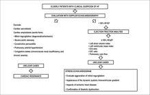
Diagnostic flowchart. HF: heart failure; HFpEF: heart failure with preserved ejection fraction; HFrEF: heart failure with reduced ejection fraction.
4.2. Peculiarities of Heart Failure Treatment in Elderly Patients
HF is prevalent among the elderly, affecting up to 20% of patients > 75 years old.11. Instituto Brasileiro de Geografia e Estatística. (IBGE). Pesquisa Nacional de Saúde (PNS). Rio de Janeiro; 2013. It is characterized by the presentation of systolic dysfunction (HFrEF) or diastolic dysfunction (HFpEF) and high mortality (2-fold risk of all-cause mortality adjusting for age and sex and 4-fold risk of cardiovascular death).180180. Vetrano DL, Lattanzio F, Martone AM, Landi F, Brandi V, Topinkova E et al. Treating heart failure in older and oldest old patients. Curr Pharm Des. 2015;21(13):1659-64.,181181. Upadhya B, Taffet GE, Cheng CP, Kitzman DW. J Heart failure with preserved ejection fraction in the elderly: scope of the problem. J Mol Cell Cardiol. 2015 Jun;83:73-87. Over the past decades, HFpEF has become the main clinical phenotype.22. Brasil. Ministério da Saúde. Sistema de informação sobre a mortalidade. Brasília; 2013.
Polypharmacy is extremely common in this context, with a strong impact on drug interactions, higher rates of adverse effects and poor adherence; however, multidisciplinary and adherence programs have been shown to be useful in this group of patients.182182. Krueger K, Botermann L, Schorr SG, Griese-Mammen N, Laufs U, Schulz M. Age-related medication adherence in patients with chronic heart failure: a systematic literature review. Int J Cardiol. 2015 Apr 1;184:728-35. Exercise training, in comparison with habitual care, in elderly HFrEF patients in New York Heart Association (NYHA) classes II and III, was shown to be safe, without an increase in mortality and hospitalization and with improvements in the walking test.183183. Chen YM, Li Y. Safety and efficacy of exercise training in elderly heart failure patients: a systematic review and meta-analysis. Int J Clin Pract. 2013;67(11):1192-8.
The objectives of pharmacological treatment of HF are: reducing mortality and hospitalization; improving functional capacity and quality of life; and including the use of ACEI, ARB, beta-blockers, and aldosterone antagonists. Elderly individuals have frequently been excluded or under-represented in studies performed on HF patients.184184. Chin KL, Skiba M, Tonkin A, Reid CM, Liew D, Krum H et al. The treatment gap in patients with chronic systolic heart failure: a systematic review of evidence-based prescribing in practice. Heart Fail Rev. 2016;21(6):675-97.
Several influential clinical trials have demonstrated the efficacy of ACEI in younger patients (average age of 60/66); however, subgroup analysis of the Heart Outcomes Prevention Evaluation (HOPE) study demonstrated a higher risk reduction in patients > 65 years old, in comparison with the younger group.184184. Chin KL, Skiba M, Tonkin A, Reid CM, Liew D, Krum H et al. The treatment gap in patients with chronic systolic heart failure: a systematic review of evidence-based prescribing in practice. Heart Fail Rev. 2016;21(6):675-97.
ARB have been little evaluated in elderly patients; however, subanalysis of the Candesartan in Heart Failure: Assessment of Reduction in Mortality and Morbidity (CHARM-Alternative) study, with 23.3% of its study population ≥ age 75, demonstrated benefits similar to those reported for the general group.185185. Anguita Sánchez M, Jiménez-Navarro M, Crespo M, Alonso-Pulpón L, de Teresa E, Castro-Beiras A et al.; OBELICA study researchers. Effect of a training program for primary care physicians on the optimization of beta-blocker treatment in elderly patients with heart failure. Rev Esp Cardiol. 2010;63(6):677-85.
Regarding beta-blockers, a recent meta-analysis of 12,719 patients did not find any differences in benefits between those defined as “elderly” in the clinical trials included and their younger counterparts. It is important to underline the fact that the oldest patient from the individual clinical trials analyzed was 71 years old.55. Morley JE, Vellas B, van Kan A, Anker SD, Bauer JM, Bernabei R et al. Frailty consensus: a call to action. J Am Med Dir Assoc. 2013;14(6):392-7. The Study of Effects of Nebivolol Intervention on Outcomes and Rehospitalization in Seniors with Heart Failure (SENIORS) demonstrated the efficacy of nebivolol in CHF patients > 70 years old. Subanalysis of the Organized Program to Initiate Lifesaving Treatment in Hospitalized Patients with Heart Failure (OPTIMIZE-HF) study provided evidence that beta-blockers may be associated with beneficial effects in patients ≥ 75 years old.186186. Flather MD, Shibata MC, Coats AJ, Van Veldhuisen DJ, Parkhomenko A, Borbola J et al.; SENIORS Investigators. Randomized trial to determine the effect of nebivolol on mortality and cardiovascular hospital admission in elderly patients with heart failure (SENIORS). Eur Heart J. 2005;26(3):215-25.,187187. Rossi JS, Flaherty JD, Fonarow GC, Nunez E, Gattis Stough W, Abraham WT et al. Influence of coronary artery disease and coronary revascularization status on outcomes in patients with acute heart failure syndromes: a report from OPTIMIZE-HF (Organized Program to Initiate Lifesaving Treatment in Hospitalized Patients with Heart Failure) Eur J Heart Fail. 2008;10(12):1215-23.
The Euro Heart Failure Survey II has suggested that the use of ACEI and/or beta-blockers is associated with a significant decrease in short-term mortality in octogenarians. The Euro HF Survey II, on the other hand, did not show improvements in mortality during 1 year with the use of beta-blockers; this is possibly related to the higher number of elderly HFpEF patients in this study.188188. Komajda M, Hanon O, Hochadel M, Lopez-Sendon JL, Follath F, Ponikowski P et al. Contemporary management of octogenarians hospitalized for heart failure in Europe: Euro Heart Failure Survey II. Eur Heart J. 2009;30(4):478-86.
In the most important studies with aldosterone antagonists, the Randomized Aldactone Evaluation Study (RALES) and the Eplerenone Post-Acute Myocardial Infarction Heart Failure Efficacy and Survival Study (EPHESUS), the average patient age was 67 and 64, respectively. Their use, in elderly patients, however, should be carefully monitored in accordance with renal dysfunction and the underlying drug interaction. In the Prospective Comparison of ARNI with ACEI to Determine Impact on Global Mortality and Morbidity in Heart Failure (PARADIGM) study, symptomatic hypotension in patients > age 75 was more frequent in the sacubitril/valsartan group (18%) than in the enalapril group (12%).189189. McMurray JJ, Packer M, Desai AS, Gong J, Lefkowitz MP, Rizkala AR et al.; PARADIGM-HF Investigators and Committees. Angiotensin-neprilysin inhibition versus enalapril in heart failure. N Engl J Med. 2014;371(11):993-1004.
In summary, current orientations recommend a therapeutic approach similar to the one applied to younger patients for HF treatment, with caution regarding interactions and tolerance.176176. Ponikowski P, Voors AA, Anker SD, Bueno H, Cleland JG, Coats AS et al.; ESC Scientific Document Group. 2016. ESC Guidelines for the diagnosis and treatment of acute and chronic heart failure: the Task Force for the diagnosis and treatment of acute and chronic heart failure of the European Society of Cardiology (ESC) Developed with the special contribution of the Heart Failure Association (HFA) of the ESC. Eur Heart J. 2016;37(27):2129-200.,190190. Edelmann F, Musial-Bright L, Gelbrich G; CIBIS-ELD Investigators and Project Multicenter Trials in the Competence Network Heart Failure. Tolerability and Feasibility of Beta-Blocker Titration in HFpEF Versus HFrEF: Insights From the CIBIS-ELD Trial. JACC Heart Fail. 2016;4(2):140-9.
4.3. General Recommendations for Elderly Heart Failure Patients
5. Arterial Hypertension in The Elderly
5.1. Diagnostic Peculiarities
A Brazilian epidemiological study titled the Multicenter Study of Elderly Patients in Outpatient Clinics of Cardiology and Geriatric Brazilian Institutions (EMI, acronym in Portuguese)191191. Taddei CF, Ramos LR, Moraes JC, Wajngarten M, Libberman A, Santos SC et al. Multicenter study of elderly patients in outpatient clinics of cardiology and geriatric Brazilian institutions. Arq Bras Cardiol. 1997;69(5):327-33. demonstrated that SAH is the main risk factor among elderly Brazilians. It is found in 65% of elderly outpatients and 80% of women > 75 years old. Aging produces vascular alterations, such as arterial stiffening, reduced elasticity and vascular compliance, reduced vasodilation capacity, increased SBP, decreased sensitivity to volume changes, slowed ventricular relaxation, increased cardiac workload, loss of myocytes, and compensatory hypertrophy.192192. Strait JB, Lakatta EG. Aging-associated cardiovascular changes and their relationship to heart failure. Heart Fail Clin. 2012;8(1):143-64. These alterations lead to peculiarities in diagnosing and treating SAH in elderly patients.
5.1.1. Peculiarities in Measuring Blood Pressure
In elderly patients, BP has high variability. It is necessary to take special care in measuring BP, owing to the possible presence of the following factors:
-
OH: defined as a drop in SBP of > 20 mmHg or in diastolic blood pressure (DBP) of > 10 mmHg, following 3 minutes in the orthostatic position. BP should be checked in the sitting, lying, and standing positions, given that atherosclerotic alterations in the carotid sinus regions may reduce baroreceptor sensitivity, leading to reduced postural reflexes and, thus, predisposing the patient to OH.33. Fried LP, Tangen CM, Walston J, Newman AB, Hirsch C, Gottdiener J et al. Frailty in older adults evidence for a phenotype. J Gerontol A Biol Sci Med Sci. 2001;56(3):M146-56. Furthermore, comorbidities, such as peripheral polyneuropathy and Parkinson’s disease, as well as the use of diuretic, antidepressant, vasodilator, and beta-blocker drugs may also lead to OH in up to 34% of elderly patients > age 75.
-
Auscultatory gap: a sitution in which, after auscultation of the first Korotkoff sound, the sound disappears completely and only reappears after the decrease in SBP, but before the beginning of the last phase of Korotkoff sounds. This leads to errors in diagnosing SBP at lower levels and false diagnoses of normotension. In order to avoid this measurement error, it is necessary to estimate systolic pressure using the radial pulse palpatory method, raising cuff pressure 20 to 30 mmHg above this point.193193. Malachias MV, Póvoa RM, Nogueira AR, Souza D, Costa LS, Magalhães ME; Sociedade Brasileira de Cardiologia. 7a Diretriz Brasileira de hipertensão arterial. Arq Bras Cardiol. 2016;107(3 supl 3):1-83.
-
Pseudo-hypertension: pseudo-hypertension may appear in elderly patients with pronounced atherosclerosis, arterial wall calcification, and vessel stiffening. In this situation, it is sufficient to inflate the cuff in order to collapse the brachial artery.193193. Malachias MV, Póvoa RM, Nogueira AR, Souza D, Costa LS, Magalhães ME; Sociedade Brasileira de Cardiologia. 7a Diretriz Brasileira de hipertensão arterial. Arq Bras Cardiol. 2016;107(3 supl 3):1-83. Osler’s maneuver is used to identify this. The maneuver consists of inflating the cuff above systolic pressure levels and, concomitantly, palpating the radial artery. If it continues to be palpable, this suggests that the artery is stiff and indicates that the index obtained by auscultation does not express the true SBP. Pseudo-arterial hypertension may also be suspected when SBP is elevated in patients who do not present injuries in target organs or in those who manifest hypotension following treatment with low doses of anti-hypertensive drugs.
-
Arterial hypertension during exercise: although BP is habitually higher during physical exercise, this increase is greater in elderly adults, due to arterial stiffness. Values for diagnosing SAH during exercise are not clear. Physically deconditioned patients respond with greater increases in BP than conditioned patients.
-
White coat hypertension: this occurs when BP increases during a clinical visit but remains normal during daily activity. This can be better evaluated by 24-hour ambulatory blood pressure monitoring (ABPM) or home blood pressure monitoring (HBPM).193193. Malachias MV, Póvoa RM, Nogueira AR, Souza D, Costa LS, Magalhães ME; Sociedade Brasileira de Cardiologia. 7a Diretriz Brasileira de hipertensão arterial. Arq Bras Cardiol. 2016;107(3 supl 3):1-83. Serial measurements may minimize this condition.
-
Masked arterial hypertension: this is the opposite of white coat hypertension, namely, pressure is high during daily activities and normal during the clinical visit.193193. Malachias MV, Póvoa RM, Nogueira AR, Souza D, Costa LS, Magalhães ME; Sociedade Brasileira de Cardiologia. 7a Diretriz Brasileira de hipertensão arterial. Arq Bras Cardiol. 2016;107(3 supl 3):1-83. This may also be evaluated by 24-h ABPM or HBPM.
-
Isolated systolic hypertension (ISH) and pulse pressure (PP): ISH and PP are cardiovascular risk factors in elderly patients.191191. Taddei CF, Ramos LR, Moraes JC, Wajngarten M, Libberman A, Santos SC et al. Multicenter study of elderly patients in outpatient clinics of cardiology and geriatric Brazilian institutions. Arq Bras Cardiol. 1997;69(5):327-33. ISH is due to lower distensibility and elasticity in the large capacitance vessels, such as the aorta, which results in increased pulse wave velocity (PWV). This increase in PWV is accompanied by an increase in reflex wave velocity, which returns from peripheral to central circulation.191191. Taddei CF, Ramos LR, Moraes JC, Wajngarten M, Libberman A, Santos SC et al. Multicenter study of elderly patients in outpatient clinics of cardiology and geriatric Brazilian institutions. Arq Bras Cardiol. 1997;69(5):327-33.,192192. Strait JB, Lakatta EG. Aging-associated cardiovascular changes and their relationship to heart failure. Heart Fail Clin. 2012;8(1):143-64. In elderly patients, the reflex wave reaches the ascending aorta during systole, leading to an even higher increase in SBP. Loss of reflex wave in protodiastole makes diastolic pressure remain equal or decrease.192192. Strait JB, Lakatta EG. Aging-associated cardiovascular changes and their relationship to heart failure. Heart Fail Clin. 2012;8(1):143-64. The final effect consists of a predominant increase in SBP, with DBP remaining normal or even low. Characteristics of ISH include SBP ≥ 140 mmHg and DBP < 90 mmHg.193193. Malachias MV, Póvoa RM, Nogueira AR, Souza D, Costa LS, Magalhães ME; Sociedade Brasileira de Cardiologia. 7a Diretriz Brasileira de hipertensão arterial. Arq Bras Cardiol. 2016;107(3 supl 3):1-83. PP is defined as the difference between SBP and DBP. This occurs due to the progressive loss of arterial elasticity, with a consequent decrease in vascular complacency. DBP tends to remain normal or even low. Limits for abnormal PP values have yet to be defined.191191. Taddei CF, Ramos LR, Moraes JC, Wajngarten M, Libberman A, Santos SC et al. Multicenter study of elderly patients in outpatient clinics of cardiology and geriatric Brazilian institutions. Arq Bras Cardiol. 1997;69(5):327-33. The Framingham study demonstrated a higher cardiovascular risk associated with higher PP, in patients between the ages of 50 and 79, as well as an important role of low DBP in this association.33. Fried LP, Tangen CM, Walston J, Newman AB, Hirsch C, Gottdiener J et al. Frailty in older adults evidence for a phenotype. J Gerontol A Biol Sci Med Sci. 2001;56(3):M146-56. In addition to the factors mentioned, target organ injuries should be investigated (eye fundus changes, LV hypertrophy, and peripheral and renal atherosclerosis), and the possibility of secondary SAH should be evaluated. The following are suspicious factors:193193. Malachias MV, Póvoa RM, Nogueira AR, Souza D, Costa LS, Magalhães ME; Sociedade Brasileira de Cardiologia. 7a Diretriz Brasileira de hipertensão arterial. Arq Bras Cardiol. 2016;107(3 supl 3):1-83.
-
Sudden onset of SAH or acute worsening
-
Abdominal murmur
-
SAH resistant to 3 or more drugs
-
Creatinine increase over 30% with the use of ACEI or ARB
-
Systemic atherosclerotic disease in smokers and patients with dyslipidemia
-
Recurrent hypertensive pulmonary edema
-
Pheochromocytoma and hyperaldosteronism should be adequately investigated with more specific exams, because, even though they are less frequent in elderly patients, once they are diagnosed and treated, they may result in the patient being cured.
-
Among secondary causes of SAH, the following stand out: aortic regurgitation (AR), hyperthyroidism, renovascular atherosclerosis, and use of drugs that increase pressure, such as non-hormonal anti-inflammatory agents, antihistamines, decongestants, corticosteroides, MAOI, and TCA.
5.1.2. Peculiarities of Clinical Laboratory Investigation
The objective of clinical laboratory investigation is to confirm that BP is increased; identify causes of SAH, target organ injuries, and associated diseases; and stratify cardiovascular risk. In addition to clinical history, cognitive tests, and physical examination including BMI and abdominal circumference, the following should be performed:
-
Resting EKG.
-
Urine examination (biochemical and sediment)
-
Blood tests: complete blood count, creatinine, blood glucose, potassium, fasting blood glucose, glycohemoglobin, total cholesterol and fractions, triglycerides, and uric acid. Blood levels of creatinine may be normal, in spite of declined renal function. This fact results from the progressive loss of muscle mass, a determining factor of creatinine production. Thus, creatinine levels > 1.5 mg/dL are considered abnormal in elderly patients. The formula most used to calculate estimated glomerular filtration rate (eGFR) is the Cockroff-Gault (mL/min): (140 – age) × weight (kg)/plasma creatinine (mg/dL) × 72,with a coefficient of 0.85 for women. Interpretation: normal renal function, > 90 mL/min; slight renal dysfunction, 60 to 90 mL/min; moderate renal dysfunction, 30 to 60 mL/min; severe renal dysfunction, < 30 mL/min.
-
ABPM and HBPM: to investigate white coat SAH and masked SAH, in cases where it is necessary to investigate episodes of arterial hypotension, or to evaluate the efficacy of SAH therapy.193193. Malachias MV, Póvoa RM, Nogueira AR, Souza D, Costa LS, Magalhães ME; Sociedade Brasileira de Cardiologia. 7a Diretriz Brasileira de hipertensão arterial. Arq Bras Cardiol. 2016;107(3 supl 3):1-83.
5.2. Treatment Peculiarities
5.2.1. Therapeutic Goals for Elderly Patients
Treating SAH in elderly patients represents a great challenge, as it involves a heterogeneous group, with multiple comorbidities, cognitive problems, risk of falling, polypharmacy, and frailty syndrome. Therapeutic goals for elderly patients should thus be individualized based on multidisciplinary team judgment, and they should consider patient preferences.193193. Malachias MV, Póvoa RM, Nogueira AR, Souza D, Costa LS, Magalhães ME; Sociedade Brasileira de Cardiologia. 7a Diretriz Brasileira de hipertensão arterial. Arq Bras Cardiol. 2016;107(3 supl 3):1-83.,194194. Beckett NS, Peters R, Fletcher AE, Staessen JA, Liu L, Dumitrascu D et al.; HYVET Study Group. Treatment of hypertension in patients 80 years of age or older. N Engl J Med. 2008;358(18):1887-98. Dose adjustments should occur every 4 weeks, in order to avoid abrupt reductions of BP. The Hypertension in the Very Elderly Trial (HYVET) randomized, placebo-controlled study194194. Beckett NS, Peters R, Fletcher AE, Staessen JA, Liu L, Dumitrascu D et al.; HYVET Study Group. Treatment of hypertension in patients 80 years of age or older. N Engl J Med. 2008;358(18):1887-98. included 3,845 patients with SBP ≥ 160 mmHg over the age of 80, with an average age of 83.6. Target blood pressure was 150/80 mmHg. They demonstrated that treatment with indapamide, with or without perindopril, was beneficial in octogenarians. In the intention-to-treat analysis, there was a 30% reduction in rate of fatal or non-fatal stroke, 39% reduction in the rate of death from stroke, a 21% reduction in death from any cause, a 23% reduction of in the rate of death from cardiovascular causes, and a 64% reduction in the rate of HF. Fewer severe adverse events occurred in the active treatment group (358 versus 448 in the placebo group). There is evidence that greatly lowering BP in elderly patients may be harmful; this fact is known as the J- or U-curve.191191. Taddei CF, Ramos LR, Moraes JC, Wajngarten M, Libberman A, Santos SC et al. Multicenter study of elderly patients in outpatient clinics of cardiology and geriatric Brazilian institutions. Arq Bras Cardiol. 1997;69(5):327-33. The recent SPRINT study149149. Wright JT Jr, Williamson JD, Whelton PK, Snyder JK, Sink KM, Rocco MV et al.; SPRINT Research Group. A randomized trial of intensive versus standard blood-pressure control. N Engl J Med. 2015;373(22):2103-16. sought to evaluate two different BP goals. In the standard group, the goal was SBP < 140 mmHg and, in the intensive treatment group, the goal was SBP < 120 mmHg. The intensive treatment group had a significant reduction in primary events (infarction, other acute coronary syndromes, stroke, HF, or death from cardiovascular causes) in comparison with the standard treatment group. Although the initial impression may be that more intensive goals may be more beneficial, it is necessary to consider that there was an increase in the number of severe adverse events, such as hypotension, syncope, electrolytic disorders, and acute renal insufficiency, in the intensive treatment group. Another important study was the ACCORD,3535. Gerstein HC, Miller ME, Byimgton RP, Goff DC Jr, Bigger JT, Buse JB et al.; Action to control cardiovascular risk in diabetes study group: effects of intensive glucose lowering in type 2 diabetes. N Engl J Med. 2008;358(24):2545-59. performed with 10,251 diabetic patients, ages 40 to 79, 4,733 of which were also randomized for BP reduction < 140 mmHg or < 120 mmHg. However, BP reduction with more intensive goals did not succeed in significantly reducing the risk of the study’s primary outcome (death from CVD, nonfatal infarction, and nonfatal stroke). Thus, to date, the III Geriatric Cardiology Guidelines recommends SBP levels ≤ 130 mmHg for elderly patients ≥ age 65, who are considered robust and who do not have frailty criteria.195195. Mancia G, Fagard R, Narkiewicz K, Redon J, Zanchetti A, Böhm M et al. 2013 ESH/ESC Guidelines for the management of arterial hypertension: the Task Force for the Management of Arterial Hypertension of the European Society of Hypertension (ESH) and of the European Society of Cardiology (ESC). Eur Heart J. 2013;34(28):2159-219.,196196. Whelton PK, Carey RM, Aronow WS, Casey DE Jr, Collins KJ, Dennison Himmelfarb C et al. 2017 ACC/AHA/AAPA/ABC/ACPM/AGS/APhA/ASH/ASPC/NMA/PCNA Guideline for the Prevention, Detection, Evaluation, and Management of High Blood Pressure in Adults: A Report of the American College of Cardiology/American Heart Association Task Force on Clinical Practice Guidelines. J Am Coll Cardiol. 2018;71(19):e127-248. For patients ≤ 80 years old, without frailty, SBP levels < 140 mmHg may be considered;195195. Mancia G, Fagard R, Narkiewicz K, Redon J, Zanchetti A, Böhm M et al. 2013 ESH/ESC Guidelines for the management of arterial hypertension: the Task Force for the Management of Arterial Hypertension of the European Society of Hypertension (ESH) and of the European Society of Cardiology (ESC). Eur Heart J. 2013;34(28):2159-219. in patients ≥ age 80 with SBP ≥ 160 mmHg, an initial reduction to SBP between 150 and 140 mmHg may be considered;77. Afilalo J, Alexander KP, Mack MJ, Maurer MS, Green P, Allen LA et al. Frailty assessment in the cardiovascular care of older adults. J Am Coll Cardiol. 2014;63(8):747-62. in fragile elderly patients or patients with multiple comorbidities, the therapeutic goal should be individualized, considering each case’s risk-benefit ratios.196196. Whelton PK, Carey RM, Aronow WS, Casey DE Jr, Collins KJ, Dennison Himmelfarb C et al. 2017 ACC/AHA/AAPA/ABC/ACPM/AGS/APhA/ASH/ASPC/NMA/PCNA Guideline for the Prevention, Detection, Evaluation, and Management of High Blood Pressure in Adults: A Report of the American College of Cardiology/American Heart Association Task Force on Clinical Practice Guidelines. J Am Coll Cardiol. 2018;71(19):e127-248.
5.2.2. Medical and Non-Medical Treatments
Salt reduction should be cautious and well accompanied by the doctor, given that the elderly patient’s diminished taste sensitivity may make food appear blander, causing the patient to eat less and thus bringing about the risk of malnutrition. It is also necessary to remember that elderly patients rarely present only one chronic disease. The evaluation of multimorbidities generally defines what the best treatment is and what drugs should be avoided in each specific case. Treatment should be initiated with low doses, and dose adjustments should be gradual. Adherence needs to be stimulated, if possible, by monthly control at the beginning of treatment and at each dose adjustment. The most commonly used drugs in elderly patients are:
-
Diuretics: thiazides and correlates (hydrochlorothiazide, chlorthalidone, indapamide) are considered first-line drugs in elderly patients without comorbidities. Their use is preferential in osteoporosis patients, as they decrease urinary excretion of calcium, and in initial phases of congestive heart failure (CHF), as they reduce preload, volume, and pulmonary congestion. Recommended doses of hydrochlorothiazide: 6.25 to 25 mg/day, maintaining efficacy and reducing adverse metabolic effects.191191. Taddei CF, Ramos LR, Moraes JC, Wajngarten M, Libberman A, Santos SC et al. Multicenter study of elderly patients in outpatient clinics of cardiology and geriatric Brazilian institutions. Arq Bras Cardiol. 1997;69(5):327-33. In most cases, diuretics are associated with therapeutic schedules. However, they should be avoided in elderly patients with incipient urinary incontinence, gout (because they increase uric acid) and prostatism.191191. Taddei CF, Ramos LR, Moraes JC, Wajngarten M, Libberman A, Santos SC et al. Multicenter study of elderly patients in outpatient clinics of cardiology and geriatric Brazilian institutions. Arq Bras Cardiol. 1997;69(5):327-33. Attention should be paid to blood glucose in elderly patients with concomitant use of thiazides and oral antidiabetics or insulin, given that thiazides may increase blood glucose and interfere with diabetes control.
-
Calcium channel blockers: they include both dihydropyridine and non-dihydropyridine derivatives. Dihydropyridine derivatives have major vasodilatory effects. The most recent generation provokes less edema. They are very commonly used in elderly SAH and symptomatic coronary disease patients. Non-dihydropyridine derivatives, especially verapamil, have fewer vasodilatory effects, and they are not usually prescribed to elderly patients, as they may alter electrical impulses of atrioventricular conduction. Verapamil may, furthermore, provoke intestinal constipation.
-
ACEI: they continue to be efficacious in elderly patients, notwithstanding the decrease in renin with aging. They decrease cardiovascular events and should be used in elderly patients with SAH and HF or asymptomatic ventricular dysfunction. Adverse effects include changes in taste, especially with captopril, which may reduce food intake, and dry cough, which limit their use. It is fundamental to check potassium, due to frequent reductions in renal function.
-
Angiotensin II receptor antagonists (ARA-II): they are effective in HF, and they have an established renal and cardiac protective action in type 2 diabetes with nefropathy.191191. Taddei CF, Ramos LR, Moraes JC, Wajngarten M, Libberman A, Santos SC et al. Multicenter study of elderly patients in outpatient clinics of cardiology and geriatric Brazilian institutions. Arq Bras Cardiol. 1997;69(5):327-33. ARA-II have a favorable tolerability profile, with few adverse effects (occasional dizziness and, rarely, hypersensitive skin reaction). They are well used in cases of ACEI intolerance.193193. Malachias MV, Póvoa RM, Nogueira AR, Souza D, Costa LS, Magalhães ME; Sociedade Brasileira de Cardiologia. 7a Diretriz Brasileira de hipertensão arterial. Arq Bras Cardiol. 2016;107(3 supl 3):1-83.
-
Beta-blockers: they are not used as initial monotherapy in elderly patients without comorbidities, due to their lower effects on BP reduction; however, in association with diuretics, they present good results. They are mainly used in elderly patients with SAH and coronary insufficiency or HF. Less liposoluble beta-blockers, such as atenolol, metoprolol, and bisoprolol, are recommended for elderly patients because they have lower risks of collateral effects on the central nervous system (depression, drowsiness, confusion, sleep disturbances).193193. Malachias MV, Póvoa RM, Nogueira AR, Souza D, Costa LS, Magalhães ME; Sociedade Brasileira de Cardiologia. 7a Diretriz Brasileira de hipertensão arterial. Arq Bras Cardiol. 2016;107(3 supl 3):1-83.
In summary, elderly patients have particularities regarding SAH diagnosis and approach. It is necessary to consider each patient’s comorbidities and particularities, including functional status, which may be determining factors for setting BP goals and for patient decision making.
6. Valvulopathies
6.1. Mitral Stenosis
6.1.1. Diagnostic Peculiarities
Mitral stenosis (MS) is rare in elderly patients (present in 6% of patients with mitral annulus calcification).197197. Desai MY, Gerstenblith G. Contemporary cardiology: cardiovascular disease in the elderly. New Jersey: Humana Press; 2010. p. 247-50.
Etiology – Sequel of rheumatic carditis or calcification of the mitral valve apparatus in patients > 85 years old.198198. Harris R. Clinical geriatric cardiology. 2nd ed. Philadelphia: JB Lippincott Company; 1970.
Symptoms – similar to those observed in non-elderly patients. Symptoms may be absent. The most frequent are dyspnea and cough, which may be accompanied by hemoptoic sputum. It may manifest as systemic embolism or AF.
Physical examination – Hyperphonetic first heart sound and apical mid-diastolic murmur with thrill may be absent. The opening snap of the mitral valve is rarely auscultated. Most patients > age 80 present AF with elevated HR which, in association with a greater anteroposterior thorax diameter, makes auscultation difficult. The more fibrosis and calcification are present in the mitral valve, the less audible the auscultatory signs of MS will be. Diagnostic suspicion may be established based on signs of pulmonary arterial hypertension (P2 hyperphonesis in the second heart sound, RV insufficiency, pulmonary and tricuspid regurgitation). In elderly pulmonary arterial hypertension patients without any other evident cause, it is important to investigate MS.199199. Cheitlin MD. Valve disease in octogenarian. In: Wenger NK. (editor). Cardiovascular disease in the octogenarian and beyond. London: Martin Dunitz; 1999. p. 255-66.–202202. Tarasoutchi F, Montera MW, Grinberg M, Piñeiro DJ, Sánchez CR, Bacelar AC et al; [Brazilian Guidelines for Valve Disease – SBC 2011 / I Guideline Inter-American Valve Disease – 2011 SIAC]. Arq Bras Cardiol. 2011;97(5 Suppl 1):1-67.
Complementary exams – EKG, chest radiography, and echocardiogram are sufficient, in most cases, to confirm diagnosis and estimate severity. The following may be found in the EKG: left atrial overload (LAO), RV hypertrophy, and AF. Chest radiography findings include: increased LA, mitral valve calcification, and posterior displacement of the barium-filled esophagus. Echocardiography data include: mitral annulus calcification (in 60% of elderly patients > age 85),44. Fried LP, Ferrucci L, Darer J, Williamson JD, Anderson G. Untangling the concepts of disability, frailty, and comorbidity: implications for improved targeting and care. J Gerontol A Biol Sci Med Sci. 2004;59(3):255-63. mitral valve area (Table 8), pulmonary BP, and status of the valvular apparatus (mobility, thickening, and subvalvular impairment).55. Morley JE, Vellas B, van Kan A, Anker SD, Bauer JM, Bernabei R et al. Frailty consensus: a call to action. J Am Med Dir Assoc. 2013;14(6):392-7.
6.1.2. Treatment Peculiarities
Clinical treatment – Patients with mild MS are generally asymptomatic, and they do not need to receive medication,203203. Nishimiura RA, Otto CM, Bonow RO, Carabello BA, Erwin JP 3rd, Guyton RA et al.; American College of Cardiology/American Heart Association Task Force on Practice Guidelines. 2014 AHA/ACC guideline for the management of patients with valvular heart disease: a report of the American College of Cardiology/American Heart Association Task Force on Practice Guidelines. J Am Coll Cardiol. 2014;63(22):e57-185. Erratum in: J Am Coll Cardiol. 2014;63(22):2489. unless they also suffer from AF. Unlike younger patients, elderly MS patients who develop AF have a higher chance of showing symptoms of HF, owing to the concomitant present of diastolic dysfunction. Thus, in cases of paroxysmal AF with hemodynamic deterioration, even if MS is mild, electrical cardioversion is indicated. Patients with MS and AF, be it permanent, persistent, or paroxysmal, should constantly use warfarin, regardless of risk scores, with the aim of keeping the international normalized ratio (INR) between 2 and 3, unless there is a formal contraindication.204204. Wann LS, Curtis AB, January CT, Ellenbogen KA, Lowe JE, Estes NA 3rd et al. 2011 ACCF/AHA/HRS focused update on the management of patients with atrial fibrilation (updating the 2006 guideline). A report of the American College of Cardiology Foundation/American Heart Association Task Force on Practice Guidelines. J Am Coll Cardiol. 2011;57(2):223-42. Although some publications recommend the use of new oral anticoagulants in this situation, these data have yet to be evaluated in comparative studies.205205. Martins RP, Galand V, Colette E, Behar N, Pavin D, Leclercq C et al. Defining nonvalvular atrial fibrillation: a quest for clarification. Am Heart J. 2016 Aug;178:161-7. The finding of LA thrombus or the occurrence of a systemic embolic event, even in the presence sinus rhythm (SR), also indicate the need for anticoagulant use. In MS of rheumatic etiology, prophylaxis for rheumatic fever is not necessary, given that elderly patients rarely have relapses of this disease.206206. Barbosa PJ, Müller RE, Latado AL, Achutti AC, Ramos AI, Weksler C et al. Diretrizes Brasileiras para Diagnóstico, Tratamento e Prevenção da Febre Reumática da Sociedade Brasileira de Cardiologia, da Sociedade Brasileira de Pediatria e da Sociedade Brasileira de Reumatologia. Arq Bras Cardiol. 2009;93(3 supl.4):1-18. Early treatment of bacterial infections is recommended with the aim of protecting the patient from the risk of infective endocarditis (IE). Chemoprophylaxis against IE in elderly MS patients is not indicated.207207. Habib G, Lancellotti P, Antunes MJ, Bongiorni MG, Casalta JP, Del Zotti F et al.; ESC Scientific Document Group. 2015 ESC Guidelines for the management of infective endocarditis: The Task Force for the Management of Infective Endocarditis of the European Society of Cardiology (ESC). Endorsed by: European Association for Cardio-Thoracic Surgery (EACTS), the European Association of Nuclear Medicine (EANM). Eur Heart J. 2015;36(44):3075-128. In symptomatic patients with moderate to severe MS, loop diuretics are the best option for controlling pulmonary or systemic congestion, and beta-blockers are indicated for reducing HR and facilitating atrial emptying. There is no evidence that the use of beta-blockers is beneficial in patients with SR who do not have elevated HR.208208. Monmeneu Menadas JV, Marín Ortuño F, Reyes Gomis F, Jordán Torrent A, García Martínez M, Bodí Peris V et al. Betablockade and exercise capacity in patients with mitral stenosis in sinus rhythm. J Heart Valve Dis. 2002;11(2):199-203. In the presence of AF with elevated ventricular response, beta-blockers are the drugs of choice for reducing HR. In cases where they are contraindicated, nondihydropyridine calcium channel blockers or digitalis may be used. In the presence of signs of RV failure with associated hepatomegaly, due to the frequent coexistence of secondary hyperaldosteronism, elevated doses of spironolactone (100 mg/day) are an option.209209. Teresa E. Tratamiento diurético de la insuficiencia cardíaca. Rev Esp Cardiol Supl. 2007;7:34F-44F. Caution is necessary with the risk of hyperkalemia.
Options for correcting MS – When evaluating an elderly patient with MS who has been indicated for intervention, the following should be considered and discussed with the patient and/or family members: etiology, whether rheumatic or degenerative; patient life expectancy; evaluation of functionality; and the presence of multimorbidities. There are 2 options for correcting rheumatic MS: percutaneous balloon mitral valvuloplasty (PBMV) or extracorporeal circulation surgery. Randomized clinical trials have shown that, in selected cases, PBMV offers immediate and long-term results similar to those of open commissurotomy.210210. Bouleti C, Iung B, Laouénan C, Himbert D, Brochet E, Messika-Zeitoun D et al. Late results of percutaneous mitral commissurotomy up to 20 years development and validation of a risk score predicting late functional results from a series of 912 patients. Circulation. 2012;125(17):2119-27. For this intervention, presence of favorable valve morphology is important. This may be evaluated by several proposed echocardiography criteria, the Wilkins and Block score being the most widely used.211211. Wilkins GT, Weyman AE, Abascal VM, Block PC, Palacios IF. Percutaneous balloon dilatation of the mitral valve: an analysis of echocardiographic variables related to outcome and the mechanism of dilatation. Br Heart J. 1988;60(4):299-308. It is, additionally, necessary to respect contraindications to this procedure (presence of LA thrombus or mitral regurgitation with more than a mild degree of severity). Unfortunately, elderly patients frequently have valve morphologies which are unfavorable for this procedure, whether the etiology be rheumatic or degenerative.212212. Chmielak Z, Klopotowski M, Demokow M, Konka M, Hoffman P, Kukuła K et al. Percutaneous mitral balloon valvuloplasty beyond 65 years of age. Cardiol J. 2013;20(1):44-51. In the latter case, owing to the fact there is no commissural fusion, as occurs in rheumatic disease, the success of PBMV is restricted, and mitral valve replacement surgery is the procedure of choice. As degenerative MS patients frequently have multimorbidities that elevate their risks, clinical treatment should be attempted initially; mitral valve replacement surgery is indicated only in cases that do not respond to clinical treatment.213213. Sud K, Agarwal S, Parashar A, Raza MQ, Patel K, Min D et al. Degenerative mitral stenosis unmet need for percutaneous interventions. Circulation. 2016;133(16):1594-604. There are reports of small series of percutaneous implants of mitral prostheses in degenerative MS patients, with relative success.214214. Guerrero M, Dvir D, Himbert D, Urena M, Eleid M et al. Transcatheter mitral valve replacement in native mitral valve disease with severe mitral annular calcification: results from the First Multicenter Global Registry. JACC Cardiovasc Interv. 2016;9(13):136-71.
6.2. Mitral Regurgitation
6.2.1. Diagnostic Peculiarities
From the etiological point of view, mitral regurgitation (MR) may be: (a) primary: when there are histological changes in the valve, for example, myxomatous degeneration, degenerative fibroelastic disease, and IE; or (b) secondary: when MR is functional and the valve is histologically normal, for example, poor leaflet coaptation with dilated cardiomyopathy. MR is common in elderly patients; the degenerative cause is the most frequent, followed by ischemia, and, less frequently, rheumatic disease and IE.215215. Singh JP, Evans JC, Levy D, Larson MG, Freed LA, Fuller DL et al. Prevalence and clinical determinants of mitral, tricuspid and aortic regurgitation (The Framingham Heart Study). Am J Cardiol. 1999;83(6):897-902. Erratum in: Am J Cardiol. 1999;84(9):1143.,216216. Bell MH, Mintz GS. Mitral valve disease in the elderly. Cardiovasc Clin. 1986;16(2):313-24. Acute MR is mainly linked to CAD by papillary muscle dysfunction or chordae tendineae rupture, with condition of acute HF.
Symptoms – symptoms of chronic MR are related to severity, rate of disease progression, pulmonary BP, presence of arrhythmias (e.g., AF), and associated diseases. The most common symptoms are stress dyspnea and fatigue.
Physical examination – The following are present: protosystolic murmur in mitral focus, variable intensity, and displaced ictus, with characteristics of volumetric overload. Thoracic deformities, which are common at this age, may modify ictus, sounds, and murmurs.102102. Gravina CF, Franken R, Wenger N, Freitas EV, Batlouni M, Rich M et al.; Sociedade Brasileira de Cardiologia. [II Guidelines of Brazilian Society of Cardiology in geriatric cardiology]. Arq Bras Cardiol. 2010;95(3 Suppl 2):e16-76.,202202. Tarasoutchi F, Montera MW, Grinberg M, Piñeiro DJ, Sánchez CR, Bacelar AC et al; [Brazilian Guidelines for Valve Disease – SBC 2011 / I Guideline Inter-American Valve Disease – 2011 SIAC]. Arq Bras Cardiol. 2011;97(5 Suppl 1):1-67.
Complementary exams – During EKG, frequent abnormalities are LAO, AF and left ventricular overload (LVO).217217. Huep JC, Gonçalves RS, Ferreira RM. A importância do eletrocardiograma, da radiografia de tórax e dos testes de capacidade funcional na avaliação das valvopatias mitrais. Rev Soc Cardiol Estado São Paulo. 2008;18(4):319-27. In the presence of ischemic MR, electrocardiographic signs of coronary insufficiency, such as electrically inactive zones and alterations in ventricular repolarization, may occur.218218. Rossi EG. Insuficiência mitral. In: Grinberg M, Sampaio RO. Doença Valvar. São Paulo: Manole; 2006. In cases of acute MR, EKG may be normal, or it may show only sinus tachycardia.217217. Huep JC, Gonçalves RS, Ferreira RM. A importância do eletrocardiograma, da radiografia de tórax e dos testes de capacidade funcional na avaliação das valvopatias mitrais. Rev Soc Cardiol Estado São Paulo. 2008;18(4):319-27.,219219. Moffa PJ, Sanches PCR, Uchida A. Eletrocardiologia: utilidade do eletrocardiograma em repouso. In: Grinberg M, Sampaio RO. Doença Valvar. São Paulo: Manole; 2006. Chest radiography aids detection of comorbidities, evaluation of pulmonary congestion, and distinction between acute and chronic cases. In cases of acute MR, the heart may have normal dimensions, and pulmonary congestion may, nevertheless, be present. In cases of chronic MR, there will be an increase in the LA and LV.217217. Huep JC, Gonçalves RS, Ferreira RM. A importância do eletrocardiograma, da radiografia de tórax e dos testes de capacidade funcional na avaliação das valvopatias mitrais. Rev Soc Cardiol Estado São Paulo. 2008;18(4):319-27.,218218. Rossi EG. Insuficiência mitral. In: Grinberg M, Sampaio RO. Doença Valvar. São Paulo: Manole; 2006.,220220. Lucarelli C. Radiologia convencional. In: Grimberg M, Sampaio RO. Doença Valvar. São Paulo: Manole; 2006. Transthoracic echocardiography is indispensable for diagnosing and evaluating degree of mitral regurgitation, chamber size, and ventricular function. The sizes of the LA and LV and measurements of pulmonary artery pressure are especially important. Identification of the cause and detailed evaluation of valvular apparatus impairment, leaflet morphology, and reflux mechanism are important for deciding whether the most adequate treatment is mitral valve replacement or plasty.202202. Tarasoutchi F, Montera MW, Grinberg M, Piñeiro DJ, Sánchez CR, Bacelar AC et al; [Brazilian Guidelines for Valve Disease – SBC 2011 / I Guideline Inter-American Valve Disease – 2011 SIAC]. Arq Bras Cardiol. 2011;97(5 Suppl 1):1-67.,221221. Vahanian A, Alfieri O, Andreotti F, Antunes MJ, Barón-Esquivias G, Baumgartner H et al.; Joint Task Force on the Management of Valvular Heart Disease of the European Society of Cardiology (ESC); European Association for Cardio-Thoracic Surgery (EACTS). Guidelines on the management of valvular heart disease (version 2012). Eur Heart J. 2012;33(19):2451-96.,222222. Nishimura RA, Otto CM, Bonow RO, Carabello BA, Erwin JP 3rd, Guyton RA et al.; ACC/AHA Task Force Members. 2014 AHA/ACC Guideline for the Management of Patients With Valvular Heart Disease: A Report of the American College of Cardiology/American Heart Association Task Force on Practice Guidelines. Circulation. 2014;129(23):e521-643. Erratum in: Circulation. 2014;130(13):e120. Circulation. 2014;129(23):e651. Transesophageal echocardiography (TEE) may be used when there are technical difficulties to acquiring an adequate echocardiography window. Cardiac catheters are indicated for diagnosis of CAD in patients referred for surgery and in cases where there are doubts regarding the severity of the lesion.102102. Gravina CF, Franken R, Wenger N, Freitas EV, Batlouni M, Rich M et al.; Sociedade Brasileira de Cardiologia. [II Guidelines of Brazilian Society of Cardiology in geriatric cardiology]. Arq Bras Cardiol. 2010;95(3 Suppl 2):e16-76.,202202. Tarasoutchi F, Montera MW, Grinberg M, Piñeiro DJ, Sánchez CR, Bacelar AC et al; [Brazilian Guidelines for Valve Disease – SBC 2011 / I Guideline Inter-American Valve Disease – 2011 SIAC]. Arq Bras Cardiol. 2011;97(5 Suppl 1):1-67.,221221. Vahanian A, Alfieri O, Andreotti F, Antunes MJ, Barón-Esquivias G, Baumgartner H et al.; Joint Task Force on the Management of Valvular Heart Disease of the European Society of Cardiology (ESC); European Association for Cardio-Thoracic Surgery (EACTS). Guidelines on the management of valvular heart disease (version 2012). Eur Heart J. 2012;33(19):2451-96.,222222. Nishimura RA, Otto CM, Bonow RO, Carabello BA, Erwin JP 3rd, Guyton RA et al.; ACC/AHA Task Force Members. 2014 AHA/ACC Guideline for the Management of Patients With Valvular Heart Disease: A Report of the American College of Cardiology/American Heart Association Task Force on Practice Guidelines. Circulation. 2014;129(23):e521-643. Erratum in: Circulation. 2014;130(13):e120. Circulation. 2014;129(23):e651. ET/ergospirometry may be used to evaluate the reproduction of symptoms and changes in tolerance to exercise. They are less used with very elderly patients with physical limitations.202202. Tarasoutchi F, Montera MW, Grinberg M, Piñeiro DJ, Sánchez CR, Bacelar AC et al; [Brazilian Guidelines for Valve Disease – SBC 2011 / I Guideline Inter-American Valve Disease – 2011 SIAC]. Arq Bras Cardiol. 2011;97(5 Suppl 1):1-67.,217217. Huep JC, Gonçalves RS, Ferreira RM. A importância do eletrocardiograma, da radiografia de tórax e dos testes de capacidade funcional na avaliação das valvopatias mitrais. Rev Soc Cardiol Estado São Paulo. 2008;18(4):319-27.,221221. Vahanian A, Alfieri O, Andreotti F, Antunes MJ, Barón-Esquivias G, Baumgartner H et al.; Joint Task Force on the Management of Valvular Heart Disease of the European Society of Cardiology (ESC); European Association for Cardio-Thoracic Surgery (EACTS). Guidelines on the management of valvular heart disease (version 2012). Eur Heart J. 2012;33(19):2451-96.,222222. Nishimura RA, Otto CM, Bonow RO, Carabello BA, Erwin JP 3rd, Guyton RA et al.; ACC/AHA Task Force Members. 2014 AHA/ACC Guideline for the Management of Patients With Valvular Heart Disease: A Report of the American College of Cardiology/American Heart Association Task Force on Practice Guidelines. Circulation. 2014;129(23):e521-643. Erratum in: Circulation. 2014;130(13):e120. Circulation. 2014;129(23):e651. Magnetic resonance and computerized tomography are not routinely used in patients with mitral disease, but they may be indicated when the severity of MR or LV function have not been adequately evaluated by echocardiogram or when there are discrepancies.221221. Vahanian A, Alfieri O, Andreotti F, Antunes MJ, Barón-Esquivias G, Baumgartner H et al.; Joint Task Force on the Management of Valvular Heart Disease of the European Society of Cardiology (ESC); European Association for Cardio-Thoracic Surgery (EACTS). Guidelines on the management of valvular heart disease (version 2012). Eur Heart J. 2012;33(19):2451-96.,222222. Nishimura RA, Otto CM, Bonow RO, Carabello BA, Erwin JP 3rd, Guyton RA et al.; ACC/AHA Task Force Members. 2014 AHA/ACC Guideline for the Management of Patients With Valvular Heart Disease: A Report of the American College of Cardiology/American Heart Association Task Force on Practice Guidelines. Circulation. 2014;129(23):e521-643. Erratum in: Circulation. 2014;130(13):e120. Circulation. 2014;129(23):e651.
6.2.2. Treatment Peculiarities
Treatment of MR should consider its etiology and severity. AF, pulmonary hypertension, and symptoms are relevant factors in the decision making process. Elderly patients > age 75 have elevated surgical risks. Surgical management in this age range will aim to improve and maintain quality of life. Thus, the symptoms present are a determining factor for surgical indication. Patients with ventricular dysfunction who are asymptomatic should continue clinical treatment.221221. Vahanian A, Alfieri O, Andreotti F, Antunes MJ, Barón-Esquivias G, Baumgartner H et al.; Joint Task Force on the Management of Valvular Heart Disease of the European Society of Cardiology (ESC); European Association for Cardio-Thoracic Surgery (EACTS). Guidelines on the management of valvular heart disease (version 2012). Eur Heart J. 2012;33(19):2451-96. Therapeutic decisions for MR should be guided by presentation (acute or chronic), clinical hemodynamic profile, and severity of symptoms. Echocardiography parameters, such as LVEF, left ventricular end-systolic diameter (LVESD), and the presence of dyspnea are indicators for surgical therapy (See the following recommendations table). Mitral plasty is the preferred surgical treatment. Currently, mitral clips are an incipient and promising alternative.221221. Vahanian A, Alfieri O, Andreotti F, Antunes MJ, Barón-Esquivias G, Baumgartner H et al.; Joint Task Force on the Management of Valvular Heart Disease of the European Society of Cardiology (ESC); European Association for Cardio-Thoracic Surgery (EACTS). Guidelines on the management of valvular heart disease (version 2012). Eur Heart J. 2012;33(19):2451-96.,223223. Bonow RO, Carabello BA, Chatterjee K, de Leon AC Jr, Faxon DP, Freed MD et al.; American College of Cardiology; American Heart Association Task Force on Practice Guidelines (Writing Committee to revise the 1998 guidelines for the management of patients with valvular heart disease); Society of Cardiovascular Anesthesiologists. ACC/AHA 2006 guidelines for the management of patients with valvular heart disease: a report of the American College of Cardiology/American Heart Association Task Force on Practice Guidelines (writing Committee to Revise the 1998 guidelines for the management of patients with valvular heart disease) developed in collaboration with the Society of Cardiovascular Anesthesiologists endorsed by the Society for Cardiovascular Angiography and Interventions and the Society of Thoracic Surgeons. J Am Coll Cardiol. 2006;48(3):e1-148. Erratum in: J Am Coll Cardiol. 2007;49(9):1014.
Treatment of acute MR – In patients with acute, severe MR, immediate surgical treatment is recommended. Some patients with moderate MR may develop hemodynamic compensation due to LV dilation, thus making lower filling pressure and normalization of cardiac output possible. In cases of chordae tendineae rupture, mitral repair is preferable to mitral replacement, and surgery may be scheduled according to the patient’s clinical and hemodynamic status.221221. Vahanian A, Alfieri O, Andreotti F, Antunes MJ, Barón-Esquivias G, Baumgartner H et al.; Joint Task Force on the Management of Valvular Heart Disease of the European Society of Cardiology (ESC); European Association for Cardio-Thoracic Surgery (EACTS). Guidelines on the management of valvular heart disease (version 2012). Eur Heart J. 2012;33(19):2451-96.,223223. Bonow RO, Carabello BA, Chatterjee K, de Leon AC Jr, Faxon DP, Freed MD et al.; American College of Cardiology; American Heart Association Task Force on Practice Guidelines (Writing Committee to revise the 1998 guidelines for the management of patients with valvular heart disease); Society of Cardiovascular Anesthesiologists. ACC/AHA 2006 guidelines for the management of patients with valvular heart disease: a report of the American College of Cardiology/American Heart Association Task Force on Practice Guidelines (writing Committee to Revise the 1998 guidelines for the management of patients with valvular heart disease) developed in collaboration with the Society of Cardiovascular Anesthesiologists endorsed by the Society for Cardiovascular Angiography and Interventions and the Society of Thoracic Surgeons. J Am Coll Cardiol. 2006;48(3):e1-148. Erratum in: J Am Coll Cardiol. 2007;49(9):1014.,224224. Borer JS, Bonow RO. Contemporary approach to aortic and mitral regurgitation. Circulation. 2003;108(20):2432-8. Medical treatment of acute MR must by implemented as a support therapy for the definitive surgical correction.221221. Vahanian A, Alfieri O, Andreotti F, Antunes MJ, Barón-Esquivias G, Baumgartner H et al.; Joint Task Force on the Management of Valvular Heart Disease of the European Society of Cardiology (ESC); European Association for Cardio-Thoracic Surgery (EACTS). Guidelines on the management of valvular heart disease (version 2012). Eur Heart J. 2012;33(19):2451-96. In the presence of severe manifestations, such as acute pulmonary edema or shock, vasoactive drugs, such as intravenous vasodilators, sodium nitroprusside, nitroglycerin, and vasopressin amines, in addition to an intra-aortic balloon for hemodynamic support, should be used up to the moment of the indicated surgical procedure.223223. Bonow RO, Carabello BA, Chatterjee K, de Leon AC Jr, Faxon DP, Freed MD et al.; American College of Cardiology; American Heart Association Task Force on Practice Guidelines (Writing Committee to revise the 1998 guidelines for the management of patients with valvular heart disease); Society of Cardiovascular Anesthesiologists. ACC/AHA 2006 guidelines for the management of patients with valvular heart disease: a report of the American College of Cardiology/American Heart Association Task Force on Practice Guidelines (writing Committee to Revise the 1998 guidelines for the management of patients with valvular heart disease) developed in collaboration with the Society of Cardiovascular Anesthesiologists endorsed by the Society for Cardiovascular Angiography and Interventions and the Society of Thoracic Surgeons. J Am Coll Cardiol. 2006;48(3):e1-148. Erratum in: J Am Coll Cardiol. 2007;49(9):1014.
Treatment of chronic MR – Patients with chronic, asymptomatic MR and normal LVEF are not indicated for medical treatment. There is no evidence that long-term treatment with vasodilators presents therapeutic benefits.221221. Vahanian A, Alfieri O, Andreotti F, Antunes MJ, Barón-Esquivias G, Baumgartner H et al.; Joint Task Force on the Management of Valvular Heart Disease of the European Society of Cardiology (ESC); European Association for Cardio-Thoracic Surgery (EACTS). Guidelines on the management of valvular heart disease (version 2012). Eur Heart J. 2012;33(19):2451-96. In symptomatic patients, treatment with ACEI, beta-blockers, such as carvedilol, and diuretics should be implemented.224224. Borer JS, Bonow RO. Contemporary approach to aortic and mitral regurgitation. Circulation. 2003;108(20):2432-8.,225225. Sampaio RO, Grinberg M, Leite JJ, Tarasoutchi F, Chalela WA, Izaki M et al. Effect of enalapril on left ventricular diameters and exercise capacity in asymptomatic or mildly symptomatic patients with regurgitation secondary to mitral valve prolapse or rheumatic heart disease. Am J Cardiol. 2005;96(1):117-21. Biventricular pacemakers in patients classified as “responders” show improvements in MR in reverse LV geometry.226226. Reuter S, Garrigue S, Barold SS, Jais P, Hocini M, Haissaguerre M et al. Comparison of characteristics in responders versus nonresponders with biventricular pacing for drug-resistant congestive heart failure. Am J Cardiol. 2002;89(3):346-50. Patients with symptomatic chronic primary MR should undergo surgical treatment, preferably plasty, regardless of LV function. Asymptomatic patients who have progressive dysfunction (LVEF < 0.60) and/or increased ventricular diameters (LVESD > 45 mm), should also be considered surgery. Indication for valve surgery in elderly patients > age 75 has not been consistently evaluated in clinical trials, it being necessary to prioritize the presence of symptoms as an indication for invasive intervention. In valve replacement surgery, bioprostheses are indicated in elderly patients owing to their lower rates of prosthetic dysfunction and to the inherent risks of anticoagulant therapy.227227. Lazam S, Vanoverschelde JL, Tribouilloy C, Grigioni F, Suri RM, Avierinos JF et al.; MIDA (Mitral Regurgitation International Database) Investigators. Twenty-year outcome after mitral repair versus replacement for severe degenerative mitral regurgitation: analysis of a large, prospective, multicenter, international registry. Circulation. 2017;135(5):410-22.,228228. Prendergast BD, De Bonis M. Valve repair: a durable surgical option in degenerative mitral regurgitation. Circulation. 2017;135(5):423-5.
Percutaneous treatment of mitral regurgitation – Percutaneous treatment of MR has been performed, particularly in Europe. In Brazil, MitraClip® is the only commercially available device, and it is used only in select cases, owing to the high cost. The use of this device is indicated in patients whose primary chronic MR is degenerative in etiology and whose surgical risks are high or prohibitive. Furthermore, patients with chronic MR secondary to ventricular dilation who are refractory to optimized clinical treatment and cardiac resynchronization may eventually benefit from this procedure. In symptomatic patients with severe MR due to degeneration of a bioprosthesis or previously implanted valve rings and prohibitive surgical risks, percutaneous mitral replacement via the valve-in-valve procedure at a specialized center is an alternative. Percutaneous mitral replacement for symptomatic patients with severe native valve MR and prohibitive surgical risks is at an advanced phase of development and should be available in Brazil in the coming years.229229. Muller DW, Farivar RS, Jansz P, Bae R, Walters D, Clarke A et al.; Tendyne Global Feasibility Trial Investigators. Transcatheter mitral valve replacement for patients with symptomatic mitral regurgitation. J Am Coll Cardiol. 2017;69(4):381-91.
6.3. Aortic Stenosis
6.3.1. Diagnostic Peculiarities
In order to diagnose AS, the most frequent valvulopathy in elderly patients, it is necessary to consider clinical history, which may be difficult in this age range due to possible cognitive and sensory alterations.
Symptoms – Patients may be asymptomatic or present dyspnea, angina pectoris, or syncope.
Physical examination – Findings may include: (a) impulsive type ictus cordis, which may be absent in elderly patients due to increased anteroposterior diameter of the rib cage; (b) the parvus et tardus pulse (reduced amplitude and longer duration time), which is characteristic of AS in younger patients, may be absent in elderly patients, due to the stiffening of arterial walls which promotes an increase in PWV, thus masking this semiological finding; (c) mid-systolic murmur in crescendo and decrescendo which radiates toward the neck and clavicles. Gallavardin’s phenomenon is frequently auscultated. This is a radiation of the AS murmur to the apical region; (d) hypophonetic second sound.
Complementary exams – EKG may present findings compatible with LAO and LVO. Chest radiography may be normal, in approximately half of elderly patients examined, or there may be aspects of hypertrophy, which may or may not present post-stenotic aortic dilatation. Echocardiography is a fundamental exam for diagnosing and classifying this valvulopathy. Three echocardiography parameters are frequently used to classify severity of AS: (a) peak aortic jet velocity; (b) mean transvalvular gradient; (c) valve area (Table 9). The ET has been indicated for asymptomatic patients with severe AS in order to verify the hemodynamic response to effort; on the other hand, its use in elderly patients should be individualized, owing to the presence of multimorbidities which may impede the procedure.
6.3.2. Treatment Peculiarities
Medical treatment – Arterial hypertension is common in elderly AS patients. It contributes to increased total afterload, in conjunction with obstruction, thus promoting LV overload. In elderly patients, it is necessary to begin antihypertensive treatment with low doses and gradually increase posology. It is necessary to be cautious when using diuretics, due to the risk of hypotension. ACEI may be advantageous due to their effect on ventricular fibrosis, and beta-blockers are appropriate in patients with CAD. Statin use is not indicated for preventing the progression of AS.203203. Nishimiura RA, Otto CM, Bonow RO, Carabello BA, Erwin JP 3rd, Guyton RA et al.; American College of Cardiology/American Heart Association Task Force on Practice Guidelines. 2014 AHA/ACC guideline for the management of patients with valvular heart disease: a report of the American College of Cardiology/American Heart Association Task Force on Practice Guidelines. J Am Coll Cardiol. 2014;63(22):e57-185. Erratum in: J Am Coll Cardiol. 2014;63(22):2489. In the presence of HF, beta-blockers should be initiated with low doses, and the same precautions should be taken in prescribing aldosterone antagonists, ACEI, and ARB, and especially with digitalis drugs, as their toxicity and therapeutic thresholds are close.203203. Nishimiura RA, Otto CM, Bonow RO, Carabello BA, Erwin JP 3rd, Guyton RA et al.; American College of Cardiology/American Heart Association Task Force on Practice Guidelines. 2014 AHA/ACC guideline for the management of patients with valvular heart disease: a report of the American College of Cardiology/American Heart Association Task Force on Practice Guidelines. J Am Coll Cardiol. 2014;63(22):e57-185. Erratum in: J Am Coll Cardiol. 2014;63(22):2489. In elderly patients, it is important to evaluate creatinine clearance in order to adjust dosages and thus avoid drug intoxication.
Surgical treatment – Indicating surgery, whether aortic valve replacement or transcatheter aortic valve implantation (TAVI), depends on a set of factors, including: severity of valve lesion; complementary exam data; evaluation of multimorbidities; risk scores, for example the STS score; and functional evaluation (frailty and cognitive function). Deciding on percutaneous implantation requires a multidisciplinary team for integrated action.203203. Nishimiura RA, Otto CM, Bonow RO, Carabello BA, Erwin JP 3rd, Guyton RA et al.; American College of Cardiology/American Heart Association Task Force on Practice Guidelines. 2014 AHA/ACC guideline for the management of patients with valvular heart disease: a report of the American College of Cardiology/American Heart Association Task Force on Practice Guidelines. J Am Coll Cardiol. 2014;63(22):e57-185. Erratum in: J Am Coll Cardiol. 2014;63(22):2489.,230230. Edwards FH, Cohen DJ, O'Brien SM, Peterson ED, Mack MJ, Shahian DM et al.; Steering Committee of the Society of Thoracic Surgeons/American College of Cardiology Transcatheter Valve Therapy Registry. Development and validation of a risk prediction model for in-hospital mortality after transcatheter aortic valve replacement. JAMA Cardiol. 2016;1(1):46-52. The first step in deciding on surgery is establishing that the patient has a severe aortic valve lesion, which, associated with the presence of symptoms, presents a high grade of recommendation. Surgical treatment may still be offered to asymptomatic patients with ventricular dysfunction (LVEF < 50%) or who have already scheduled another cardiac surgery.203203. Nishimiura RA, Otto CM, Bonow RO, Carabello BA, Erwin JP 3rd, Guyton RA et al.; American College of Cardiology/American Heart Association Task Force on Practice Guidelines. 2014 AHA/ACC guideline for the management of patients with valvular heart disease: a report of the American College of Cardiology/American Heart Association Task Force on Practice Guidelines. J Am Coll Cardiol. 2014;63(22):e57-185. Erratum in: J Am Coll Cardiol. 2014;63(22):2489. In relation to the risks of surgical procedures, patients are classified as low risk: STS < 4%, without frailty, without comorbidity; intermediate risk: STS 4% to 8%, mild frailty, affected organic system; high risk: STS > 8%, moderate to severe frailty, more than 2 affected organic systems; prohibitive risk: pre-operative risk > 50% in 1 year, 3 affected organic systems, or extreme frailty.203203. Nishimiura RA, Otto CM, Bonow RO, Carabello BA, Erwin JP 3rd, Guyton RA et al.; American College of Cardiology/American Heart Association Task Force on Practice Guidelines. 2014 AHA/ACC guideline for the management of patients with valvular heart disease: a report of the American College of Cardiology/American Heart Association Task Force on Practice Guidelines. J Am Coll Cardiol. 2014;63(22):e57-185. Erratum in: J Am Coll Cardiol. 2014;63(22):2489.,231231. Rolfson DB, Majumdar SR, Tsuyuki RT, Tahir A, Rockwood K. Validity and reliability of the Edmonton Frail Scale. Age Ageing. 2006;35(5):526-9. In most cases, the decision is complex, making it necessary to involve family and the medical and multidisciplinary team and, above all, to respect the patient’s own wishes. When the benefits are considered less than the risks, palliative care may be the patient’s best option.
6.4. Aortic Regurgitation
6.4.1. Diagnostic Peculiarities
AR is less common in elderly patients than AS and MR.
Etiology – The most common causes of chronic AR in elderly patients are ascending aorta dilation due to SAH, primary aortic disease, calcified valve disease, and, rarely, atrioventricular block (AVB). Another cause is rheumatic cardiac disease (especially in developing countries).232232. Enriquez-Sarano M, Tajik AJ. Clinical practice: aortic regurgitation. N Engl J Med. 2004;351(15):1539-46.
Symptoms – Chronic AR evolves slowly and insidiously in most cases, with very low morbidity during the asymptomatic phase. After this phase, some patients present progression of the regurgitant lesion, with subsequent LV dilation, systolic dysfunction, and, eventually, HF.233233. Gaasch WH, Sundaram M, Meyer TE. Managing asymptomatic patients with chronic aortic regurgitation. Chest. 1997;111(6):1702-9. Mortality rates for patients with severe AR with NYHA class II symptoms are approximately 6% yearly and almost 25% in patients in NYHA classes III or IV.234234. Bonow RO, Lakatos E, Maron BJ, Epstein SE. Serial long-term assessment of the natural history of asymptomatic patients with chronic aortic regurgitation and normal left ventricular systolic function. Circulation. 1991;84(4):1625-35.
Physical examination – The murmur is diastolic, decrescendo, blowing, and high frequency, and it is best heard in the left sternal border or in aortic focus. Its severity is more related to duration of murmur than to intensity. The ictus is dislocated, revealing LV volumetric overload, and its dimension is related to lesion severity. Peripheral alterations, which are characteristics of severity in young patients (increased PP, arterial neck pulsation, and systolic pulsation in the head), may be exacerbated in elderly patients, given that alterations resulting from the loss of elasticity of the great arteries may accentuate them.
Complementary exams – EKG is not very specific in AR, and the routine finding is LVO in cases with long duration. Chest radiography helps detect comorbidities, evaluate pulmonary congestion, and distinguish between acute and chronic cases. Acute cases present pulmonary congestion and normal or slightly enlarged cardiac area. Chronic cases present increased cardiac area secondary to LV dilation. Ascending aorta dilation, on the other hand, suggests that the AR is secondary to aneurysmal dilatation of the aorta. Echocardiography is the pillar of serial monitoring and evaluation of chronic AR patients. It is useful for confirming diagnosis, evaluating cause and valve morphology, estimating lesion severity, and evaluating LV dimensions, mass, and systolic function, as well as aortic root dimensions.203203. Nishimiura RA, Otto CM, Bonow RO, Carabello BA, Erwin JP 3rd, Guyton RA et al.; American College of Cardiology/American Heart Association Task Force on Practice Guidelines. 2014 AHA/ACC guideline for the management of patients with valvular heart disease: a report of the American College of Cardiology/American Heart Association Task Force on Practice Guidelines. J Am Coll Cardiol. 2014;63(22):e57-185. Erratum in: J Am Coll Cardiol. 2014;63(22):2489. For patients with suspected moderate or severe AR, cardiovascular magnetic resonance (CMR) provides precise quantification of regurgitant volume and fraction, in addition to precise measurements of LV volumes and function. CMR is particularly useful when the degree of LV dilatation in echocardiography seems to be greater than what would be expected. Cardiac catheterization should be performed routinely in all patients referred for surgical correction or coronary disease evaluation, or when clinical and laboratory tests are unclear or divergent regarding AR severity.203203. Nishimiura RA, Otto CM, Bonow RO, Carabello BA, Erwin JP 3rd, Guyton RA et al.; American College of Cardiology/American Heart Association Task Force on Practice Guidelines. 2014 AHA/ACC guideline for the management of patients with valvular heart disease: a report of the American College of Cardiology/American Heart Association Task Force on Practice Guidelines. J Am Coll Cardiol. 2014;63(22):e57-185. Erratum in: J Am Coll Cardiol. 2014;63(22):2489.
6.4.2. Treatment Peculiarities
In cases of severe acute AR, surgical treatment should be implemented as early as possible, especially if there are signs and symptoms of low cardiac output. In these cases, clinical treatment is inferior to surgical treatment. Inotropic drugs and vasodilators may aid clinical control while the patient is waiting for surgery.203203. Nishimiura RA, Otto CM, Bonow RO, Carabello BA, Erwin JP 3rd, Guyton RA et al.; American College of Cardiology/American Heart Association Task Force on Practice Guidelines. 2014 AHA/ACC guideline for the management of patients with valvular heart disease: a report of the American College of Cardiology/American Heart Association Task Force on Practice Guidelines. J Am Coll Cardiol. 2014;63(22):e57-185. Erratum in: J Am Coll Cardiol. 2014;63(22):2489.,235235. Lancelotti P, Rosenhek R, Pibarot P, Iung B, Otto CM, Tornos P et al. ESC Working Group on Valvular Heart Disease position paper--heart valve clinics: organization, structure, and experiences. Eur Heart J. 2013;34(21):1597-606.
Clinical treatment – Clinical treatment of AR patients with vasodilators is applied to those with associated SAH and those with severe symptomatic AR and high surgical risks, especially owing to comorbidities, in order to alleviate symptoms. They are not routinely recommended for patients with mild, moderate, or severe asymptomatic chronic AR and normal systolic function.203203. Nishimiura RA, Otto CM, Bonow RO, Carabello BA, Erwin JP 3rd, Guyton RA et al.; American College of Cardiology/American Heart Association Task Force on Practice Guidelines. 2014 AHA/ACC guideline for the management of patients with valvular heart disease: a report of the American College of Cardiology/American Heart Association Task Force on Practice Guidelines. J Am Coll Cardiol. 2014;63(22):e57-185. Erratum in: J Am Coll Cardiol. 2014;63(22):2489.,235235. Lancelotti P, Rosenhek R, Pibarot P, Iung B, Otto CM, Tornos P et al. ESC Working Group on Valvular Heart Disease position paper--heart valve clinics: organization, structure, and experiences. Eur Heart J. 2013;34(21):1597-606. Studies have not demonstrated the efficacy of these drugs in slowing surgical indication in AR patients, and they do not substitute surgery when it is indicated.236236. Evangelista A, Tornos P, Sambola A, Permanyer-Miralda G, Soler-Soler J. Long-term vasodilator therapy in patients with severe aortic regurgitation. N Engl J Med. 2005;353(13):1342-9.
Surgical treatment – Patients with severe symptomatic AR, as well as some asymptomatic patients, have reduced quality of life and life expectancy without surgical treatment. Selecting the appropriate moment for and type of procedure is paramount for a satisfactory surgical result; it is, naturally, necessary to observe and respect functionality and associated comorbidities in this group of patients.235235. Lancelotti P, Rosenhek R, Pibarot P, Iung B, Otto CM, Tornos P et al. ESC Working Group on Valvular Heart Disease position paper--heart valve clinics: organization, structure, and experiences. Eur Heart J. 2013;34(21):1597-606. Surgical treatment is indicated for patients with severe symptomatic AR or for asymptomatic patients with reduced LVEF or significant LV dilatation.203203. Nishimiura RA, Otto CM, Bonow RO, Carabello BA, Erwin JP 3rd, Guyton RA et al.; American College of Cardiology/American Heart Association Task Force on Practice Guidelines. 2014 AHA/ACC guideline for the management of patients with valvular heart disease: a report of the American College of Cardiology/American Heart Association Task Force on Practice Guidelines. J Am Coll Cardiol. 2014;63(22):e57-185. Erratum in: J Am Coll Cardiol. 2014;63(22):2489.,235235. Lancelotti P, Rosenhek R, Pibarot P, Iung B, Otto CM, Tornos P et al. ESC Working Group on Valvular Heart Disease position paper--heart valve clinics: organization, structure, and experiences. Eur Heart J. 2013;34(21):1597-606. There has recently been some speculation regarding aortic valve repair for this pathology, given that complications resulting from anticoagulant use in patients who receive mechanical prostheses are not uncommon. Scientific studies have demonstrated that valve repair is an independent predictor of better survival, with a great reduction in the need for reoperation.237237. de Meester C, Pasquet A, Gerber BL, Vancraeynest D, Noirhomme P, El Khoury G et al. Valve repair improves the outcome of surgery for chronic severe aortic regurgitation: a propensity score analysis. J Thorac Cardiovasc Surg. 2014;148(5):1913-20. Few centers, however, have the experience necessary to perform this procedure, and, in elderly patients, thickened, deformed, or calcified leaflets are common findings, which complicate the procedure.203203. Nishimiura RA, Otto CM, Bonow RO, Carabello BA, Erwin JP 3rd, Guyton RA et al.; American College of Cardiology/American Heart Association Task Force on Practice Guidelines. 2014 AHA/ACC guideline for the management of patients with valvular heart disease: a report of the American College of Cardiology/American Heart Association Task Force on Practice Guidelines. J Am Coll Cardiol. 2014;63(22):e57-185. Erratum in: J Am Coll Cardiol. 2014;63(22):2489.
Percutaneous treatment – Percutaneous aortic valve implantation is an effective option for AR patients with moderate or high risks for conventional valve replacement surgery. The use of TAVI is still off-label for AR patients, but studies have demonstrated that it is feasible and will be able to be a treatment alternative.238238. Testa L, Latib A, Rossi ML, De Marco F, De Carlo M, Fiorina C et al. CoreValve implantation for severe aortic regurgitation: a multicentre registry. EuroIntervention. 2014;10(6):739-45.
6.5. Infective Endocarditis
6.5.1. Diagnostic Peculiarities
IE, which was previously prevalent in young and middle-aged patients, owing to its association with rheumatic valve disease, has progressively increased in the elderly population.239239. Hogevik H, Olaison L, Anderson R, Lindberg J, Alestig K. Epidemiologic aspects of infective endocarditis in an urban population. A 5-years prospective study. Medicine (Baltimore). 1995;74(6):324-39. In Europe and the United States, more than half of cases occur in patients > age 60. Diagnosis of IE in elderly patients may be more difficult owing to the fact that signs and symptoms such as mental confusion, fatigue, weight loss, and murmur may be attributed to age itself. The forms in which IE is present in elderly patients, such as clinical signs of stroke, HF, pneumonia, and abdominal pain, may also confuse the initial diagnosis. In some case registries, fever appears in only 2% of cases in elderly patients, in comparison with 90% of patients < age 60. Other not very specific symptoms, such as anorexia, weight loss, arthralgia, dyspnea, and headache, similarly appear in elderly patients. Classic peripheral signs of IE such as Osler’s nodes, Roth spots, and petechiae, are less frequent in elderly patients, being found in 1% to 14% of cases.240240. Terpenning MS, Buggy BP, Kauffman CA. Infective endocarditis: clinical features in young and elderly patients. Am J Med. 1987;83(4):626-34.
Laboratory and echocardiography data – hemogram may be normal or present leucocytosis, with the frequent presence of normochromic, normocytic anaemia. Erythrocyte sedimentation rate (ESR) may be elevated in 90% of cases. Positive rheumatoid factor is found in 50% of cases, and the majority of patients have proteinuria and microscopic hematuria.241241. Selton-suty C, Hoen B, GrentzingerA, Houplon P, Maignan M, Juillière Y et al. Clinical and bacteriological characteristics of endocarditis in the elderly. Heart. 1997;77(3):260-3. Blood cultures: at least 3 blood samples should be collected during the first 24 hours, with intervals of less than 15 minutes between samples, and they must be collected before beginning antibiotic therapy, given that antibiotic use is the leading cause of failure to identify the germ responsible for endocarditis. In the most developed countries, blood cultures reach 80% to 95% positivity. Echocardiogram: with the advent of echocardiography in the 1980’s,242242. Stewart JA, Silimperi D, Harris P, Wise NK, Fraker TD Jr, Kisslo JA. Echocardiographic documentation of vegetative lesions in infective endocarditis: clinical implications. Circulation. 1980;61(2):374-80. the probability of diagnosing IE has increased, given that it is used to confirm the presence of vegetations, which are one of the 3 diagnostic pillars of IE, along with identification of the germ in blood culture and signs of affected valves, such as murmurs. In elderly patients, the sensitivity and specificity of transthoracic echocardiography is lower owing to the higher frequency of calcified lesions and valve prostheses, as well as the presence of obesity and thoracic deformities.243243. Pedersen WR, Walker M, Olson JD, Gobel F, Lange HW, Daniel JA et al. Value of transesophageal echocardiography as an adjunct to transthoracic echocardiography in evaluation of native and prosthetic valve endocarditis. Chest. 1991;100(2):351-6. TEE improves diagnostic accuracy, and it may be performed in elderly patients as safely as in younger patients.
Diagnostic criteria – In various cases of IE, diagnosis is uncertain due to the impossibility of demonstrating the existence of vegetations and to unspecific clinical manifestations, resulting in diagnostic errors. The Duke criteria, modified by Li et al.244244. Li JS, Sexton DJ, Mick N, Nettles R, Fowler VG Jr, Ryan T et al. Proposed modifications to the Duke criteria for the diagnosis of infective endocarditis. Clin Infect Dis. 2000;30(4):633-8. (Table 10), are the most widely used to establish IE diagnosis. Nevertheless, IE diagnosis is a difficult process, but the inclusion of clinical, laboratory, and echocardiography data reduces the chance of error.
6.5.2. Treatment Peculiarities
As the population ages, IE affects more and more elderly individuals. More than a third of IE patients in Western countries are over age 70.245245. Slipczuk L, Codolosa JN, Davila CD, Romero-Corral A, Yun J, Pressman GS et al. Infective endocarditis epidemiology over five decades: a systematic review. PLoS One. 2013;8(12):e82665. Erratum in: PLoS One. 2014;9(10):e111564. Mortality in elderly patients is also higher when compared to the general population.246246. Durante-Mangoni E, Bradley S, Selton-Suty C, Tripodi MF, Barsic B, Bouza E et al.; International Collaboration on Endocarditis Prospective Cohort Study Group. Current features of infective endocarditis in elderly patients: results of the International Collaboration on Endocarditis Prospective Cohort Study. Arch Intern Med. 2008;168(19):2095-103. Aging is a heterogeneous process, and it is always recommended to use AGA, which considers nutritional, functional, and cognitive status to better define prognosis as well as treatment options for this population.247247. High KP, Bradley S, Loeb M, Palmer R, Quagliarello V, Yoshikawa T. A new paradigm for clinical investigation of infectious syndromes in older adults: assessment of functional status as a risk factor and outcome measure. Clin Infect Dis. 2005;40(1):114-22. The majority of elderly IE patients have multimorbidities, and the most common entryways for bacteria are the digestive and urinary tracts. Furthermore, these patients have predisposing factors, such as AS, valve prostheses, and intracardiac devices.248248. Remadi JP, Nadji G, Goissen T, Zomvuama NA, Sorel C, Tribouilloy C. Infective endocarditis in elderly patients: clinical characteristics and outcome. Eur J Cardiothorac Surg. 2009;35(1):123-9. In defining treatment, the international literature makes no considerations regarding age and its consequences for treatment choice.248248. Remadi JP, Nadji G, Goissen T, Zomvuama NA, Sorel C, Tribouilloy C. Infective endocarditis in elderly patients: clinical characteristics and outcome. Eur J Cardiothorac Surg. 2009;35(1):123-9.–250250. Gould FK, Denning DW, Elliott TS, Foweraker J, Perry JD, Prendergast BD et al.; Working Party of the British Society for Antimicrobial Chemotherapy. Guidelines for the diagnosis and antibiotic treatment of endocarditis in adults: a report of the Working Party of the British Society for Antimicrobial Chemotherapy. J Antimicrob Chemother. 2012;67(2):269-89. Erratum in: J Antimicrob Chemother. 2012;67(5):1304. AGA data and the presence of frailty syndrome are factors which should be considered in deciding on a proposed treatment.207207. Habib G, Lancellotti P, Antunes MJ, Bongiorni MG, Casalta JP, Del Zotti F et al.; ESC Scientific Document Group. 2015 ESC Guidelines for the management of infective endocarditis: The Task Force for the Management of Infective Endocarditis of the European Society of Cardiology (ESC). Endorsed by: European Association for Cardio-Thoracic Surgery (EACTS), the European Association of Nuclear Medicine (EANM). Eur Heart J. 2015;36(44):3075-128.,240240. Terpenning MS, Buggy BP, Kauffman CA. Infective endocarditis: clinical features in young and elderly patients. Am J Med. 1987;83(4):626-34.,250250. Gould FK, Denning DW, Elliott TS, Foweraker J, Perry JD, Prendergast BD et al.; Working Party of the British Society for Antimicrobial Chemotherapy. Guidelines for the diagnosis and antibiotic treatment of endocarditis in adults: a report of the Working Party of the British Society for Antimicrobial Chemotherapy. J Antimicrob Chemother. 2012;67(2):269-89. Erratum in: J Antimicrob Chemother. 2012;67(5):1304.,251251. Baddour LM, Wilson WR, Bayer AS, Fowler VG Jr, Tleyjeh IM, Rybak MJ et al.; American Heart Association Committee on Rheumatic Fever, Endocarditis, and Kawasaki Disease of the Council on Cardiovascular Disease in the Young, Council on Clinical Cardiology, Council on Cardiovascular Surgery and Anesthesia, and Stroke Council. Infective endocarditis in adults: diagnosis, antimicrobial therapy, and management of complications: a scientific statement for healthcare professionals from the American Heart Association. Circulation. 2015;132(15):1435-86. Erratum in: Circulation. 2015;132(17):e215. Circulation. 2016;134(8):e113. Table 11 shows examples of possible adaptations for elderly patients. The majority of elderly patients have decreased renal function; nephrotoxic antibiotics should, thus, be used carefully and, in some cases, even avoided in this population.252252. Forestier E, Fraisse T, Roubaud-Baudron C, Selton-Suty C, Pagani L. Managing infective endocarditis in the elderly: new issues for an old disease. Clin Interv Aging. 2016 Sep 2;11:1199-206. Treatment of IE often entails prolonged hospital stay, which is associated with functional and cognitive decline in the elderly population. The use of outpatient parenteral antibiotic therapy should be encouraged in this population, thus avoiding the complications of prolonged hospital stay; this requires that the patient’s infection be controlled and the clinical situation stabilized, in addition to long-term venous access. In the event of difficult venous access, the subcutaneous or even the oral route may be considered, depending on the antibiotic in use.252252. Forestier E, Fraisse T, Roubaud-Baudron C, Selton-Suty C, Pagani L. Managing infective endocarditis in the elderly: new issues for an old disease. Clin Interv Aging. 2016 Sep 2;11:1199-206. Regarding surgical treatment, the indications are the same as in the general population (severe valvular lesion with HF, large vegetation with a risk of systemic embolism, and uncontrolled infection); in this context, however, AGA becomes more important in deciding on surgical treatment due to the fact that the risks of existing multimorbidities may interfere with the planned procedures. In these cases, a careful risk-benefit assessment of the procedures must be performed in an individualized manner.253253. Afilalo J, Mottillo S, Eisenberg MJ, Alexander KP, Noiseux N, Perrault LP et al. Addition of frailty and disability to cardiac surgery risk scores identifies elderly patients at high risk of mortality or major morbidity. Circ Cardiovasc Qual Outcomes. 2012;5(2):222-8. When surgery is indicated, the decision should be made in a multidisciplinary fashion, and, when possible, it should involve the opinion of an infectologist, cardiologist, cardiac surgeon, anesthesiologist, and geriatrician, in order to define patients who may or may not benefit from a surgical procedure with the highest possible accuracy.252252. Forestier E, Fraisse T, Roubaud-Baudron C, Selton-Suty C, Pagani L. Managing infective endocarditis in the elderly: new issues for an old disease. Clin Interv Aging. 2016 Sep 2;11:1199-206.
Adaptations of the 2015 guidelines for elderly patients in accordance with comorbidities and functional status252252. Forestier E, Fraisse T, Roubaud-Baudron C, Selton-Suty C, Pagani L. Managing infective endocarditis in the elderly: new issues for an old disease. Clin Interv Aging. 2016 Sep 2;11:1199-206.
7. Cardiac Arrhythmias
Arrhythmias and conduction disorders are common in elderly patients, and they are an important cause of emergency room visits and hospitalization in this age group.11. Instituto Brasileiro de Geografia e Estatística. (IBGE). Pesquisa Nacional de Saúde (PNS). Rio de Janeiro; 2013. Structural alterations in the cardiovascular system, which are promoted by aging and are associated with a higher incidence of comorbidities such as LVH, CAD, degenerative valvulopathy, SAH, LV dysfunction, and pulmonary disease, in addition to polypharmacy, are responsible for the increased prevalence of arrhythmias in this population.254254. Moya A, Sutton R, Ammirati F, Blanc JJ, Brignole M, Dahm JB et al. Task Force for the Diagnosis and Management of Syncope; European Society of Cardiology (ESC); European Heart Rhythm Association (EHRA); Heart Failure Association (HFA); Heart Rhythm Society (HRS). Guidelines for the diagnosis and management of syncope (version 2009). Eur Heart J. 2009;30(21):2631-71.–258258. Galizia G, Abete P, Mussi C, Noro G, Morrione A, Langellotto A et al. Role of early symptoms in assessment of syncope in elderly people: results from the Italian group for the study of syncope in the elderly. J Am Geriatr Soc. 2009;57(1):18-23. Clinical evaluation should be meticulous, as many elderly patients have atypical manifestations such as unexplained falls, intermittent mental confusion, thromboembolic events, and syncope; some are even asymptomatic and are casually detected during routine EKG.257257. Soteriades ES, Evans JC, Larson MG, Chen MH, Chen L, Benjamin EJ et al. Incidence and prognosis of syncope. N Engl J Med. 2002;347(12):878-85. Multimorbidities, frailty syndrome, and impaired functionality and cognitive function interfere with the management of arrhythmias in this group, which should be individualized.
This section will discuss the diagnostic and treatment peculiarities of the main cardiac arrhythmias in elderly patients.
7.1. Syncope and Bradyarrhythmias
7.1.1. Syncope and its Differential Diagnoses in Elderly Patients
Syncope has a multifactorial etiology in elderly patients. Postural hypotension, also known as orthostatic hypotension (OH) is common, secondary to medication use and severe arrhythmias. It has an average prevalence of 6%, increasing exponentially with age.254254. Moya A, Sutton R, Ammirati F, Blanc JJ, Brignole M, Dahm JB et al. Task Force for the Diagnosis and Management of Syncope; European Society of Cardiology (ESC); European Heart Rhythm Association (EHRA); Heart Failure Association (HFA); Heart Rhythm Society (HRS). Guidelines for the diagnosis and management of syncope (version 2009). Eur Heart J. 2009;30(21):2631-71. It has a recurrence rate of 25% to 30% per year, in the first 2 years.255255. Ungar A, Galizia G, Morrione A, Mussi C, Noro G, Ghirelli L et al. Two-year morbidity and mortality in elderly patients with syncope. Age Ageing. 2011;40(6):696-702. It is an independent predictor of morbimortality, reduced functional capacity and institutionalization,257257. Soteriades ES, Evans JC, Larson MG, Chen MH, Chen L, Benjamin EJ et al. Incidence and prognosis of syncope. N Engl J Med. 2002;347(12):878-85. as well as a frequent cause of hospital admission. Cardiogenic syncope has the worst prognoses, accounting for up to 20% of cases in elderly patients.258258. Galizia G, Abete P, Mussi C, Noro G, Morrione A, Langellotto A et al. Role of early symptoms in assessment of syncope in elderly people: results from the Italian group for the study of syncope in the elderly. J Am Geriatr Soc. 2009;57(1):18-23. Bradyarrhythmias (sinus node disease or advanced AVB) are commonly related to syncope in elderly patients. Tachyarrhythmias manifest with syncope less frequently; they are “on-off” type manifestations, with sudden onset and without short-duration prodromes. They are unrelated to orthostatic position and characterized by fast recovery. It is worthwhile to remember that AS is a possible cause of effort-induced syncope in elderly patients. The following are considered to be predictors of cardiogenic syncope, according to the Evaluation of Guidelines in Syncope Study 2 (EGSYS-2) score: EKG abnormalities, structural heart disease, palpitations before syncope, syncope during effort or in the supine position, absence of autonomic prodromes, and absence of triggering or precipitating factors (≥ 3 points suggest cardiogenic syncope).259259. Del Rosso A, Ungar A, Maggi R, Giada F, Petix NR, De Santo T et al. Clinical predictors of cardiac syncope at initial evaluation in patients referred urgently to a general hospital: the EGSYS score. Heart. 2008;94(12):1620-6. The presence of dyspnea before syncope also suggests cardiogenic etiology.258258. Galizia G, Abete P, Mussi C, Noro G, Morrione A, Langellotto A et al. Role of early symptoms in assessment of syncope in elderly people: results from the Italian group for the study of syncope in the elderly. J Am Geriatr Soc. 2009;57(1):18-23. Syncope due to postural hypotension is common in dehydrated patients and patients with diminished intravascular volume. Its prevalence increases with age, varying from 6% in population studies to 70% in hospitalized, institutionalized, or Parkinson’s disease patients.260260. Huff JS, Decker WW, Quinn JV, Perron AD, Napoli AM, Peeters S et al. Clinical policy: critical issues in the evaluation and management of adult patients presenting to the emergency department with syncope Ann Emerg Med. 2007;49(4):431-44. In patients with dementia, 48% of syncope episodes occur due to OH.261261. Ungar A, Mussi C, Ceccofiglio A, Bellelli G, Nicosia F, Bo M et al. Etiology of syncope and unexplained falls in elderly adults with dementia: syncope and dementia (SYD) study. J Am Geriatr Soc. 2016;64(8):1567-73. Syncope episodes up to 2 hours after a meal should lead to a diagnosis of postprandial hypotension. Neuromediated syncope is common in elderly patients, of which the most prevalent types are situational (associated with urination, defecation, coughing, and carotid sinus hypersensitivity).258258. Galizia G, Abete P, Mussi C, Noro G, Morrione A, Langellotto A et al. Role of early symptoms in assessment of syncope in elderly people: results from the Italian group for the study of syncope in the elderly. J Am Geriatr Soc. 2009;57(1):18-23.,259259. Del Rosso A, Ungar A, Maggi R, Giada F, Petix NR, De Santo T et al. Clinical predictors of cardiac syncope at initial evaluation in patients referred urgently to a general hospital: the EGSYS score. Heart. 2008;94(12):1620-6. The presence of nausea, blurred vision, and sweating suggests a non-cardiogenic cause (OH or neurocardiogenic).258258. Galizia G, Abete P, Mussi C, Noro G, Morrione A, Langellotto A et al. Role of early symptoms in assessment of syncope in elderly people: results from the Italian group for the study of syncope in the elderly. J Am Geriatr Soc. 2009;57(1):18-23. Neurological syncope, due to preexisting bilateral vertebrobasilar insufficiency is often accompanied by symptoms such as vertigo and ataxia, and it has a lower prevalence. It is also necessary to consider syncope an atypical manifestation of severe diseases such as AMI, which occurs in 3% of elderly patients > age 65262262. Woon VC, Lim KH. Acute myocardial infarction in the elderly--the differences compared with the young. Singapore Med J. 2003;44(8):414-8. and is common in patients > age 85 with prevalence reaching 20%,263263. Bayer AJ, Chadha JS, Farag RR, Pathy MS. Changing presentation of myocardial infraction with increasing old age. J Am Geriatr Soc. 1986;34(4):263-6. as well as of pulmonary thromboembolism (PTE) (24% of elderly patients > age 65)264264. Timmons S, Kingston M, Hussain M, Kelly H, Liston R. Pulmonary embolism: differences in presentation between older and younger patients. Age Ageing. 2004;32(6):601-5. and acute aortic dissection (5% to 10%).265265. Nallamothu BK, Mehta RH, Saint S, Llovet A, Bossone E, Cooper JV et al. Syncope in acute aortic dissection: diagnostic, prognostic, and clinical implications. Am J Med. 2002;113(6):468-71.
7.1.1.1. Stratifying Risk of Death
The San Francisco Syncope Rules (SFS) are simple rules that evaluate risk of adverse events in syncope patients. It has 74% to 98% sensitivity and 56% specificity.266266. Martin TP, Hanusa BH, Kapoor WN. Risk stratification of patients with syncope. Ann Emerg Med. 1997;29(4):459-66. The low specificity is owing to the fact that it is not very specific for cardiogenic syncope, but it makes it possible to discharge low-risk patients and hospitalize more severe cases. The following mnemonic device is used for the SFS:
C – History of CHF.
H – Hematocrit < 30%.
E – EKG abnormalities.
S – Shortness of breath.
S – SBP at admission < 90 mmHg.
(A) – Age > 75.
In a patient with syncope, any one of these findings is considered a high risk for events such as death, AMI, arrhythmia, PTE, stroke, subarachnoid hemorrhage, or emergency room re-admission and hospitalization related to a new syncope episode. When age is included, sensitivity increases to 100%, while specificity is reduced.
In conjunction with the SFS, the Short-Term Prognosis of Syncope (STePS) Study is another useful score,267267. Costantino G, Perego F, Dipaola F, Borella M, Galli A, Cantoni G et al.; STePS Investigators. Short- and long-term prognosis of syncope, risk factors, and role of hospital admission results from the STePS (Short-Term Prognosis of Syncope) study. J Am Coll Cardiol. 2008;51(3):276-83. which evaluates the risk of events 10 days after a syncope episode. It includes only 4 independent risk factors:
-
EKG abnormalities (the best predictor).
-
Concomitant trauma.
-
Absence of prodromes.
-
Male sex.
Predictors of poor long-term (1-year) prognosis include: EKG abnormalities, ventricular arrhythmia, HF, and age > 45. The 1-year event rate (severe arrhythmia or death) varies from 0% for patients with none of the 4 risk factors to 27% in patients with ≥ 3 factors. We may, thus, consider a high risk of short-term (7 to 10 days) and long-term (1 year) events for elderly patients who have syncope and:
-
Are male.
-
Do not have prodromes and have syncope with concomitant trauma.
-
Have dyspnea or sustained hypotension associated with the syncopal event.
-
Have previous diagnosis of HF and/or ventricular arrhythmias.
-
Have altered EKG at admission.
7.1.1.2. General Recommendations
Elderly patients with unexplained recurrent falls, which are not witnessed by third parties and which are associated with trauma, should be interpreted as possible cardiogenic syncope. Investigation should occur in a hospital environment for episodes which occurred < 1 week prior, with trauma or in patients with known heart disease. Patients with a single episode, which occurred > 1 week prior, without trauma, may be investigated as outpatients. All elderly patients > age 75 with previous heart disease diagnosis and abnormal EKG should be investigated in a hospital environment, due to the high probability of cardiogenic syncope. The flowchart in Figure 2 suggests investigation routes, based on risk stratification, clinical history, and physical examination, which will define investigation strategy and treatment.

7.1.2. Diagnostic Peculiarities of Bradyarrhythmias
First-degree AVB have a prevalence of 6% to 8% in individuals ≥ age 70, and, like Mobitz I second-degree AVB, they are not predictive of cardiovascular events. Mobitz II second-degree AVB and third-degree AVB, on the other hand, have worse prognoses and require treatment. Extreme bradycardia (< 35 bpm), sinus pauses > 2 seconds, and advanced AVB are associated with structural heart disease, and they are frequently symptomatic. The association of bradycardia induced by negative chronotropic drugs, acetylcholinesterase or anticholinesterase inhibitors (rivastigmine, donepezil, and galantamine) and central alpha-blockers used for prostatic symptoms or SAH is common. Many cases are asymptomatic and are casually diagnosed during a routine check-up, especially in sedentary patients or patients with functional limitations.268268. Di Marco JP, Philbrick JT. Use of ambulatory electrocardiographic (Holter) monitoring. Ann Intern Med. 1990;113(1):53-68. Common symptoms include non-rotatory dizziness, effort-induced dyspnea or fatigue, caused by chronotropic deficit. The classic “on-off” syncope (Stokes-Adams syndrome) caused by total or intermittent high-degree AVB is an alert symptom.269269. Bigger JT, Rolnitzky LM, Leahey EB, LaPook JD. Duration of recording; activity protocol. In: Wenger NK, Nock MB and Ringquist I. Chicago: Year Book Medical. Publishers Inc; 1981. p. 87-102. Diagnosis may be performed via 12-derivation EKG, 24-h Holter, loop event monitor, and electrophysiological studies (EPS). Holter is indicated for bradycardia patients who have daily symptoms. Event monitors (implantable or portable) are indicated for detecting symptoms which occur rarely, but which have significant hemodynamic impairment and prolonged duration and which place the elderly patient’s life at risk.270270. Zeldis SM, Levine BJ, Michelson EL, Morganroth J. Cardiovascular complaints: Correlation with cardiac arrhythmias on 24-hour electrocardiographic monitoring. Chest. 1980;78:456-61.–274274. Kinlay S, Leitch JW, Neil A, Chapman BL, Hardy DB, Fletcher PJ. Cardiac event recorders yield more diagnoses and are more cost- effective than 48 – hour Holter monitoring in patients with palpitations. A controlled clinical trial. Ann Intern Med. 1996;124(1 Pt 1):16-20. In cases of effort-induced symptoms, treadmill ET may clarify diagnostic suspicion (chronotropic incompetence or advanced degree of AVB). EPS is indicated for patients with inconclusive 24-h Holter or loop monitor results and unexplained recurrent syncope.
7.1.3. Treatment Peculiarities
Treating syncope – Syncope treatment in elderly patients must be multifactorial, with an approach that covers various components which may be involved in the syncopal episode. Cases of cardiogenic syncope in no way differ from the approach used in younger patients. Treatment of baseline heart disease is in accordance with specific recommendations, respecting the elderly patient’s specificities.254254. Moya A, Sutton R, Ammirati F, Blanc JJ, Brignole M, Dahm JB et al. Task Force for the Diagnosis and Management of Syncope; European Society of Cardiology (ESC); European Heart Rhythm Association (EHRA); Heart Failure Association (HFA); Heart Rhythm Society (HRS). Guidelines for the diagnosis and management of syncope (version 2009). Eur Heart J. 2009;30(21):2631-71. It is necessary to avoid hypovolemia and substitute vasodilatadory medications which may promote OH, by accentuating the dysautonomic response, such as beta-blockers with alpha and beta blocking action, calcium channel blockers, and central alpha-blockers. Centrally acting drugs (tricyclics, fluoxetine, aceprometazine, haloperidol, L-dopa, et al.) are also associated with risk of syncope and should be substituted.275275. Cherin P, Colvez A, Deville de Periere G, Sereni D. Risk of syncope in the elderly and consumption of drugs: a case-control study. J Clin Epidemiol.1997;50(3):313-20. The non-pharmacological measures commonly prescribed to treat neuromediated syncope have conflicting results in the elderly population, and they also present difficulties in adherence. Not limiting sodium intake and stimulating water intake are effective, but with low adhesion.276276. Volkert D, Kreuel K, Stehle P. Fluid intake of community-living, independent elderly in German--a nationwide, representative study. J Nutr Health Aging. 2005;9(5):305-9. Avoiding heavy meals and meals in hot environments, as well as standing up immediately after a meal, may reduce the occurrence of postprandial hypotension. Classical medical treatment of neuromediated syncope has also not been shown to be effective in the elderly.277277. Chen LY, Shen WK. Neurocardiogenic syncope: latest pharmacological therapies. Expert Opin Pharmacother. 2006;7(9):1151-62. Regarding drugs, fludrocortisone has proven efficacy in this age range, at the expense of more collateral effects, mainly edema, hypokalemia, metabolic alkalosis, weight gain, and supine hypertension.277277. Chen LY, Shen WK. Neurocardiogenic syncope: latest pharmacological therapies. Expert Opin Pharmacother. 2006;7(9):1151-62.,278278. Hood R. Syncope in the elderly. Clin Geriatr Med. 2007;23(2):351-61. Treatment of cardioinhibitory syncope with pacemakers was shown to reduce syncope recurrence in a randomized clinical trial carried out in the elderly population (5% versus 61% recurrence in the pacemaker and control groups, respectively, p = 0.00000).279279. Sutton R, Brignole M, Menozzi C, Raviele A, Alboni P, Giani P et al. Dual-chamber pacing in treatment of neurally-mediated tilt-positive cardioinhibitory syncope. Pacemaker versus no therapy: a multicentre randomized study. The Vasovagal Syncope International Study (VASIS) Investigators. Circulation. 2000;102(3):294-9.
Treating bradyarrhythmias – Treatment of bradyarrhythmias in the elderly follows the same recommendations as in younger patients.280280. Epstein AE, DiMarco JP, Ellenbogen KA, Estes NA 3rd, Freedman RA, Gettes LS et al.; American College of Cardiology/American Heart Association Task Force on Practice Guidelines (Writing Committee to Revise the ACC/AHA/NASPE 2002 Guideline Update for Implantation of Cardiac Pacemakers and Antiarrhythmia Devices); American Association for Thoracic Surgery; Society of Thoracic Surgeons. ACC/AHA/HRS 2008 guidelines for device-based therapy of cardiac rhythm abnormalities: a report of the American College of Cardiology/American Heart Association Task Force on Practice Guidelines (Writing Committee to Revise the ACC/AHA/NASPE 2002 Guideline Update for Implantation of Cardiac Pacemakers and Antiarrhythmia Devices). Circulation. 2008;117(21):e350-408. Erratum in: Circulation. 2009; 120(5):e34-5,281281. Scanavacca MI, de Brito FS, Maia I, Hachul D, Gizzi J, Lorga A et al. [Guidelines for the evaluation and treatment of patients with cardiac arrhythmias]. Arq Bras Cardiol. 2002;79 Suppl 5:1-50. The suspension of negative chronotropic drugs is fundamental. In patients with symptomatic sinus bradycardia, resting HR < 40 bpm, or symptomatic pauses, indicating definitive pacemaker implant reduces symptoms and improves quality of life, but it does not interfere with prognosis.282282. Lamas GA, Knight JD, Sweeney MO, Mianulli M, Jorapur V, Khalighi K et al. Impact of rate-modulated pacing on quality of life and exercise capacity: evidence from the Advanced Elements of Pacing Randomized Controlled Trial (ADEPT). Heart Rhythm. 2007;4(9):1125-32.,283283. Gammage M, Schofield S, Rankin I, Bennett M, Coles P, Pentecost B. Benefit of single setting rate responsive ventricular pacing compared with fixed rate demand pacing in elderly patients. Pacing Clin Electrophysiol. 1991;14(2 Pt 1):174-80. In patients with sinus bradycardia and dementia who need to initiate cholinesterase inhibitors, this may aggravate their bradyarrhythmia, the effect being dose dependent. Indication for a pacemaker in these patients should be individualized, as there is no evidence regarding the efficacy of this approach. In patients with advanced AVB, indication for a definitive pacemaker is associated with reduced mortality and should follow the same indications as in younger patients.280280. Epstein AE, DiMarco JP, Ellenbogen KA, Estes NA 3rd, Freedman RA, Gettes LS et al.; American College of Cardiology/American Heart Association Task Force on Practice Guidelines (Writing Committee to Revise the ACC/AHA/NASPE 2002 Guideline Update for Implantation of Cardiac Pacemakers and Antiarrhythmia Devices); American Association for Thoracic Surgery; Society of Thoracic Surgeons. ACC/AHA/HRS 2008 guidelines for device-based therapy of cardiac rhythm abnormalities: a report of the American College of Cardiology/American Heart Association Task Force on Practice Guidelines (Writing Committee to Revise the ACC/AHA/NASPE 2002 Guideline Update for Implantation of Cardiac Pacemakers and Antiarrhythmia Devices). Circulation. 2008;117(21):e350-408. Erratum in: Circulation. 2009; 120(5):e34-5,281281. Scanavacca MI, de Brito FS, Maia I, Hachul D, Gizzi J, Lorga A et al. [Guidelines for the evaluation and treatment of patients with cardiac arrhythmias]. Arq Bras Cardiol. 2002;79 Suppl 5:1-50.
General recommendations – With relation to treating syncope and bradyarrhythmias in the elderly, multiprofessional evaluation is important regarding the functional aspect and prognosis of comorbidities. Generally speaking, there is no specificity regarding the treatment efficacy of interventions with respect to bradyarrhythmias, and the same treatment recommendations used for younger patients should be followed. It is necessary to be attentive to non-cardiovascular use of drugs with negative chronotropic properties, as they may aggravate preexisting bradycardia.
7.2. Tachyarrhythmias in Elderly Patients
7.2.1. Diagnostic Peculiarities
Supraventricular tachyarrhythmias (SVT) – SVT are frequent in elderly patients, and their prevalence increases with age. The most common in this age range are: atrial tachycardia, flutter, and AF.284284. Berry C, Rankin AC, Brady AJ. Bradycardia and tachycardia occurring in older people: an introduction. Br J Cardiol. 2004;11(1):61-4. Atrial extrasystoles (AES) in patients ages 60 to 86 have an approximate prevalence of 80%, and supraventricular paroxysmal tachycardia (SVPT) has a prevalence from 10% to 15%. In individuals ≥ age 80, the prevalence of AES may reach 100%, and that of SVPT is from 25% to 30%. Effort-induced atrial arrhythmias in patients > age 80 reach a prevalence of > 10%.285285. Arrhythmias and conduction disturbances. In: The Merck Manual of Geriatrics. [Internet]. [Cited in 2018 Jan 10]. Available from: http://www.merck.com/mrkshared/mm_geriatrics/sec11/ch91.jsp.
http://www.merck.com/mrkshared/mm_geriat...
,286286. Fleg JL, Kennedy HL. Cardiac arrhythmias in a healthy elderly population: detection by 24-hour ambulatory electrocardiography. Chest. 1982;81(3):302-7. In spite of their high prevalence, SVT (with the exception of AF) are not associated with increased morbimortality.285285. Arrhythmias and conduction disturbances. In: The Merck Manual of Geriatrics. [Internet]. [Cited in 2018 Jan 10]. Available from: http://www.merck.com/mrkshared/mm_geriatrics/sec11/ch91.jsp.
http://www.merck.com/mrkshared/mm_geriat...
,286286. Fleg JL, Kennedy HL. Cardiac arrhythmias in a healthy elderly population: detection by 24-hour ambulatory electrocardiography. Chest. 1982;81(3):302-7. AES and non-sustained SVT (duration < 30 seconds) are not very symptomatic, observed with palpitation, “lightheadedness,” dizziness, neck pounding, and “shortness of breath.” Occasionally, dyspnea, chest pain, and syncope may occur, especially in patients with acute sustained arrhythmias, significant diastolic dysfunction, severe AS, HF, or CAD. The higher the HR, the less tolerated the arrhythmia, as a consequence of reduced cardiac output, which results in manifestations of cerebral and myocardial ischemia, arterial hypotension, and pulmonary congestion.287287. Andrén B, Lind L, Hedenstierna G, Lithell H. Impaired systolic and diastolic function and ventricular arrhythmia are common in normotensive healthy elderly men with left ventricular hypertrophy. Coron Artery Dis. 1999;10(2):111-7.
Some arrhythmias are peculiar in elderly patients:288288. Saviolli Neto FS, Batlouni M, Guedes Mdo C, Armaganijan D, Faludi AA. [Cardiac arrhythmia in healthy elderly subjects: detection by dynamic electrocardiography]. Arq Bras Cardiol. 1988;51(5):373-5.,289289. Chandra VS, Purday JP, Macmillan NC, Taylor DJ. Rhythm disorders in healthy elderly people. Geriatr Cardiovasc Med. 1988;1:263-6.
-
Atrial tachycardia with AVB: presents high atrial frequency associated with slow ventricular response due to an AVB. Digitalis toxicity and hypokalemia are common causes.
-
Multifocal atrial tachycardia: is common in the presence of COPD.285285. Arrhythmias and conduction disturbances. In: The Merck Manual of Geriatrics. [Internet]. [Cited in 2018 Jan 10]. Available from: http://www.merck.com/mrkshared/mm_geriatrics/sec11/ch91.jsp.
http://www.merck.com/mrkshared/mm_geriat... ,287287. Andrén B, Lind L, Hedenstierna G, Lithell H. Impaired systolic and diastolic function and ventricular arrhythmia are common in normotensive healthy elderly men with left ventricular hypertrophy. Coron Artery Dis. 1999;10(2):111-7. Treatment focuses on the baseline disease, considering pre-fibrillatory rhythm. -
Accelerated junctional rhythm: digitalis toxicity and inferior wall AMI are the most common causes in elderly patients.285285. Arrhythmias and conduction disturbances. In: The Merck Manual of Geriatrics. [Internet]. [Cited in 2018 Jan 10]. Available from: http://www.merck.com/mrkshared/mm_geriatrics/sec11/ch91.jsp.
http://www.merck.com/mrkshared/mm_geriat... ,287287. Andrén B, Lind L, Hedenstierna G, Lithell H. Impaired systolic and diastolic function and ventricular arrhythmia are common in normotensive healthy elderly men with left ventricular hypertrophy. Coron Artery Dis. 1999;10(2):111-7. Diagnosis is suggested for regular bradycardic rhythm, in the presence of AF. -
Atrial flutter: habitually indicates structural heart disease. CAD and COPD are the most common causes in elderly patients. Elderly patients with atrial flutter have a higher chance of degeneration to AF; they are at a high risk for thromboembolic events, and they should receive a similar approach to AF cases.
Ventricular tachyarrhythmias – Ventricular extrasystoles are common in the elderly, with an incidence of 70% to 90%.284284. Berry C, Rankin AC, Brady AJ. Bradycardia and tachycardia occurring in older people: an introduction. Br J Cardiol. 2004;11(1):61-4.,287287. Andrén B, Lind L, Hedenstierna G, Lithell H. Impaired systolic and diastolic function and ventricular arrhythmia are common in normotensive healthy elderly men with left ventricular hypertrophy. Coron Artery Dis. 1999;10(2):111-7.,288288. Saviolli Neto FS, Batlouni M, Guedes Mdo C, Armaganijan D, Faludi AA. [Cardiac arrhythmia in healthy elderly subjects: detection by dynamic electrocardiography]. Arq Bras Cardiol. 1988;51(5):373-5. They do not generally produce symptoms, unless they are very frequent. Symptomology is variable; patients may perceive repetitive heart beats or the sensation that their “heart is going to stop,” due to compensatory pauses. They are associated with risk of death in the presence of structural heart diseases. Treating arrhythmia in an isolated manner, however, does not reduce risk in elderly patients with CAD.286286. Fleg JL, Kennedy HL. Cardiac arrhythmias in a healthy elderly population: detection by 24-hour ambulatory electrocardiography. Chest. 1982;81(3):302-7.,289289. Chandra VS, Purday JP, Macmillan NC, Taylor DJ. Rhythm disorders in healthy elderly people. Geriatr Cardiovasc Med. 1988;1:263-6. Pre-syncope, syncope, low output, pulmonary congestion, behavior disorder, and disorientation are frequent clinical manifestations of poor prognosis. Ventricular tachycardia (VT) is frequently associated with structural heart disease. LVH is an important determinant of ventricular arrhythmia,287287. Andrén B, Lind L, Hedenstierna G, Lithell H. Impaired systolic and diastolic function and ventricular arrhythmia are common in normotensive healthy elderly men with left ventricular hypertrophy. Coron Artery Dis. 1999;10(2):111-7. as well as HF, which increases the incidence of VT from 2% to 4%, in patients without HF, to 20% to 80%.287287. Andrén B, Lind L, Hedenstierna G, Lithell H. Impaired systolic and diastolic function and ventricular arrhythmia are common in normotensive healthy elderly men with left ventricular hypertrophy. Coron Artery Dis. 1999;10(2):111-7. In these patients, the presence of complex ventricular arrhythmia is associated with an increase in total mortality, cardiac mortality, and sudden death. The worse the ventricular dysfunction, the more complex and severe the ventricular arrhythmia will be. Thus, patients with LV dysfunction or LVH with complex ventricular arrhythmia should be considered at a high risk of sudden death, even if they are asymptomatic. In elderly patients without heart disease, the finding of tachyarrhythmias on Holter has no prognostic implications.286286. Fleg JL, Kennedy HL. Cardiac arrhythmias in a healthy elderly population: detection by 24-hour ambulatory electrocardiography. Chest. 1982;81(3):302-7.
Based on these premises, with respect to diagnostic evaluation of tachyarrhythmias in elderly patients, these Guidelines recommend:
7.2.2. Treatment Peculiarities
Treatment principles for tachyarrhythmias in the elderly are similar to those in younger patients; however, treatment is more frequently influenced by the presence of baseline heart diseases such as CAD, LV dysfunction, LVH, and comorbidities such as chronic renal insufficiency (CRI) and COPD.290290. Tresch DD. Evaluation and management of cardiac arrhythmias in the elderly. Med Clin North Am. 2001;85(2):527-50. Non-sustained atrial arrhythmias (supraventricular extrasystoles and atrial tachycardias), generally, do not require treatment. In most cases, they are associated with baseline respiratory diseases, whose treatment, associated with avoiding stimulants such as caffeine, cigarettes, soft drinks, black tea, and fast-acting beta-agonist drugs, is normally sufficient to reduce the number of events and symptoms. Otherwise, the use of calcium channel blockers in patients with COPD (contraindicated in cases of LV dysfunction) or beta-blockers (in low doses and selectively, such as bisoprolol or metoprolol), in patients without contraindication, may be indicated. SVPT is usually caused by reentrant mechanisms, and may be interrupted by vagal maneuvers, such as the Valsalva maneuver, coughing, and vomiting. Due to the risk of arterial embolism, carotid sinus massage should be avoided in all elderly patients unless the presence of significant carotid disease has been excluded. If the attempted vagal maneuvers do not succeed in reversing arrhythmia, chemical cardioversion should be attempted. The first-choice drug should initially be adenosine, with electrocardiographic monitoring. Second-line drugs are calcium channel blockers (verapamil, diltiazem), if LV function is normal, and beta-blockers, in the presence of CAD. Digoxin should be restricted to patients with depressed LV function. In cases that do not respond to first- and second-line agents, class III antiarrhythmic drugs (amiodarone or sotalol) should be used. Beta-blockers and calcium channel blockers are equally effective in maintaining SR and avoiding recurrence of arrhythmia290290. Tresch DD. Evaluation and management of cardiac arrhythmias in the elderly. Med Clin North Am. 2001;85(2):527-50. (Table 12). In the event of hypotension, signs of low cerebral blood flow, pulmonary congestion, or chest angina, electric cardioversion should be performed at 50 to 75 J. Catheter ablation for treating sustained SVPT whose mechanism is nodal reentrant or an accessory pathway is as effective in elderly patients as it is in younger patients, with a success rate of > 95%.291291. Ojeda LAL. Arritmias cardiacas en los ancianos. Arch Cardiol (Mexico). 2002;72(supl):S106-S110.–295295. Kihel J, Da Costa A, Kihel A, Roméyer-Bouchard C, Thévenin J, Gonthier R et al. Long-term efficacy and safety of radiofrequency ablation in elderly patients with atrioventricular nodal re-entrant tachycardia. Europace. 2006;8(6):416-20. Elderly patients have a higher risk of complications such as perforation, vascular lesion, renal insufficiency, a higher tendency to develop AF, and thromboembolic events after the procedure. Nevertheless, larger complications occur in < 3% of elderly patients.292292. Dreifus LS, Pollak SJ. Ablation therapy of supraventricular tachycardia in elderly persons. Am J Geriatr Cardiol. 2005;14(1):20-5.,293293. Haghjoo M, Arya A, Heidari A, Fazelifar AF, Sadr-Ameli MA. Electrophysiologic characteristics and results of radiofrequency catheter ablation in elderly patients with atrioventricular nodal reentrant tachycardia. J Electrocardiol. 2007;40(2):208-13. It should be considered the treatment of choice for patients with frequent episodes (> 2 events/year, in spite of medical treatment) or patients with contraindications to the previously cited drugs, such as sinus bradycardia, hypotension, broncospasms, and severe LV dysfunction, as well as for patients who do not wish to undergo medical treatment.
Drugs used to treat SVPT in elderly patients290290. Tresch DD. Evaluation and management of cardiac arrhythmias in the elderly. Med Clin North Am. 2001;85(2):527-50.
General recommendations – Treatment of tachyarrhythmias in elderly patients: treatment of tachyarrhythmias in elderly patients, especially those between the ages of 65 and 75, should be similar to that in younger patients. In patients > age 75, individualization of conduct is recommended with multiprofessional evaluation that takes into consideration not only age, but also comorbidities, cognitive function, functional capacity, patient preferences, and severity of symptoms.296296. Page RL, Joglar JA, Caldwell MA, Calkins H, Conti JB, Deal BJ et al. 2015 ACC/AHA/HRS Guideline for the Management of Adult Patients With Supraventricular Tachycardia: A Report of the American College of Cardiology/American Heart Association Task Force on Clinical Practice Guidelines and the Heart Rhythm Society. J Am Coll Cardiol. 2016;67(13):e27-115.,297297. Priori SG, Blomström-Lundqvist C, Mazzanti A, Blom N, Borggrefe M, Camm J et al.; ESC Scientific Document Group. 2015 ESC Guidelines for the management of patients with ventricular arrhythmias and the prevention of sudden cardiac death: The Task Force for the Management of Patients with Ventricular Arrhythmias and the Prevention of Sudden Cardiac Death of the European Society of Cardiology (ESC). Endorsed by: Association for European Paediatric and Congenital Cardiology (AEPC). Eur Heart J. 2015;36(41):2793-867.
7.3. Atrial Fibrillation
7.3.1. Diagnostic Peculiarities
AF is the most common persistent arrhythmia in elderly patients.298298. Kannel WB, Abbott RD, Savage DD, McNamara PM. Epidemiologic features of chronic atrial fibrillation: the Framingham study. N Engl J Med. 1982;306(17):1018-22. Its prevalence and incidence double every decade after age 60, affecting as many as 8% to 10% of patients > age 80 and 27% of patients > age 90.287287. Andrén B, Lind L, Hedenstierna G, Lithell H. Impaired systolic and diastolic function and ventricular arrhythmia are common in normotensive healthy elderly men with left ventricular hypertrophy. Coron Artery Dis. 1999;10(2):111-7.–301301. Dayer M, Hardman SM. Special problems with antiarrhythmic drugs in the elderly: safety, tolerability, and efficacy. Am J Geriatr Cardiol. 2002;11(6):370-5. It may occur isolatedly as a consequence of morphological and electrophysiological alterations inherent in aging of the atrial myocardium and sinus node, known as “isolated AF” or “lone atrial fibrillation.” Truly isolated AF is, however, rare in elderly patients.302302. Berry C, Rae A, Taylor J, Brady AJ. Atrial fibrillation in the elderly. Br J Cardiol. 2003;10:373-8. In general, it is associated with structural heart diseases: CAD, SAH, mitral valvulopathy, and HF.303303. Cintra FD, Leite RP, Storti LJ, Bittencourt LA, Poyares D, Castro LD et al. Sleep apnea and nocturnal cardiac arrhythmia: a populational study. Arq Bras Cardiol. 2014;103(5):368-74. Subclinical hyperthyroidism triples the risk of AF.300300. Vlietstra RE. Optimal investigation of the elderly and very elderly patient with atrial fibrillation: what must be done? Am J Geriatr Cardiol. 2002;11(6):376-9. Patients with clinical hyperthyroidism may present episodes of paroxysmal AF. Other causes of AF in elderly patients include: obstructive sleep apnea-hypopnea syndrome (commonly called paroxysmal AF),303303. Cintra FD, Leite RP, Storti LJ, Bittencourt LA, Poyares D, Castro LD et al. Sleep apnea and nocturnal cardiac arrhythmia: a populational study. Arq Bras Cardiol. 2014;103(5):368-74. sinus node disease, and dilated cardiomyopathy, which are generally associated with AF with low ventricular response. Special attention should be paid to sinus node disease represented by tachycardia-bradycardia syndrome, where recurring paroxysmal AF is observed with a sudden stop followed by a long or asystolic pause, which is a frequent cause of unexplained syncope in the elderly. After adjusting for coexisting CVD, mortality in patients with AF is 1.5 to 1.9 times higher that in patients of the same age without AF.299299. Cheitlin MD, Zipes DP. Cardiovascular Disease in the Elderly. In: Braunwald heart disease. a textbook of cardiovascular medicine. 6th ed. New York: Elsevier; 2001. p. 2019-37. This higher rate of mortality is mainly due to the 4- to 5-fold increase in the occurrence of stroke, a risk which proportionally increases after age 50 (< 1.5% in patients < age 50 and approximately 23.5% in patients > age 80).304304. Podrid PJ. Atrial fibrillation in the elderly. Cardiol Clin. 1999;17(1):173-88.,305305. Ruigómez A, Johansson S, Wallander MA, Edvardsson N, García Rodríguez LA. Risk of cardiovascular and cerebrovascular events after atrial fibrillation diagnosis. Int J Cardiol. 2009;136(2):186-92. Diagnosis of AF in elderly patients is initially made by physical examination, anamnesis, and EKG. As many as 20% of AF diagnoses in elderly patients occur casually, during clinical visits, in patients without complaints, especially in cases of permanent AF and ventricular response < 100 bpm, which occurs on account of concomitant AV nodal disease or use of beta-blockers.287287. Andrén B, Lind L, Hedenstierna G, Lithell H. Impaired systolic and diastolic function and ventricular arrhythmia are common in normotensive healthy elderly men with left ventricular hypertrophy. Coron Artery Dis. 1999;10(2):111-7. The most frequent symptoms in elderly patients are: dyspnea, asthenia, dizziness, easy fatigue, decreased tolerance to exercise, sweating, polyuria, syncope, and palpitation. Permanent AF is related to silent thromboembolic events which, associated with chronic decreased cerebral blood flow and cerebrovascular alterations inherent in aging, are responsible for cognitive and motor impairments, such as slowing, motor incoordination, and dementia, which are initially discrete, but progressive and which may go unnoticed and delay diagnosis.306306. Aronow WS. Management of the older person with atrial fibrillation. J Gerontol A Biol Sci Med Sci. 2002;57(6):M352-63.
7.3.2. Treatment Peculiarities
Treatment of AF in elderly patients does not differ from that in younger patients. Oral anticoagulation (unless contraindicated) and the elimination of precipitating or reversible factors that induce paroxysmal AF or loss of ventricular frequency control in patients with persistent or permanent AF are the bases of AF treatment in elderly patients.307307. Desai Y, El-Chami MF, Leon AR, Merchant MF. Management of atrial fibrillation in elderly adults. J Am Geriatr Soc. 2017;65(1):185-93.,308308. Patel PA, Ali N, Hogarth A, Tayebjee MH. Management strategies for atrial fibrillation. J R Soc Med. 2017;110(1):13-22. The decision to control HR or SR should be individualized; however, as an initial routine strategy, rhythm control has no benefits over HR control in asymptomatic patients in this age range.309309. Van Gelder IC, Rienstra M, Crijns HJ, Olshansky B. Rate control in atrial fibrillation. Lancet. 2016;388(10046):818-28.,310310. Piccini JP, Fauchier L. Rhythm control in atrial fibrillation. Lancet. 2016;388(10046):829-40.
7.3.2.1. Heart Rate Control
Lenient strategies for HR control (target baseline HR < 110 bpm) is as effective for controlling symptoms as restrictive HR control (target resting HR < 80 bpm), except in cases of ventricular dysfunction, where caution is necessary to avoid significant bradycardias (HR < 50 bpm).311311. Mulder BA, Van Veldhuisen DJ, Crijns HJ, Tijssen JG, Hillege HL, Hillege HL, et al; RACE II Investigators. Lenient vs. strict rate control in patients with atrial fibrillation and heart failure: a post-hoc analysis of the RACE II Study. Eur J Heart Fail. 2013;15(11):1311-8.,312312. Van Gelder IC, Groenveld HF, Crijns HJ, Tuininga YS, Tijssen JG, Alings AM, et al; RACE II Investigators. Lenient versus strict rate control in patients with atrial fibrillation. N Engl J Med. 2010;362(15):1363-73. Beta-blockers, used alone, manage to adequately control HR in 42% of elderly patients,312312. Van Gelder IC, Groenveld HF, Crijns HJ, Tuininga YS, Tijssen JG, Alings AM, et al; RACE II Investigators. Lenient versus strict rate control in patients with atrial fibrillation. N Engl J Med. 2010;362(15):1363-73. and they should be the first-choice drug for this purpose. Combination with non-dihydropyridine calcium channel blockers should be used cautiously and only in patients without LV dysfunction. Attention should be paid to the condition worsening or to constipation appearing with their use, notably with verapamil, in addition to bradycardia and inferior member edema. Digoxin is less effective when used alone for controlling HR during effort. It is an acceptable choice for physically inactive patients, patients > age 80, and patients in whom other treatments have been ineffective or are contraindicated, and it should be used with due caution.311311. Mulder BA, Van Veldhuisen DJ, Crijns HJ, Tijssen JG, Hillege HL, Hillege HL, et al; RACE II Investigators. Lenient vs. strict rate control in patients with atrial fibrillation and heart failure: a post-hoc analysis of the RACE II Study. Eur J Heart Fail. 2013;15(11):1311-8.,313313. Magalhães LP, Figueiredo MJ, Cintra FD, Saad EB, Kuniyishi RR, Teixeira RA, et al. II Diretrizes brasileiras de fibrilação atrial. Arq Bras Cardiol. 2016;106 (4 Supl. 2):1-22. In cases of tachycardia-bradycardia syndrome and in patients who do not tolerate pharmacological HR control, pacemaker implant or atrioventricular node ablation followed by pacemaker implant may be indicated.314314. Marshall HJ, Harris ZI, Griffith MJ, Gammage MD. Atrioventricular nodal ablation and implantation of mode switching dual chamber pacemakers: effective treatment for drug refractory paroxysmal atrial fibrillation. Heart. 1998;79(6):543-7.,315315. Ganesan AN, Brooks AG, Roberts-Thomson KC, Lau DH, Kalman JM, Sanders P. Role of AV nodal ablation in cardiac resynchronization in patients with coexistent atrial fibrillation and heart failure: a systematic review. J Am Coll Cardiol. 2012;59(8):719-26. Rhythm control should be reserved for specific circumstances, particularly when symptoms cannot be contained by HR control, given that it is related to a higher number of hospitalizations due to the collateral effects of antiarrhythmic drugs (AAD) and the complications of invasive procedures, mainly in persistent AF with long duration. The strategy of rhythm control does not dispense with anticoagulation.316316. Olshansky B, Rosenfeld LE, Warner AL, Solomon AJ, O’Neill G, Sharma A et al.; AFFIRM Investigators. The atrial fibrillation follow-up investigation of rhythm management (AFFIRM) study: approaches to control rate in atrial fibrillation. J Am Coll Cardiol. 2004;43(7):1201-8. Control may be via AAD, electric cardioversion, or interventional procedures. Electric cardioversion restores SR and is indicated for acute cases of AF that do not respond to pharmacological therapy and that have hemodynamic instability. The basis for choosing AAD either for chemical cardioversion or for maintenance of rhythm depends on the baseline heart disease and the comorbidities, taking the occurrence of major collateral effects into consideration, due to decreased physiological function and the interactions between multiple medications common in elderly patients. Propafenone, sotalol, and amiodarone may be used for patients with minimal or no structural heart disease, bearing in mind that there is a higher risk of collateral and proarrhythmic effects in elderly patients when using propafenone and sotalol. For patients with structural heart disease (LVH with interventricular septum > 12 mm or coronary disease), sotalol or amiodarone are indicated. Amiodarone is reserved for elderly patients with reduced HF and LVEF.317317. January CT, Wann LS, Alpert JS, Calkins H, Cigarroa JE, Cleveland JC Jr et al.; ACC/AHA Task Force Members. 2014 AHA/ACC/HRS Guideline for the Management of Patients With Atrial Fibrillation: Executive Summary: a Report of the American College of Cardiology/American Heart Association Task Force on Practice Guidelines and the Heart Rhythm Society. Circulation. 2014;130(23):2071-104. Catheter ablation may be useful in healthy elderly patients who are symptomatic, without many comorbidities, without underlying heart disease, with AF paroxysms, and patients who are refractory to treatment or patients who do not wish to use AAD and who have no renal dysfunction. This procedure should be performed in a center with a great deal of experience.318318. Calkins H, Kuck KH, Cappato R, Brugada J, Camm AJ, Chen SA et al. 2012 HRS/EHRA/ECAS expert consensus statement on catheter and surgical ablation of atrial fibrillation: recommendations for patient selection, procedural techniques, patient management and follow-up, definitions, endpoints, and research trial design: a report of the Heart Rhythm Society (HRS) Task Force on Catheter and Surgical Ablation of Atrial Fibrillation. Developed in partnership with the European Heart Rhythm Association (EHRA), a registered branch of the European Society of Cardiology (ESC) and the European Cardiac Arrhythmia Society (ECAS); and in collaboration with the American College of Cardiology (ACC), American Heart Association (AHA), the Asia Pacific Heart Rhythm Society (APHRS), and the Society of Thoracic Surgeons (STS). Endorsed by the governing bodies of the American College of Cardiology Foundation, the American Heart Association, the European Cardiac Arrhythmia Society, the European Heart Rhythm Association, the Society of Thoracic Surgeons, the Asia Pacific Heart Rhythm Society, and the Heart Rhythm Society. Heart Rhythm. 2012;9(4):632-96.
7.3.3. Oral Anticoagulants in Elderly Atrial Fibrillation Patients
The most feared complication in AF is thromboembolic events, notably stroke, whose incidence and severity increase with age.319319. Lip GY, Skjøth F, Nielsen PB, Larsen TB. Non-valvular atrial fibrillation patients with none or one additional risk factor of the CHA2DS2-VASc score. A comprehensive net clinical benefit analysis for warfarin, aspirin, or no therapy. Thromb Haemost. 2015;114(4):826-34. It is the cause of up to 25% of strokes in elderly patients.320320. Hart RG, Pearce LA, Aguilar MI. Meta-analysis: antithrombotic therapy to prevent stroke in patients who have nonvalvular atrial fibrillation. Ann Intern Med. 2007;146(12):857-67. Oral anticoagulant therapy reduces the risk of stroke in elderly patients with non-valvular AF by 64%. It is thus superior to aspirin, which reduces the risk by only 22%, and is no longer recommended for stroke prevention in AF patients.319319. Lip GY, Skjøth F, Nielsen PB, Larsen TB. Non-valvular atrial fibrillation patients with none or one additional risk factor of the CHA2DS2-VASc score. A comprehensive net clinical benefit analysis for warfarin, aspirin, or no therapy. Thromb Haemost. 2015;114(4):826-34.,320320. Hart RG, Pearce LA, Aguilar MI. Meta-analysis: antithrombotic therapy to prevent stroke in patients who have nonvalvular atrial fibrillation. Ann Intern Med. 2007;146(12):857-67. Double antiplatelet aggregation has not demonstrated benefits for preventing thromboembolic events in patients with AF and is not recommended.321321. Connolly S, Pogue J, Hart R, Pfeffer M, Hohnloser S, Chrolavicius S et al.; ACTIVE Writing Group of the ACTIVE Investigators. Clopidogrel plus aspirin versus oral anticoagulation for atrial fibrillation in the Atrial fibrillation Clopidogrel Trial with Irbesartan for prevention of Vascular Events (ACTIVE W): a randomised controlled trial. Lancet. 2006;367(9526):1903-12. The risk of thromboembolism in AF may be calculated using risk factor scores.322322. Piccini JP, Hammill BG, Sinner MF, Jensen PN, Hernandez AF, Heckbert SR et al. Incidence and prevalence of atrial fibrillation and associated mortality among Medicare beneficiaries: 1993-2007. Circ Cardiovasc Qual Outcomes. 2012;5(1):85-93. For evaluation of thromboembolic risk, the congestive heart failure, hypertension, age ≥ 75, diabetes mellitus, prior stroke, or transient ischemic attack (CHADS2) score has been the most used. Its variables are age (≥ 75) and the presence of comorbidities (HF, SAH, diabetes mellitus, and previous history of thromboembolism). Thromboembolism is worth 2 points, and the other variables are worth 1. Anticoagulation is indicated for patients with scores ≥ 2, as they are at a high risk of events.323323. Gage BF, Waterman AD, Shannon W, Boechler M, Rich MW, Radford MJ. Validation of clinical classification schemes for predicting stroke: results from the National Registry of Atrial Fibrillation. JAMA. 2001;285(22):2864-70. In 2010, the CHA2DS2-VASc score was proposed, considering a higher risk for female patients over age 65 and patients with peripheral arterial disease (1 point for the following variables: HF, hypertension, age between 65 and 74, diabetes mellitus, and peripheral arterial disease; 2 points for age 75 or over and previous thromboembolic event), resulting in higher scores for more elderly patients, women, and peripheral arterial disease patients. These Guidelines, following the recommendations of the most recent guidelines324324. Steffel J, Verhamme P, Potpara TS, Albaladejo P, Antz M, Desteghe L et al. ESC Scientific Document Group. The 2018 European Heart Rhythm Association Practical Guide on the use of non-vitamin K antagonist oral anticoagulants in patients with atrial fibrillation. European Heart Journal. 2018;39(16):1330-1393.,325325. January CT, Wann LS, Calkins H, Field ME, Chen LY, Furie KL et al. 2019 AHA/ACC/HRS Focused Update of the 2014 AHA/ACC/HRS Guideline for the Management of Patients With Atrial Fibrillation. Heart Rhythm. 2019. for treating AF, recommend the use of the CHA2DS2-VASc clinical score for defining start of anticoagulation in men with scores of 2 or more and women with score of 3 or more. In low-risk patients (men with scores of 0 and women with scores of 1), we recommend the use of echocardiography parameters, such as increased LA and auricular flow velocity, the presence of moderate to accentuated spontaneous contrast, or LA/auricular thrombus as an additional stratification for CHA2DS2-VASc. If a patient presents any one of these findings, anticoagulation is indicated.326326. Lee JM, Shim J, Uhm JS, Kim YJ, Lee HJ, Pak HN et al. Impact of increased orifice size and decreased flow velocity of left atrial appendage on stroke in nonvalvular atrial fibrillation. Am J Cardiol. 2014;113(6):963-9.,327327. Gupta DK, Shah AM, Giugliano RP, Ruff CT, Antman EM, Grip LT et al.; Effective aNticoaGulation with factor xA next GEneration in AF-Thrombolysis In Myocardial Infarction 48 Echocardiographic Study Investigators. Left atrial structure and function in atrial fibrillation: ENGAGE AF-TIMI 48. Eur Heart J. 2014;35(22):1457-65. After defining the risk of a thromboembolic event, it becomes necessary to stratify the risk of bleeding, before beginning anticoagulant therapy. The most used risk score for bleeding during anticoagulation is the HAS-BLED, where a score > 3 indicates a high risk of hemorrhage due to oral anticoagulants and includes, in addition to age range (> 65), variables such as SAH with SBP > 160 mmHg (1 point), renal or hepatic dysfunction (1 point each), prior history of stroke (1 point), bleeding (1 point), labile INR (1 point), and drug or alcohol use (1 point each).328328. Lip GY. Stroke and bleeding risk assessment in atrial fibrillation: when, how, and why? Eur Heart J. 2013;34(14):1041-9. Data on the isolated influence of age on the risk of bleeding are conflicting; for this reason, age should not be used to contraindicate anticoagulation.329329. Ansell J, Hirsh J, Dalen D, Bussey H, Anderson D, Poller L et al. Managing oral anticoagulant therapy. Chest. 2001;119(1 Suppl):22S-38S. Vitamin K antagonists, especially warfarin, are the pillar of oral anticoagulation in patients with AF, significantly reducing stroke and mortality attributed to AF.330330. Ruff CT, Giugliano RP, Braunwald E, Hoffman EB, Deenadayalu N, Ezekowitz MD et al. Comparison of the efficacy and safety of new oral anticoagulants with warfarin in patients with atrial fibrillation: a meta-analysis of randomised trials. Lancet. 2014;383(9921):955-62. Variability of INR with warfarin use depends not only on the dose used, but also on other medications and certain types of foods.331331. Sharma M, Cornelius VR, Patel JP, Davies JG, Molokhia M. Efficacy and harms of direct oral anticoagulants in the elderly for stroke prevention in atrial fibrillation and secondary prevention of venous thromboembolism: systematic review and meta-analysis. Circulation. 2015;132(3):194-204. In an observational study, labile INR has been described in 21.3% of patients ages 40 to 89,328328. Lip GY. Stroke and bleeding risk assessment in atrial fibrillation: when, how, and why? Eur Heart J. 2013;34(14):1041-9.,329329. Ansell J, Hirsh J, Dalen D, Bussey H, Anderson D, Poller L et al. Managing oral anticoagulant therapy. Chest. 2001;119(1 Suppl):22S-38S. according to which the risk of INR ≥ 5 increases by 15% with each 10-year increment. As a result of this greater risk, it is necessary to monitor INR in elderly patients (especially those > age 75) more regularly and at more frequent intervals (grade of recommendation I, level of evidence B). These Guidelines recommend the use of low initial doses for elderly patients < age 85 (3 to 4 mg) and 2.5 mg for elderly patients ≥ age 85, patients with frailty syndrome, malnutrition, or hepatic disease and moderate to advanced renal insufficiency (creatinine clearance < 30 mL/min). The INR is at 3 days, with a new dose at 7 days, if there is dose adjustment, and 14 days, if the dose remains stable. It is weekly during the first 90 days in patients with greater risks, whatever they may be, > age 85, frailty, hepatic or renal insufficiency, history of falls, cognitive impairment, low level of education, and initial treatment. In other patients, evaluation of INR may occur every 15 days during the first 90 days of treatment, and may be monthly afterwards, in cases with stable INR. Oral anticoagulation with warfarin is, thus, safe in elderly patients, provided that precautions in indication and follow-up are respected. Warfarin is the least expensive oral anticoagulant, and its antagonist (vitamin K) is widely available to reverse the drug’s anticoagulant effect.
Recently, non-vitamin K antagonist oral anticoagulants have become available with the advantages of not requiring constant monitoring of blood coagulation and presenting fewer drug interactions. They include direct thrombin inhibitors (dabigatran) and direct inhibitors of factor Xa (rivaroxaban, apixaban, and edoxaban). A meta-analysis of the main randomized clinical trials with non-vitamin K antagonist oral anticoagulants330330. Ruff CT, Giugliano RP, Braunwald E, Hoffman EB, Deenadayalu N, Ezekowitz MD et al. Comparison of the efficacy and safety of new oral anticoagulants with warfarin in patients with atrial fibrillation: a meta-analysis of randomised trials. Lancet. 2014;383(9921):955-62. has shown a significantly lower risk of stroke or systemic embolism compared with warfarin (relative risk [RR] = 0.81, 95% confidence interval [95% CI] = 0.73 to 0.91), as well as a lower risk of intracranial bleeding (RR = 0.48, 95% CI = 0.39 to 0.59), but not of major bleeding (RR = 0.86, HF 95% = 0.73 to 1.00). Findings were similar to those described in a second meta-analysis331331. Sharma M, Cornelius VR, Patel JP, Davies JG, Molokhia M. Efficacy and harms of direct oral anticoagulants in the elderly for stroke prevention in atrial fibrillation and secondary prevention of venous thromboembolism: systematic review and meta-analysis. Circulation. 2015;132(3):194-204. with participants ≥ age 75. Notwithstanding the clear benefit of non-vitamin K antagonist oral anticoagulants, as well as the fact that they are safer regarding intracranial bleeding, this complication has relatively low rates (< 1%/year) even with warfarin (0.76% to 0.85% with warfarin and 0.26% to 0.49% with non-vitamin K antagonists).331331. Sharma M, Cornelius VR, Patel JP, Davies JG, Molokhia M. Efficacy and harms of direct oral anticoagulants in the elderly for stroke prevention in atrial fibrillation and secondary prevention of venous thromboembolism: systematic review and meta-analysis. Circulation. 2015;132(3):194-204. The new oral anticoagulants are, thus, the safest option for anticoagulation in elderly patients with higher risks of bleeding, patients with difficulties in adhering to INR monitoring, patients using multiple medications, or patients who individually opt for them. It is, nonetheless, necessary to adjust doses according to renal function and age (< or > 75)330330. Ruff CT, Giugliano RP, Braunwald E, Hoffman EB, Deenadayalu N, Ezekowitz MD et al. Comparison of the efficacy and safety of new oral anticoagulants with warfarin in patients with atrial fibrillation: a meta-analysis of randomised trials. Lancet. 2014;383(9921):955-62.,331331. Sharma M, Cornelius VR, Patel JP, Davies JG, Molokhia M. Efficacy and harms of direct oral anticoagulants in the elderly for stroke prevention in atrial fibrillation and secondary prevention of venous thromboembolism: systematic review and meta-analysis. Circulation. 2015;132(3):194-204. (grade of recommendation I, level of evidence B). Until recently, there were some concerns due to the lack of a specific antidote for reversing the anticoagulant effects of non-vitamin K antagonists; idarucizumab, however, has been introduced and was recently approved for use in humans in order to reverse the effects of dabigatran.332332. Pollack CV Jr, Reilly PA, Eikelboom J, Glund S, Verhamme P, Bernstein RA et al. Idarucizumab for dabigatran reversal. N Engl J Med. 2015;373(6):511-20.
7.3.3.1. General Recommendations
-
Unless there are formal contraindications to anticoagulation, elderly AF patients should begin anticoagulation, if their CHADS2VCAS2 scores are ≥ 2 for men and ≥ 3 for women (grade of recommendation I, level of evidence A).324324. Steffel J, Verhamme P, Potpara TS, Albaladejo P, Antz M, Desteghe L et al. ESC Scientific Document Group. The 2018 European Heart Rhythm Association Practical Guide on the use of non-vitamin K antagonist oral anticoagulants in patients with atrial fibrillation. European Heart Journal. 2018;39(16):1330-1393.,325325. January CT, Wann LS, Calkins H, Field ME, Chen LY, Furie KL et al. 2019 AHA/ACC/HRS Focused Update of the 2014 AHA/ACC/HRS Guideline for the Management of Patients With Atrial Fibrillation. Heart Rhythm. 2019. If the CHADS2VCAS2 score is < 2 for men or < 3 for women and LA size is > 5.0 cm (or area indexed by body surface > 30 mm/m2) on transthoracic echocardiography, anticoagulation should also be initiated (grade of recommendation IIa, level of evidence B). Elderly patients < age 65, with CHADS2VCAS2 = 0 for men or 1 for women should only start anticoagulation if LA > 5.0 cm or in the presence of moderate to severe spontaneous contrast or thrombus on transesophageal echocardiography (grade of recommendation IIa, level of evidence B).
-
The HAS-BLED score is recommended to evaluate risk of bleeding during anticoagulation (grade of recommendation I, level of evidence B). Elderly patients are considered at higher risks if they are > age 85, are fragile, have renal or hepatic insufficiency, have moderate to severe cognitive impairment, or have low levels of education, as well as during the first 90 days of treatment with anticoagulants. In these patients, anticoagulation is recommended with dose adjustment and more regular follow-up; however, it should not be contraindicated (grade of recommendation I, level of evidence C).
-
In parallel, the risk of hemorrhagic complications may further be reduced by controlling SAH and the risk of falls, as well as by paying attention to the introduction of new drugs in association with antiplatelet medications and antibiotics, which may interfere with serum levels or increase the risk of bleeding.
-
For warfarin patients, INR is recommended 5 to 7 days after beginning antibiotic therapy (grade of recommendation I, level of evidence C).
-
Regarding the choice of anticoagulant, current evidence demonstrates that direct oral anticoagulants (DOAC) are preferable to warfarin, except in patients with moderate to severe mitral stenosis and patients with valve prostheses (grade of recommendation I, level of evidence A).324324. Steffel J, Verhamme P, Potpara TS, Albaladejo P, Antz M, Desteghe L et al. ESC Scientific Document Group. The 2018 European Heart Rhythm Association Practical Guide on the use of non-vitamin K antagonist oral anticoagulants in patients with atrial fibrillation. European Heart Journal. 2018;39(16):1330-1393.,325325. January CT, Wann LS, Calkins H, Field ME, Chen LY, Furie KL et al. 2019 AHA/ACC/HRS Focused Update of the 2014 AHA/ACC/HRS Guideline for the Management of Patients With Atrial Fibrillation. Heart Rhythm. 2019. These Guidelines, however, also recommend warfarin use, in situations of availability or preference, owing to the fact that it is an oral drug that is well known, inexpensive, and widely available to patients through the public system in Brazil, as well as to the fact that it has an antagonist (vitamin K) available to reverse its anticoagulant effect (grade of recommendation I, level of evidence A).
-
DOAC are a safe option for anticoagulation in elderly patients with higher risks of bleeding, in patients with difficulties in adhering to INR monitoring, patients using multiple medications, or patients who individually opt for them. It is, nonetheless, necessary to adjust doses according to renal function and age330330. Ruff CT, Giugliano RP, Braunwald E, Hoffman EB, Deenadayalu N, Ezekowitz MD et al. Comparison of the efficacy and safety of new oral anticoagulants with warfarin in patients with atrial fibrillation: a meta-analysis of randomised trials. Lancet. 2014;383(9921):955-62.,331331. Sharma M, Cornelius VR, Patel JP, Davies JG, Molokhia M. Efficacy and harms of direct oral anticoagulants in the elderly for stroke prevention in atrial fibrillation and secondary prevention of venous thromboembolism: systematic review and meta-analysis. Circulation. 2015;132(3):194-204. (grade of recommendation I, level of evidence A). Rivaroxaban and edoxaban are the DOAC of choice given their use practicality (taken once a day). In patients with dyspepsia, dabigatran should be avoided (grade of recommendation I, level of evidence B). No DOAC have been tested with severe renal insufficiency.324324. Steffel J, Verhamme P, Potpara TS, Albaladejo P, Antz M, Desteghe L et al. ESC Scientific Document Group. The 2018 European Heart Rhythm Association Practical Guide on the use of non-vitamin K antagonist oral anticoagulants in patients with atrial fibrillation. European Heart Journal. 2018;39(16):1330-1393. For this reason, these Guidelines do not recommend using them in patients with creatinine clearance < 30 ml/min, in which case warfarin is preferable (grade of recommendation I, level of evidence B).
Erratum
May 2019 Issue, vol. 112 (5), pages 649-705
In the “Updated Geriatric Cardiology Guidelines of the Brazilian Society of Cardiology 2019”, with DOI: https://doi.org/10.5935/abc.20190086, published in the journal Arquivos Brasileiros de Cardiologia, the name of the author Felipe Costa Fuchs was included on page 649, in the authors of chapter 6; on page 650, in the update authors; and on page 652, in the declaration of potential conflict of interest.
-
Development:The Department of Geriatric Cardiology of the Brazilian Society of Cardiology (Departamento de Cardiogeriatria da Sociedade Brasileira da Cardiologia) and the Brazilian Geriatrics and Gerontology Society (Sociedade Brasileira de Geriatria e Gerontologia)
-
Norms and Guidelines Council:Fernando Bacal, Leandro Ioschpe Zimerman, Paulo Ricardo Avancini Caramori, and Pedro A. Lemos
-
Norms and Guidelines Coordinator:Ludhmila Abrahão Hajjar
-
Editors:Gilson Soares Feitosa-Filho, José Maria Peixoto, José Elias Soares Pinheiro
-
These updated should be cited as:Feitosa-Filho GS, Peixoto JM, Pinheiro JES, Afiune Neto A, Albuquerque ALT, Cattani AC et al. Updated Geriatric Cardiology Guidelines of the Brazilian Society of Cardiology – 2019. Arq Bras Cardiol. 2019; 112(5):649-705.
-
Note:The purpose of these Guidelines is to inform. They do not substitute the clinical judgment of doctors who, in final analysis, must determine which treatments are appropriate for their patients.
-
Erratum
May 2019 Issue, vol. 112 (5), pages 649-705In the “Updated Geriatric Cardiology Guidelines of the Brazilian Society of Cardiology – 2019”, with DOI: https://doi.org/10.5935/abc.20190086, published in the journal Arquivos Brasileiros de Cardiologia, the name of the author Felipe Costa Fuchs was included on page 649, in the authors of chapter 6; on page 650, in the update authors; and on page 652, in the declaration of potential conflict of interest.
Referências
-
1Instituto Brasileiro de Geografia e Estatística. (IBGE). Pesquisa Nacional de Saúde (PNS). Rio de Janeiro; 2013.
-
2Brasil. Ministério da Saúde. Sistema de informação sobre a mortalidade. Brasília; 2013.
-
3Fried LP, Tangen CM, Walston J, Newman AB, Hirsch C, Gottdiener J et al. Frailty in older adults evidence for a phenotype. J Gerontol A Biol Sci Med Sci. 2001;56(3):M146-56.
-
4Fried LP, Ferrucci L, Darer J, Williamson JD, Anderson G. Untangling the concepts of disability, frailty, and comorbidity: implications for improved targeting and care. J Gerontol A Biol Sci Med Sci. 2004;59(3):255-63.
-
5Morley JE, Vellas B, van Kan A, Anker SD, Bauer JM, Bernabei R et al. Frailty consensus: a call to action. J Am Med Dir Assoc. 2013;14(6):392-7.
-
6Collard RM, Boter H, Schoevers RA, Oude Voshaar RC. Prevalence of frailty in community-dwelling older persons: a systematic review. J Am Geriatr Soc. 2012;60(8):1487-92.
-
7Afilalo J, Alexander KP, Mack MJ, Maurer MS, Green P, Allen LA et al. Frailty assessment in the cardiovascular care of older adults. J Am Coll Cardiol. 2014;63(8):747-62.
-
8Puts MT, Toubasi S, Andrew MK, Ashe MC, Ploeg J, Atkinson E et al. Interventions to prevent or reduce the level of frailty in community-dwelling older adults: a scoping review of the literature and international policies. Age Ageing. 2017;46(3):383-92.
-
9Rockwood K, Mitnitski A. Frailty in relation to the accumulation of deficits. J Gerontol A Biol Sci Med Sci. 2007;62(7):722-7.
-
10Demougeot L, van Kan GA, Vellas B, de Souto Barreto P. Frailty detection with the Gérontopôle Frailty Screening Tool (GFST). J Frailty Aging. 2013;2(3):150-2.
-
11Abellan van Kan G, Rolland YM, Morley JE, Vellas B. Frailty: toward a clinical definition. J Am Med Dir Assoc. 2008;9(2):71-2.
-
12Steverink N, Slaets JPJ, Schuurmans H, van Lis M. Measuring frailty. development and testing of the Groningen Frailty Indicator (GFI). Gerontologist. 2001;41:236-7.
-
13Santiago LM, Luz LL, Mattos IE, Gobbens RJ. Adaptação transcultural do instrumento Tilburg Frailty Indicator (TFI) para a população brasileira. Cad Saúde Pública. 2012;28(9):1795-801.
-
14Raîche M, Hébert R, Dubois MF. PRISMA-7: a case-finding tool to identify older adults with moderate to severe disabilities. Arch Gerontol Geriatr. 2008;47(1):9-18.
-
15Saliba D, Elliott M, Rubenstein LZ, Solomon DH, Young RT, Kamberg CJ et al. The Vulnerable Elders Survey: a tool for identifying vulnerable older people in the community. J Am Geriatr Soc. 2001;49(12):1691-9.
-
16Fabrício-Wehbe SC, Cruz IR, Haas VJ, Diniz MA, Dantas RA, Rodrigues RA. Adaptação cultural e validade da Edmonton Frail Scale - EFS em uma amostra de idosos brasileiros. Rev Lat Am Enferm. 2009;17(6):1330-6.
-
17Chen MA. Frailty and cardiovascular disease: potential role of gait speed in surgical risk stratification in older adults. J Geriatr Cardiol. 2015;12(1):44-56.
-
18Alfredsson J, Stebbins A, Brennan JM, Matsouaka R, Afilalo J, Peterson ED et al. Gait speed predicts 30-day mortality after transcatheter aortic valve replacement: results from the Society of Thoracic Surgeons / American College of Cardiology Transcatheter Valve Therapy Registry. Circulation. 2016;133(14):1351-9.
-
19Ramos LR, Toniolo J, Cendoroglo MS, Garcia JT, Najas MS, Perracini M et al. Two-year follow-up study of elderly residents in S. Paulo, Brazil: methodology and preliminary results. Rev Saúde Pública. 1998;32(5):397-407.
-
20Smith SM, Soubhi H, Fortin M, Hudon C, O'Dowd T. Managing patients with multimorbidity: systematic review of interventions in primary care and community settings. BMJ. 2012 Sep 3;345:e5205.
-
21Studenski S, Perera S, Patel K, Rosano C, Faulkner K, Inzitari M et al. Gait speed and survival in older adults. JAMA. 2011;305(1):50-8.
-
22Freitas EV, Costa EF, Galera CS. Avaliação geriátrica ampla. In: In: Freitas EV, Py L. Tratado de geriatria e gerontologia. 4. ed. Rio de Janeiro: Guanabara Koogan; 2016. p. 152-66.
-
23Wajngarten M. Geriatric cardiology: a subspecialty or a need? Arq Bras Cardiol. 2006;87(3):e 8-9.
-
24Lucchetti G, Novaes PH, Lucchetti AL. Polifarmácia e adequação do uso de medicamentos. In: Freitas EV, Py L. Tratado de geriatria e gerontologia. 4. ed. Rio de Janeiro: Guanabara Koogan; 2016. p. 1024-30.
-
25Bavishi C, Bangalore S, Messerli FH. Outcomes of intensive blood pressure lowering in older hypertensive patients. J Am Coll Cardiol. 2017;69(5):486-93.
-
26Bansilal S, Castellano JM, Garrido E, Wei HG, Freeman A, Spettell C et al. Assessing the impact of medication adherence on long-term cardiovascular outcomes. J Am Coll Cardiol. 2016;68(8):789-801.
-
27American Geriatrics Society 2015 Beers Criteria Update Expert Panel. American Geriatrics Society 2015 updated Beers criteria for potentially inappropriate medication use in older adults. J Am Geriatr Soc. 2015;63(11):2227-46.
-
28American Geriatrics Society 2015 Updated Beers Criteria for potentially inappropriate medication use in older adults. [Internet]. [Cited in 2016 Sep 10]. Available from: https://www.guidelinescentral.com/summaries/American-geriatrics-society-2015
» https://www.guidelinescentral.com/summaries/American-geriatrics-society-2015 -
29Shehab N, Lovegrove MC, Geller AI, Rose KO, Weidle NJ, Budnitz DS. US Emergency Department Visits for Outpatient Adverse Drug Events, 2013-2014. JAMA. 2016;316(20):2115-25.
-
30Betz JK, Katz DF, Peterson PN, Borne RT, Al-Khatib SM, Wang Y et al. Outcomes among older patients receiving implantable cardioverter-defibrillators for secondary prevention: from the NCDR ICD Registry. J Am Coll Cardiol. 2017;69(3):265-74.
-
31Kirkman MS, Briscoe VJ, Clark N, Florez H, Haas LB, Halter JB et al. Diabetes in older adults. Diabetes Care. 2012;35(12):2650-64.
-
32American Diabetes Association. 11. Older adults. Diabetes Care. 2017;40(Suppl 1):S99-104.
-
33Turner RC, Holman RR, Cull CA, Stratton IM, Matthews, DR, Frighi V et al.; UK Prospective Diabetes Study Group. Intensive blood-glucose control with sulphonylureas or insulin compared with conventional treatment and risk of complications in patients with type 2 diabetes (UKPDS33). Lancet. 1998;353(9131):837-53.
-
34Holman RR, Paul SK, Bethel MA, Matthews DR, Neil HA. 10-year follow-up of intensive glucose control in type 2 diabetes. N Engl J Med. 2008;359(15):1577-89.
-
35Gerstein HC, Miller ME, Byimgton RP, Goff DC Jr, Bigger JT, Buse JB et al.; Action to control cardiovascular risk in diabetes study group: effects of intensive glucose lowering in type 2 diabetes. N Engl J Med. 2008;358(24):2545-59.
-
36Milleer ME, Bonds DE, Gerstein HC, Seaquist ER, Bergenstal RM, Calles-Escandon J et al.; ACCORD Investigators. The effects of baseline characteristics, glycaemia treatment approach, and glycated haemoglobin concentration on the risk of severe hypoglycaemia: post hoc epidemiological analysis of the ACCORD study: BMJ. 2010 Jan 8;340:b5444.
-
37Patel A, Mac Mahon S, Chalmers J, Neal B, Billot L, Woodward M et al.; ADVANCE Collaborative Group. Intensive blood glucose control and vascular outcomes in patients with type 2 diabetes. N Engl J Med. 2008;358(24):2560-72.
-
38Duckworth W, Abraira C, Moritz TE, Reda D, Emanuele N, Reaven PD et al; VADT Investigators. Glucose control and vascular complications in veterans with type 2 diabetes. N Engl J Med. 2009;360(2):129-39. Erratum in: N Engl J Med. 2009;361(10):1028.
-
39Currie CJ, Peters JR, Tynan A, Evans M, Heine RJ, Bracco OL et al. Survival as a function of HbA(1c) in people with type 2 diabetes: a retrospective cohort study. Lancet. 2010;375(9713):481-9.
-
40Huang ES, Liu JY, Moffer HH, John PM, Karter AJ. Glycemic control, complications, and death in older diabetic patients: the Diabetes and Aging Study. Diabetes Care. 2011;34(6):1329-36.
-
41Sociedade Brasileira de Diabetes. Atualização Brasileira sobre Diabetes. Síndrome metabólica. Rio de Janeiro: Diagraphic; 2005.
-
42Little D’A. A review of smoking in the elderly. Geriatrics & Aging. 2002;5(9):11-4.
-
43Siegel D, Kuller L, Lazarus NB, Black D, Feigal D, Hughes G et al. Predictors of cardiovascular events and mortality in the Systolic Hypertension in the Elderly Program Pilot Project. Am J Epidemiol. 1987;126(3):385-99.
-
44Kawachi I, Colditz GA, Speltzer FE, Manson JE, Stampfer MJ, Willett WC et al. A prospective study of passive smoking and coronary artery disease. Circulation. 1997;95(10):2374-9.
-
45Jajich CL, Ostfeld AM, Freeman DH Jr. Smoking and CHD: mortality in the elderly. JAMA. 1984;252(20):2831-4.
-
46World Health Organization (WHO). Tobacco knowledge summaries: tobacco and dementia. Geneva; 2014.
-
47Dwyer J. Exposure to environmental tobacco smoke and coronary risk. Circulation. 1997;96(5):1367-9.
-
48Hall SM, Humfleet GL, Gorecki JA, Muñoz RF, Reus VI, Prochaska JJ. Older versus younger treatment-seeking smokers: differences in smoking behavior, drug and alcohol use, and psychosocial and physical functioning. Nicotine Tob Res. 2008;10(3):463-70.
-
492008 PHS Guideline Update Panel, Liaisons, and Staff. Treating tobacco use and dependence: 2008 Update U.S. Public Health Service Clinical Practice Guideline executive summary. Respir Care. 2008;53(9):1217-22.
-
50Bratzler DW, Oehlert WH, Austelle A. Smoking in the elderly – it´s never too late to quit. J Okla State Med Assoc. 2002;95(3):185-91.
-
51Buckland A, Connolly MJ. Age-related differences in smoking cessation advice and support given to patients hospitalized with smoking-related illness. Age Ageing. 2005;34(6):639-42.
-
52Andrews JO, Heath J, Graham-Garcia J. Management of tobacco dependence in older adults: using evidence-based strategies J Gerontol Nurs. 2004;30(12):13-24.
-
53Tait RJ, Hulse GK, Waterreus A, Flicker L, Lautenschlager NT, Jamrozik K et al. Effectiveness of a smoking cessation intervention in older adults. Addiction. 2007;102(1):148-55.
-
54Burton LC, Paglia MJ, German PS, Shapiro S, Damiano AM. The effect among older persons of a general preventive visit on three health behaviors: smoking, excessive alcohol drinking, and sedentary lifestyle. The Medicare Preventive Services Research Team. Prev Med. 1995;24(5):492-7.
-
55Morgan GD, Noll EL, Orleans CT, Rimer BK, Amfoh K, Bonney G. Reaching midlife and older smokers: tailored interventions for routine medical care. Prev Med. 1996;25(3):346-54.
-
56World Health Organization (WHO). The challenge of obesity in the WHO European region and the strategies for response. Denmark: World Health Organization; 2007. p. 1-59.
-
57Eiben G, Dey DK, Rothenberg E, Steen B, Björkelund C, Bengtsson C et al. Obesity in 70-year-old Swedes: secular changes over 30 years. Int J Obes (Lond). 2005,29(7):810-7.
-
58McTigue K, Larson JC, Valoski A, Burke G, Kotchen J, Lewis CE et al. Mortality and cardiac and vascular outcomes in extremely obese women. JAMA. 2006;296(1):79-86.
-
59Freedman DM, Ron E, Ballard-Barbash R, Doody MM, Linet MS. Body mass index and all-cause mortality in a nationwide US cohort. Int J Obes (Lond). 2006;30(5):822-9.
-
60Pischon T, Boeing H, Hoffmann K, Bergmann M, Schulze MB, Overvad K et al. General and abdominal adiposity and risk of death in Europe. N Engl J Med. 2008;359(20):2105-20. Erratum in: N Engl J Med. 2010;362(25):2433.
-
61Janssen I, Katzmarzyk PT, Ross R. Body mass index is inversely related to mortality in older people after adjustment for waist circumference. J Am Geriatr Soc. 2005;53(12):2112-8.
-
62Grabowski DC, Ellis JE. High body mass index does not predict mortality in older people: analysis of the Longitudinal Study of Aging. J Am Geriatr Soc. 2001;49(7):968-79.
-
63Guallar-Castillon P, Balboa-Castillo T, Lopez-Garcia E, León-Muñoz LM, Gutiérrez-Fisac JL, Banegas JR et al. BMI, waist circumference, and mortality according to health status in the older adult population of Spain. Obesity (Silver Spring). 2009;17(12):2232-38.
-
64Zamboni M, Mazzali G, Zoico E, Harris TB, Meigs JB, Di Francesco V et al. Health consequences of obesity in the elderly: a review of four unresolved questions. Int J Obes (Lond). 2005;29(9):1011-29.
-
65Flegal KM, Kit BK, Orpana H, Graubard BI. Association of all-cause mortality with overweight and obesity using standard body mass index categories: a systematic review and meta-analysis. JAMA. 2013;309(1):71-82.
-
66McGee DL; Diverse Populations Collaboration. Body mass index and mortality: a meta-analysis based on person-level data from twenty-six observational studies. Ann Epidemiol. 2005;15(2):87-97.
-
67Janssen I, Mark AE. Elevated body mass index and mortality risk in the elderly. Obes Rev. 2007;8(1):41-59.
-
68World Health Organization (WHO). Consultation on obesity. Geneva; 1999. (WHO technical report series; 894).
-
69Martínez-González MA, García-Arellano A, Toledo E, Bes-Rastrollo M, Bulló M, Corella D et al. Obesity indexes and total mortality among elderly subjects at high cardiovascular risk: The PREDIMED Study. PLoS One. 2014;9(7):e103246.
-
70de Hollander EL, Bemelmans WJ, Boshuizen HC, Friedrich N, Wallaschofski H, Guallar-Castillón P et al; WC elderly collaborators. The association between waist circumference and risk of mortality considering body mass index in 65- to 74-year-olds: a meta-analysis of 29 cohorts involving more than 58 000 elderly persons. Int J Epidemiol. 2012;41(3):805-17.
-
71Dhana K, Koolhaas CM, van Rossum EF, Ikram MA, Hofman A, Kavousi M et al. Metabolically healthy obesity and the risk of cardiovascular disease in the elderly population. PLoS One. 2016;11(4):e0154273.
-
72Thompson PD, Arena R, Riebe D, Pescatello LS; American College of Sports Medicine. ACSM new preparticipation health screening recommendations from ACSMs Guidelines for Exercise Testing and Prescription. Ninth edition. Curr Sports Med Rep. 2013;12(4):215-7.
-
73Whaley MH, Brubaker PH, Otto RM, Armstrong LE. ACSM’S guidelines for exercise testing & prescription. Philadelphia: Lippincott Williams & Wilkins; 2006.
-
74Canada Public Health Agency of Canada. Physical activity tips for older adults (65 years and older). [Internet]. [Cited in 2017 Dec 10]. Available from: https://www.canada/ca/content/dam/phac-aspx/migration/
» https://www.canada/ca/content/dam/phac-aspx/migration/ -
75US Department of Health and Physical Activity Guidelines Advisory Committee. Report 2008. [Internet]. [Cited in 2017 Dec 10]. Available from: https://health.gov/paguidelines/report/pdf/committeeReport.pdf
» https://health.gov/paguidelines/report/pdf/committeeReport.pdf -
76Gravina, CF, Crespan SM, Araújo N. Envelhecimento e risco cardiovascular. In: Sociedade de Cardiologia do Estado de São Paulo (SOCESP). Tratando de Cardiologia da Socesp. São Paulo: Manole; 2009.
-
77Janssen I, Jolliffe CJ. Influence of physical activity on mortality in elderly with coronary artery disease. Med Sci Sports Exerc. 2006;38(3):418-7.
-
78World Health Organization (WHO). Physical activity and older adults. [Cited in 2017 Feb 16]. Available from: http://www.who.int/dietpshysicalactivity/factsheet-olderadults/en
» http://www.who.int/dietpshysicalactivity/factsheet-olderadults/en -
79Meneghello RS, Araújo CG, Stein R, Mastrocolla LE, Albuquerque PF, Serra SM et al; Sociedade Brasileira de Cardiologia. [III Guidelines of Sociedade Brasileira de Cardiologia on the exercise test]. Arq Bras Cardiol. 2010;95(5 Suppl 1):1-26.
-
80Freitas EV, Py L. Tratado de geriatria e gerontologia. 4. ed. Rio de Janeiro: Guanabara Koogan; 2016. p. 667-74.
-
81Price GM, Uauy R, Breeze E, Bulpitt CJ, Fletcher AE. Weight, shape and mortality risk in older persons: elevated waist-hip ratio, not high body mass index, is associated with a greater risk of death. Am J Clin Nutr. 2006;84(2):449-60.
-
82Costa RC, Carrera MA. Ergometria, ergoespirometria, cintilografia e ecocardiografia de esforço. São Paulo: Atheneu; 2007. p. 59-64.
-
83Moreira MC, Montenegro ST, Paola AA. Livro texto da Sociedade Brasileira de Cardiologia. 2. ed. Barueri (SP): Manole; 2015. p.128.65.
-
84American College of Sports Medicine. ACSM’s Guidelines for exercise testing & prescription. 7. ed. Baltimore: Lippincott Williams & Wilkins; 2006.
-
85Katzel LI, Blumenthal J, Sorkin JD, Goldberg AP. Dyslipoproteinemia. In: Halter JB, Ouslander JG, Tinetti ME et al. (editors). Hazzard’s geriatric medicine and gerontology. 6. ed. New York: McGraw-Hill; 2009. p. 1325-42.
-
86Long SB, Blaha MJ, Blumenthal RS, Michos ED. Clinical utility of rosuvastatin and other statins for cardiovascular risk reduction among the elderly. Clin Interv Aging. 2011;6:27-35.
-
87Shepherd J, Blauw GJ, Murphy MB, Bollen EL, Buckley BM, Cobbe SM et al. PROSPER study group. PROspective Study of Pravastatin in the Elderly at Risk. Pravastatin in elderly individuals at risk of vascular disease (Prosper): a randomised controlled trial. Lancet. 2002;360(9346):1623-30.
-
88Reiner Z, Catapano AL, De Backer G, Graham I, Taskinen MR, Wiklund O et al.; European Association for Cardiovascular Prevention & Rehabilitation, ESC Committee for Practice Guidelines (CPG) 2008-2010 and 2010-2012 Committees. ESC/EAS Guidelines for the management of dyslipidaemias: The Task Force for the management of dyslipidaemias of the European Society of Cardiology (ESC) and the European Atherosclerosis Society (EAS). Eur Heart J. 2011;32(14):1769-818.
-
89Nasser FJ, Almeida MM, Silva LS, Almeida RG, Barbirato GB, Mendlowicz MV et al. Psychiatric disorders and cardiovascular system: heart-brain interaction. Int J Cardiovasc Sci. 2016;29(1):65-75.
-
90Pająk A, Jankowski P, Kotseva K, Heidrich J, de Smedt D, De Bacquer D et al.; EUROASPIRE Study Group. Depression, anxiety, and risk factor control in patients after hospitalization for coronary heart disease: the EUROASPIRE III Study. Eur J Prev Cardiol. 2013;20(2):331-40.
-
91Whooley MA. To screen or not to screen? Depression in patients with cardiovascular disease. J Am Coll Cardiol. 2009;54(10):891-3.
-
92Gale CR, Batty GD, Osborn DP, Tynelius P, Rasmussen F. Mental disorders across the adult life course and future coronary heart disease: evidence for general susceptibility. Circulation. 2014;129(2):186-93. Erratum in: Circulation. 2015;131(20):e501.
-
93Gustad LT, Laugsand LE, Janszky I, Dalen H, Bjerkeset O. Symptoms of anxiety and depression and risk of acute myocardial infarction: the HUNT 2 study. Eur Heart J. 2014;35(21):1394-403.
-
94Lichtman JH, Froelicher ES, Blumenthal JA, Carney RM, Doering LV, Frasure-Smith N et al.; American Heart Association Statistics Committee of the Council on Epidemiology and Prevention and the Council on Cardiovascular and Stroke Nursing. Depression as a risk factor for poor prognosis among patients with acute coronary syndrome: systematic review and recommendations: a scientific statement from the American Heart Association. Circulation. 2014;129(12):1350-69.
-
95Messerli-Bürgy N, Molloy GJ, Poole L, Wikman A, Kaski JC, Steptoe A. Psychological coping and recurrent major adverse cardiac events following acute coronary syndrome. Br J Psychiatry. 2015;207(3):256-61.
-
96Yekehtaz H, Farokhnia M, Akhondzadeh S. Cardiovascular considerations in antidepressant therapy: an evidence-based review. J Tehran Heart Cent. 2013;8(4):169-76.
-
97Noordam R, Aarts N, Leening MJ, Tiemeier H, Franco OH, Hofman A et al. Use of antidepressants and the risk of myocardial infarction in middle-aged and older adults: a matched case-control study. Eur J Clin Pharmacol. 2016;72(2):211-8.
-
98Pizzi C, Rutjes AW, Costa GM, Fontana F, Mezzetti A, Manzoli L. Meta-analysis of selective serotonin reuptake inhibitors in patients with depression and coronary heart disease. Am J Cardiol. 2011;107(7):972-9.
-
99Hare DL, Toukhsati SR, Johansson P, Jaarsma T. Depression and cardiovascular disease: a clinical review. Eur Heart J. 2014;35(21):1365-72.
-
100Chobanian AV, Bakris GL, Black HR, Cushman WC, Green LA, Izzo JL Jr et al.; National Heart, Lung, and Blood Institute Joint National Committee on Prevention, Detection, Evaluation, and Treatment of High Blood Pressure; National High Blood Pressure Education Program Coordinating Committee. The Seventh Report of the Joint National Committee on Prevention, Detection, Evaluation, and Treatment of High Blood Pressure: the JNC 7 report. JAMA. 2003;289(19):2560-72. Erratum in: JAMA 2003;290(2):197.
-
101Kim SY, Guevara JP, Kim KM, Choi HK, Heitjan DF, Albert DA. Hyperuricemia and coronary heart disease: a systematic review and meta-analysis. Arthritis Care Res (Hoboken). 2010;62(2):170-80.
-
102Gravina CF, Franken R, Wenger N, Freitas EV, Batlouni M, Rich M et al.; Sociedade Brasileira de Cardiologia. [II Guidelines of Brazilian Society of Cardiology in geriatric cardiology]. Arq Bras Cardiol. 2010;95(3 Suppl 2):e16-76.
-
103Shepherd J, Blauw GJ, Murphy MB, Bollen EL, Buckley BM, Cobbe SM et al. PROSPER study group. PROspective Study of Pravastatin in the Elderly at Risk. Pravastatin in elderly individuals at risk of vascular disease (Prosper): a randomised controlled trial. Lancet. 2002;360(9346):1623-30.
-
104Monteiro Júnior FC, Mandarino N, Salgado JV, Lages JS, Salgado Filho N. Vitamin D deficiency: a new cardiovascular risk fator? Rev Bras Cardiol. 2014;27(5):356-65.
-
105Martins D, Wolf M, Pan D, Zadshir A, Tareen N, Thadhani R et al. Prevalence of cardiovascular risk factors and the serum levels of 25-hydroxyvitamin D in the United States: data from the Third National Health and Nutrition Examination Survey. Arch Intern Med. 2007;167(11):1159-65.
-
106Anderson TJ, Grégoire J, Hegele RA, Couture P, Mancini GB, McPherson R et al. 2012 update of the Canadian Cardiovascular Society guidelines for the diagnosis and treatment of dyslipidemia for the prevention of cardiovascular disease in the adult. Can J Cardiol. 2013;29(2):151-67.
-
107Wang L, Song Y, Manson JE, Pilz S, März W, Michaëlsson K et al. Circulating 25-hydroxy-vitamin D and risk of cardiovascular disease. Circ Cardiovasc Qual Outcomes. 2012;5(6):819-29.
-
108Bautista-Niño PK, Portilla-Fernandez E, Vaughan DE, Danser AH, Roks AJ. DNA damage: a main determinant of vascular aging. Int J Mol Sci. 2016;17(5). pii: E748.
-
109Kuller LH, Arnold AM, Psaty BM, Robbins JA, O'Leary DH, Tracy RP et al. 10-year follow-up of subclinical cardiovascular disease and risk of coronary heart disease in the Cardiovascular Health Study. Arch Intern Med. 2006;166(1):71-8.
-
110Mozaffarian D, Furberg CD, Psaty BM, Siscovick D. Physical activity and incidence of atrial fibrillation in older adults: the cardiovascular health study. Circulation. 2008;118(8):800-7.
-
111Wang TJ, Gona P, Larson MG, Tofler GH, Levy D, Newton-Cheh C et al. Multiple biomarkers for the prediction of first major cardiovascular events and death. N Engl J Med. 2006;355(25):2631-9.
-
112Rich MW, Chyun DA, Skolnick AH, Alexander KP, Forman DE, Kitzman DW et al. Knowledge gaps in cardiovascular care of older adults: a scientific statement from the American Heart Association, American College of Cardiology, and American Geriatrics Society: executive summary. J Am Geriatr Soc. 2016;64(11):2185-92.
-
113Erbel R, Aboyans V, Boileau C, Bossone E, Bartolomeo RD, Eggebrecht H et al.; ESC Committee for Practice Guidelines. 2014 ESC Guidelines on the diagnosis and treatment of aortic diseases. Document covering acute and chronic aortic diseases of the thoracic and abdominal aorta of the adult. The Task Force for the Diagnosis and Treatment of Aortic Diseases of the European Society of Cardiology (ESC). Eur Heart J. 2014;35(41):2873-926.
-
114Boodhwani M, Andelfinger G, Leipsic J, Lindsay T, McMurtry MS, Therrien J et al.; Canadian Cardiovascular Society. Canadian Cardiovascular Society position statement on the management of thoracic aortic disease. Can J Cardiol. 2014;30(6):577-89.
-
115Rooke TW, Hirsch AT, Misra S, Sidawy AN, Beckman JA, Findeiss L et al.; American College of Cardiology Foundation Task Force; American Heart Association Task Force. Management of patients with peripheral artery disease (Compilation of 2005 and 2011 ACCF/AHA Guideline Recommendations). A report of the American College of Cardiology Foundation/American Heart Association Task Force on Practice Guidelines. J Am Coll Cardiol. 2013;61(14):1555-70.
-
116Hiratzka LF, Bakris GL, Beckman JA, Bersin RM, Carr VF, Casey DE Jr et al.; American College of Cardiology Foundation/American Heart Association Task Force on Practice Guidelines; American Association for Thoracic Surgery; American College of Radiology; American Stroke Association; Society of Cardiovascular Anesthesiologists; Society for Cardiovascular Angiography and Interventions; Society of Interventional Radiology; Society of Thoracic Surgeons; Society for Vascular Medicine. 2010 ACCF/AHA/AATS/ACR/ASA/SCA/SCAI/SIR/STS/SVM Guidelines for the diagnosis and management of patients with thoracic aortic disease. A Report of the American College of Cardiology Foundation/American Heart Association Task Force on Practice Guidelines, American Association for Thoracic Surgery, American College of Radiology, American Stroke Association, Society of Cardiovascular Anesthesiologists, Society for Cardiovascular Angiography and Interventions, Society of Interventional Radiology, Society of Thoracic Surgeons, and Society for Vascular Medicine. J Am Coll Cardiol. 2010;55(14):e27-129. Erratum in: J Am Coll Cardiol. 2013;62(11):1039-40.
-
117Brott TG, Hobson RW 2nd, Howard G, Roubin GS, Clark WM, Brooks W et al.; CREST Investigators. Stenting versus endarterectomy for treatment of carotid-artery stenosis. N Engl J Med. 2010;363(1):11-23. Erratum in: N Engl J Med. 2010;363(2):198. N Engl J Med. 2010;363(5):498.
-
118Brott TG, Howard G, Roubin GS, Meschia JF, Mackey A, Brooks W et al.; CREST Investigators. Long-term results of stenting versus endarterectomy for carotid-artery stenosis. N Engl J Med. 2016;374(11):1021-31.
-
119Rosenfield K, Matsumura JS, Chaturvedi S, Riles T, Ansel GM, Metzger DC et al.; ACT I Investigators. Randomized trial of stent versus surgery for asymptomatic carotid stenosis. N Engl J Med. 2016;374(11):1011-20.
-
120Spence JD, Naylor AR. Endarterectomy, stenting, or neither for asymptomatic carotid-artery stenosis. N Engl J Med. 2016;374(11):1087-8.
-
121Naylor AR. Why is the management of asymptomatic carotid disease so controversial? Surgeon. 2015;13(1):34-43.
-
122Brott TG, Halperin JL, Abbara S, Bacharach JM, Barr JD, Bush RL et al. 2011ASA/ACCF/AHA/AANN/AANS/ACR/ASNR/CNS/SAIP/SCAI/SIR/SNIS/SVM/SVS Guideline on the management of patients with extracranial carotid and vertebral artery disease: a report of the American College of Cardiology Foundation/American Heart Association Task Force on Practice Guidelines, and the American Stroke Association, American Association of Neuroscience Nurses, American Association of Neurological Surgeons, American College of Radiology, American Society of Neuroradiology, Congress of Neurological Surgeons, Society of Atherosclerosis Imaging and Prevention, Society for Cardiovascular Angiography and Interventions, Society of Interventional Radiology, Society of NeuroInterventional Surgery, Society for Vascular Medicine, and Society for Vascular Surgery. J Am Coll Cardiol. 2011;57(8):e16-94. doi: 10.1016/j.jacc.2010.11.006. Erratum in: J Am Coll Cardiol. 2012;60(6):566. J Am Coll Cardiol. 2011;57(23):2379.
» https://doi.org/10.1016/j.jacc.2010.11.006 -
123Tendera M, Aboyans V, Bartelink ML, Baumgartner I, Clément D, Collet JP et al.; ESC Committee for Practice Guidelines. ESC Guidelines on the diagnosis and treatment of peripheral artery diseases: document covering atherosclerotic disease of extracranial carotid and vertebral, mesenteric, renal, upper and lower extremity arteries: the Task Force on the Diagnosis and Treatment of Peripheral Artery Diseases of the European Society of Cardiology (ESC). Eur Heart J. 2011;32(22):2851-906.
-
124Rich MW, Chyun DA, Skolnick AH, Alexander KP, Forman DE, Kitzman DW et al.; American Heart Association Older Populations Committee of the Council on Clinical Cardiology, Council on Cardiovascular and Stroke Nursing, Council on Cardiovascular Surgery and Anesthesia, and Stroke Council; American College of Cardiology; and American Geriatrics Society. Knowledge Gaps in Cardiovascular Care of the Older Adult Population: A Scientific Statement From the American Heart Association, American College of Cardiology, and American Geriatrics Society. J Am Coll Cardiol. 2016;67(20):2419-40.
-
125Finlayson EV, Birkmayer JD. Operative mortality with elective surgery in older adults. Eff Clin Pract. 2001;4(4):172-7. Erratum in: Eff Clin Pract. 2001;4(5):235.
-
126Herrera AP, Snipes AS, King DW, Torres-Vigil I, Goldberg DS, Weinberg AD. Disparate inclusion of older adults in clinical trials: priorities and opportunities for police and practice change. Am J Public Health. 2010;100 Suppl 1:S105-12.
-
127Baquero GA, Rich MW. Perioperative care in older adults. J Geriatr Cardiol. 2015;12(5):465-9.
-
128Conselho Federal de Medicina (CFM). Determinações para consentimento informado. [Internet]. [Citado em 2017 dez 10]. Disponível em: https://portal.cfm.org.br/index/php?
» https://portal.cfm.org.br/index/php? -
129Wijeysundera DN, Duncan D, Nkonde-Price C, Virani SS, Washam JB, Fleischmann KE et al. Perioperative beta blockade in noncardiac surgery: a systematic review for the 2014 ACC/AHA guideline on perioperative cardiovascular evaluation and management of patients undergoing noncardiac surgery: a report of the American College of Cardiology/American Heart Association Task Force on practice guidelines. J Am Coll Cardiol. 2014;64(22):2406-25.
-
130Levine GN, Bates ER, Blankenship JC, Bailey SR, Bittl JA, Cercek B et al.; American College of Cardiology Foundation; American Heart Association Task Force on Practice Guidelines; Society for Cardiovascular Angiography and Interventions. ACCF/AHA/SCA guideline for percutaneous coronary intervention: report of the American College of Cardiology Foundation/American Heart Association Task Force on Practice Guidelines and the Society for Cardiovascular Angiography and Interventions. J Am Coll Cardiol. 2011;58(24):e44-122.
-
131Sociedade Brasileira de Imunizações (SBIm). Guia de vacinação [internet]. [Citado em 2018 jan 10]. Disponível em http://sbim.org.br/images/files/guia-geriatria-sbim-sbgg-3a-ed-2016-2017-160525-web.pdf
» http://sbim.org.br/images/files/guia-geriatria-sbim-sbgg-3a-ed-2016-2017-160525-web.pdf -
132World Health Organization (WHO). Planning and implementing palliative care services: a guide for programme managers. Geneva; 2016.
-
133Carvalho RT, Parsons HA. Manual de cuidados paliativos da Academia Nacional de Cuidados Paliativos (ANCP). 2. ed. Porto Alegre: Meridional; 2012.
-
134Fine, P. MacLow C. Hospice referral and care: practical guidance for clinicians. [Internet]. [Cited in 2017 Dec 10]. Available from: http://cme.medscape.com/viewarticle/487401
» http://cme.medscape.com/viewarticle/487401 -
135Wei JY, Gersh BJ. Heart disease in the elderly. Curr Probl Cardiol. 1987;12(1):1-65.
-
136Molander U, Dey DK, Sundh V, Steen B. ECG abnormalities in the elderly: prevalence, time and generation trends and association with mortality. Aging Clin Exp Res. 2003;15(6):488-93.
-
137Diamond GA, Forrester JS. Analysis of probability as an aid in the clinical diagnosis of coronary-artery disease. N Engl J Med. 1979;300(24):1350-8.
-
138Katsikis A, Theodorakos A, Kouzoumi A, Papaioannou S, Drosatos A, Koutelou M. Prognostic value of the Duke treadmill score in octogenarians undergoing myocardial perfusion imaging. Atherosclerosis. 2014;236(2):373-80.
-
139Rai M, Baker WL, Parker MW, Heller GV. Meta-analysis of optimal risk stratification in patients > 65 years of age. Am J Cardiol. 2012;110(8):1092-9.
-
140Oliveira JL, Góes TJ, Santana TA, Silva IS, Travassos TF, Teles LD et al. Exercise stress echocardiography in the identification of coronary artery disease in the elderly with chronotropic incompetence. Arq Bras Cardiol. 2007;89(2):100-6, 111-8.
-
141Tota-Maharaj R, Blaha MJ, McEvoy JW, Blumenthal RS, Muse ED, Budoff MJ, et al. Coronary artery calcium for the prediction of mortality in young adults < 45 years old and elderly adults > 75 years old. Eur Heart J. 2012;33(23):2955-62.
-
142Chan W, Liew C, Chin S, Sim KH. Feasibility and accuracy of coronary imaging in elderly patients using the 64-row muItidetector computed tomography: a correlation study with conventionaI coronary angiography. J Geriatr Cardiol. 2006;3(1):9-14.
-
143de Lorgeril M, Salen P, Martin JL, Monjaud I, Delaye J, Mamelle N. Mediterranean diet, traditional risk factors, and the rate of cardiovascular complications after myocardial infarction: final report of the Lyon Diet Heart Study. Circulation. 1999;99(6):779-85.
-
144Sacks FM, Svetkey LP, Vollmer WM, Appel LJ, Bray GA, Harsha D et al.; DASH-Sodium Collaborative Research Group. Effects on blood pressure of reduced dietary sodium and the Dietary Approaches to Stop Hypertension (DASH) diet. DASH-Sodium Collaborative Research Group. N Engl J Med. 2001;344(1):3-10.
-
145Estruch R, Ros E, Salas-Salvado J, Covas MI, Corella D, Arós F et al.; PREDIMED Study Investigators. Primary prevention of cardiovascular disease with a Mediterranean diet. N Engl J Med. 2013;368(14):1279-90. Erratum in: N Engl J Med. 2014;370(9):886.
-
146Akin I, Tölg R, Hochadel M, Bergmann MW, Khattab AA, Schneider S et al. DES.DE (German Drug-Eluting Stent) Study Group. No evidence of “obesity paradox” after treatment with drug-eluting stents in a routine clinical practice: results from the prospective multicenter German DES.DE (German Drug-Eluting Stent) Registry. JACC Cardiovasc Interv. 2012;5(2):162-9.
-
147Gnanasekaran G, Biedenbender R, Davidson HE, Gravenstein S. Vaccinations for the older adult. Clin Geriatr Med. 2016;32(3):609-25.
-
148Hermanson B, Omenn GS, Kronmal RA, Gersh BJ. Beneficial six-year outcome of smoking cessation in older men and women with coronary artery disease. Results from the CASS registry. N Engl J Med. 1988;319(21):1365-9.
-
149Wright JT Jr, Williamson JD, Whelton PK, Snyder JK, Sink KM, Rocco MV et al.; SPRINT Research Group. A randomized trial of intensive versus standard blood-pressure control. N Engl J Med. 2015;373(22):2103-16.
-
150Dai X, Busby-Whitehead J, Forman DE, Alexander KP. Stable ischemic heart disease in the older adults. J Geriatr Cardiol. 2016;13(2):109-14.
-
151Pfisterer M, Buser P, Osswald S, Allemann U, Amann W, Angehrn W et al.; Trial of Invasive versus Medical therapy in Elderly patients (TIME) Investigators. Outcome of elderly patients with chronic symptomatic coronary artery disease with an invasive versus optimized medical treatment strategy: one-year results of the randomized TIME trial. JAMA. 2003;289(9):1117-23.
-
152Metha RH, Rathore SS, Radford MJ, Wang Y, Krumholz HM. Acute myocardial infarction in the elderly: differences by age. J Am Col Cardiol. 2001;38(3):736-41.
-
153Alexander KP, Newby LK, Cannor CP, Armstrong PW, Gibler WB, Rich MW et al.; American Heart Association Council on Clinical Cardiology; Society of Geriatric Cardiology. Acute coronary care in the elderly: part I: Non-ST-segment-elevation acute coronary syndromes: a scientific statement for healthcare professionals from the American Heart Association Council on Clinical Cardiology: in collaboration with the Society of Geriatric Cardiology. Circulation. 2007;115(19):2549-69.
-
154Alexander KP, Newby LK, Cannor CP, Armstrong PW, Gibler WB, Rich MW et al.; American Heart Association Council on Clinical Cardiology; Society of Geriatric Cardiology. part II: ST-segment-elevation myocardial infarction: a scientific statement for healthcare professionals from the American Heart Association Council on Clinical Cardiology: in collaboration with the Society of Geriatric Cardiology. Circulation. 2007;115(19):2570-89.
-
155Saunderson CE, Brogan RA, Simms AD, Sutton G, Batin PD, Gale CP. Acute coronary syndrome management in older adults: guidelines, temporal changes and challenges. Age Ageing. 2014;43(4):450-5.
-
156Piegas LS, Timerman A, Feitosa GS, Nicolau JC, Mattos LAP, Andrade MD et al.; Sociedade Brasileira de Cardiologia. V Diretriz da Sociedade Brasileira de Cardiologia sobre tratamento do infarto agudo do miocárdio com supradesnível do segmento ST. Arq Bras Cardiol. 2015;105(2):1-105.
-
157Nicolau JC, Timerman A, Marin-Neto JA, Piegas LS, Barbosa CJ, Franci A; Sociedade Brasileira de Cardiologia. [Guidelines of Sociedade Brasileira de Cardiologia for unstable angina and non-ST-segment elevation myocardial infarction (II edition, 2007) 2013-2014 update]. Arq Bras Cardiol. 2014;102(3 Suppl 1):1-61.
-
158Zhang SJ, Wang Q, Cui YJ, Wu W, Zhao QH, Xu Y, et al. High-sensitivity cardiac troponin T in geriatric inpatients Arch Gerontol Geriatr. 2016 Jul-Aug;65:111-5.
-
159Dai X, Busby-Whitehead J, Alexander KP. Acute coronary syndrome in older adults J Geriatr Cardiol. 2016;13(2):101-8.
-
160Antman EM, Cohen M, Bernink PJ, McCabe CH, Horacek T, Papuchis G, et al. The TIMI risk score for unstable angina/non-ST elevation MI: a method for prognostication and therapeutic decision making. JAMA. 2000;284(7):835-42.
-
161Backus BE, Six AJ, Kelder JH, Gibler WB, Moll FL, Doevendans PA. Risk scores for patients with chest pain: evaluation in the emergency department. Curr Cardiol Rev. 2011;7(1):2-8.
-
162Angeli F, Cavallini C, Verdecchia P, Morici N, Del Pinto M, Petronio AS et al. A risk score for predicting 1-year mortality in patients ≥ 75 years of age presenting with non-ST-elevation acute coronary syndrome. Am J Cardiol. 2015;116(2):208-13.
-
163Roe MT, Chen AY, Thomas L, Wang TY, Alexander KP, Hammill BG et al. Predicting long-term mortality in older patients after non-ST-segment elevation myocardial infarction: the CRUZADE long-term mortality model and risk score. Am Heart J. 2011;162(5):875-83.
-
164Ekerstad N, Swahn E, Janzon M, Alfredsson J, Löfmark R, Lindenberger M et al. Frailty is independently associated with short-term outcomes for elderly patients with non-ST-segment elevation myocardial infarction. Circulation. 2011;124(22):397-2404.
-
165Ariza-Sole A, Formiga F, Lorente V, Sánchez-Salado JC, Sánchez-Elvira G, Roura G et al. Efficacy of bleeding risk scores in elderly patients with acute coronary syndromes. Rev Esp Cardiol (Engl Ed) 2014;67(6):463-70.
-
166Decourcelle V, Marecahux S, Pinçon C, Barrailler S, Le Jemtel TH, Ennezat PV. Impact of functional decline on outcome in elderly patients with acute coronary syndromes. Am J Crit Care. 2013;22(1):e1-11.
-
167Moretti C, Quadri G, D`Ascenzo F, Bertaina M, Giusto F, Marra S et al. THE STORM (acute coronary syndrome in patients end of life and risk assesment ) study. Emerg Med J. 2016;33(1):10-6.
-
168O’Gara PT, Kushner FG, Ascheim DD, Casey DE Jr, Chung MK, de Lemos JA et al.; American College of Cardiology Foundation/American Heart Association Task Force on Practice Guidelines. 2013 ACCF/AHA guideline for the management of ST-elevation myocardial infarction: a report of the American College of Cardiology Foundation/American Heart Association Task Force on Practice Guidelines. Circulation. 2013;127(4):e362-425.
-
169Roffi M, Patrono C, Collet JP, Mueller C, Valgimigli M, Andreotti F et al. 2015 ESC Guidelines for the management of acute coronary syndromes in patients presenting without persistent ST-segment elevation. Task Force for the Management of Acute Coronary Syndromes in Patients Presenting without Persistent ST-Segment Elevation of the European Society of Cardiology (ESC). Eur Heart J. 2016;37(3):267-315.
-
170Steg PG, James SK, Atar D, Badano LP, Blomstrom-Lundqvist C, Borger MA et al.; Task Force on the management of ST-segment elevation acute myocardial infarction of the European Society of Cardiology (ESC). ESC Guidelines for the management of acute myocardial infarction in patients presenting with ST-segment elevation. Eur Heart J. 2012;33(20):2569-619.
-
171Gale CP, Cattle BA, Woolston A, Baxter PD, West TH, Simms AD et al. Resolving inequalities in care? Reduced mortality in the elderly after acute coronary syndromes. The Myocardial Ischaemia National Audit Project 2003–2010. Eur Heart J. 2012;33(5):630-9.
-
172Deedwania P, Stone PH, Bairey Merz CN, Cosin-Aguilar J, Koylan N, Luo D et al. Effects of intensive versus moderate lipid-lowering therapy on myocardial ischemia in older patients with coronary heart disease: results of the Study Assessing Goals in the Elderly (SAGE). Circulation. 2007;115(6):700-7.
-
173Carro A, Kaski JC. Myocardial infarction in the elderly. Aging Dis. 2011;2(2):116-37.
-
174Tegn N, Abdelnoor M, Aaberge L, Endresen K, Smith P, Aakhus S et al.; After Eighty study investigators. Invasive versus conservative strategy in patients aged 80 years or older with non ST elevation myocardial infarction or unstable angina pectoris (after Eighty Study); an open-label randomised trial. Lancet. 2016;387(10023):1057-65.
-
175Bader F, Atallah B, Brennan LF, Rimawi RH, Khalil ME. Heart failure in the elderly: ten peculiar management considerations. Heart Fail Rev. 2017;22(2):219-28.
-
176Ponikowski P, Voors AA, Anker SD, Bueno H, Cleland JG, Coats AS et al.; ESC Scientific Document Group. 2016. ESC Guidelines for the diagnosis and treatment of acute and chronic heart failure: the Task Force for the diagnosis and treatment of acute and chronic heart failure of the European Society of Cardiology (ESC) Developed with the special contribution of the Heart Failure Association (HFA) of the ESC. Eur Heart J. 2016;37(27):2129-200.
-
177Yancy CW, Jessup M, Bozkurt B, Butler J, Casey DE Jr, Drazner MH et al.; American College of Cardiology Foundation/American Heart Association Task Force on Practice Guidelines. 2013 ACCF/AHA guideline for the management of heart failure: a report of the American College of Cardiology Foundation/American Heart Association Task Force on Practice Guidelines. Circulation. 2013;128(16):e240-327.
-
178Rickenbacher P, Kaufmann BA, Maeder MT, Bernheim A, Goetschalckx K, Pfister O et al.; TIMI-CHF Investigators. Heart failure with mid-range ejection fraction: a distinct clinical entity? Insights from the Trial of Intensified versus standard Medical therapy in Elderly patients with Congestive Heart Failure (TIMI-CHF). Eur J Heart Fail. 2017;19(12):1586-96.
-
179Bocchi EA, Marcondes-Braga FG, Bacal F, Ferraz AS, Albuquerque D, Rodrigues Dde A et al. [Updating of the Brazilian guideline for chronic heart failure – 2012]. Arq Bras Cardiol. 2012;98(1 Suppl 1):1-33.
-
180Vetrano DL, Lattanzio F, Martone AM, Landi F, Brandi V, Topinkova E et al. Treating heart failure in older and oldest old patients. Curr Pharm Des. 2015;21(13):1659-64.
-
181Upadhya B, Taffet GE, Cheng CP, Kitzman DW. J Heart failure with preserved ejection fraction in the elderly: scope of the problem. J Mol Cell Cardiol. 2015 Jun;83:73-87.
-
182Krueger K, Botermann L, Schorr SG, Griese-Mammen N, Laufs U, Schulz M. Age-related medication adherence in patients with chronic heart failure: a systematic literature review. Int J Cardiol. 2015 Apr 1;184:728-35.
-
183Chen YM, Li Y. Safety and efficacy of exercise training in elderly heart failure patients: a systematic review and meta-analysis. Int J Clin Pract. 2013;67(11):1192-8.
-
184Chin KL, Skiba M, Tonkin A, Reid CM, Liew D, Krum H et al. The treatment gap in patients with chronic systolic heart failure: a systematic review of evidence-based prescribing in practice. Heart Fail Rev. 2016;21(6):675-97.
-
185Anguita Sánchez M, Jiménez-Navarro M, Crespo M, Alonso-Pulpón L, de Teresa E, Castro-Beiras A et al.; OBELICA study researchers. Effect of a training program for primary care physicians on the optimization of beta-blocker treatment in elderly patients with heart failure. Rev Esp Cardiol. 2010;63(6):677-85.
-
186Flather MD, Shibata MC, Coats AJ, Van Veldhuisen DJ, Parkhomenko A, Borbola J et al.; SENIORS Investigators. Randomized trial to determine the effect of nebivolol on mortality and cardiovascular hospital admission in elderly patients with heart failure (SENIORS). Eur Heart J. 2005;26(3):215-25.
-
187Rossi JS, Flaherty JD, Fonarow GC, Nunez E, Gattis Stough W, Abraham WT et al. Influence of coronary artery disease and coronary revascularization status on outcomes in patients with acute heart failure syndromes: a report from OPTIMIZE-HF (Organized Program to Initiate Lifesaving Treatment in Hospitalized Patients with Heart Failure) Eur J Heart Fail. 2008;10(12):1215-23.
-
188Komajda M, Hanon O, Hochadel M, Lopez-Sendon JL, Follath F, Ponikowski P et al. Contemporary management of octogenarians hospitalized for heart failure in Europe: Euro Heart Failure Survey II. Eur Heart J. 2009;30(4):478-86.
-
189McMurray JJ, Packer M, Desai AS, Gong J, Lefkowitz MP, Rizkala AR et al.; PARADIGM-HF Investigators and Committees. Angiotensin-neprilysin inhibition versus enalapril in heart failure. N Engl J Med. 2014;371(11):993-1004.
-
190Edelmann F, Musial-Bright L, Gelbrich G; CIBIS-ELD Investigators and Project Multicenter Trials in the Competence Network Heart Failure. Tolerability and Feasibility of Beta-Blocker Titration in HFpEF Versus HFrEF: Insights From the CIBIS-ELD Trial. JACC Heart Fail. 2016;4(2):140-9.
-
191Taddei CF, Ramos LR, Moraes JC, Wajngarten M, Libberman A, Santos SC et al. Multicenter study of elderly patients in outpatient clinics of cardiology and geriatric Brazilian institutions. Arq Bras Cardiol. 1997;69(5):327-33.
-
192Strait JB, Lakatta EG. Aging-associated cardiovascular changes and their relationship to heart failure. Heart Fail Clin. 2012;8(1):143-64.
-
193Malachias MV, Póvoa RM, Nogueira AR, Souza D, Costa LS, Magalhães ME; Sociedade Brasileira de Cardiologia. 7a Diretriz Brasileira de hipertensão arterial. Arq Bras Cardiol. 2016;107(3 supl 3):1-83.
-
194Beckett NS, Peters R, Fletcher AE, Staessen JA, Liu L, Dumitrascu D et al.; HYVET Study Group. Treatment of hypertension in patients 80 years of age or older. N Engl J Med. 2008;358(18):1887-98.
-
195Mancia G, Fagard R, Narkiewicz K, Redon J, Zanchetti A, Böhm M et al. 2013 ESH/ESC Guidelines for the management of arterial hypertension: the Task Force for the Management of Arterial Hypertension of the European Society of Hypertension (ESH) and of the European Society of Cardiology (ESC). Eur Heart J. 2013;34(28):2159-219.
-
196Whelton PK, Carey RM, Aronow WS, Casey DE Jr, Collins KJ, Dennison Himmelfarb C et al. 2017 ACC/AHA/AAPA/ABC/ACPM/AGS/APhA/ASH/ASPC/NMA/PCNA Guideline for the Prevention, Detection, Evaluation, and Management of High Blood Pressure in Adults: A Report of the American College of Cardiology/American Heart Association Task Force on Clinical Practice Guidelines. J Am Coll Cardiol. 2018;71(19):e127-248.
-
197Desai MY, Gerstenblith G. Contemporary cardiology: cardiovascular disease in the elderly. New Jersey: Humana Press; 2010. p. 247-50.
-
198Harris R. Clinical geriatric cardiology. 2nd ed. Philadelphia: JB Lippincott Company; 1970.
-
199Cheitlin MD. Valve disease in octogenarian. In: Wenger NK. (editor). Cardiovascular disease in the octogenarian and beyond. London: Martin Dunitz; 1999. p. 255-66.
-
200Sharma S, Maron DJ, Figueiredo VM, Pressman GS, Talavera F, Selpazuk L et al. Mitral annular calcification. [Internet]. [Cited in 2017 Jan 30]. Available from: https://emedicine.medscape.com/article/1967024-overview
» https://emedicine.medscape.com/article/1967024-overview -
201Meneghelo ZM. Doença valvar no idoso. In: Borges JL. ed. Manual de Cardiogeriatria. São Paulo: Lemos Editorial; 2012. p. 138-41.
-
202Tarasoutchi F, Montera MW, Grinberg M, Piñeiro DJ, Sánchez CR, Bacelar AC et al; [Brazilian Guidelines for Valve Disease – SBC 2011 / I Guideline Inter-American Valve Disease – 2011 SIAC]. Arq Bras Cardiol. 2011;97(5 Suppl 1):1-67.
-
203Nishimiura RA, Otto CM, Bonow RO, Carabello BA, Erwin JP 3rd, Guyton RA et al.; American College of Cardiology/American Heart Association Task Force on Practice Guidelines. 2014 AHA/ACC guideline for the management of patients with valvular heart disease: a report of the American College of Cardiology/American Heart Association Task Force on Practice Guidelines. J Am Coll Cardiol. 2014;63(22):e57-185. Erratum in: J Am Coll Cardiol. 2014;63(22):2489.
-
204Wann LS, Curtis AB, January CT, Ellenbogen KA, Lowe JE, Estes NA 3rd et al. 2011 ACCF/AHA/HRS focused update on the management of patients with atrial fibrilation (updating the 2006 guideline). A report of the American College of Cardiology Foundation/American Heart Association Task Force on Practice Guidelines. J Am Coll Cardiol. 2011;57(2):223-42.
-
205Martins RP, Galand V, Colette E, Behar N, Pavin D, Leclercq C et al. Defining nonvalvular atrial fibrillation: a quest for clarification. Am Heart J. 2016 Aug;178:161-7.
-
206Barbosa PJ, Müller RE, Latado AL, Achutti AC, Ramos AI, Weksler C et al. Diretrizes Brasileiras para Diagnóstico, Tratamento e Prevenção da Febre Reumática da Sociedade Brasileira de Cardiologia, da Sociedade Brasileira de Pediatria e da Sociedade Brasileira de Reumatologia. Arq Bras Cardiol. 2009;93(3 supl.4):1-18.
-
207Habib G, Lancellotti P, Antunes MJ, Bongiorni MG, Casalta JP, Del Zotti F et al.; ESC Scientific Document Group. 2015 ESC Guidelines for the management of infective endocarditis: The Task Force for the Management of Infective Endocarditis of the European Society of Cardiology (ESC). Endorsed by: European Association for Cardio-Thoracic Surgery (EACTS), the European Association of Nuclear Medicine (EANM). Eur Heart J. 2015;36(44):3075-128.
-
208Monmeneu Menadas JV, Marín Ortuño F, Reyes Gomis F, Jordán Torrent A, García Martínez M, Bodí Peris V et al. Betablockade and exercise capacity in patients with mitral stenosis in sinus rhythm. J Heart Valve Dis. 2002;11(2):199-203.
-
209Teresa E. Tratamiento diurético de la insuficiencia cardíaca. Rev Esp Cardiol Supl. 2007;7:34F-44F.
-
210Bouleti C, Iung B, Laouénan C, Himbert D, Brochet E, Messika-Zeitoun D et al. Late results of percutaneous mitral commissurotomy up to 20 years development and validation of a risk score predicting late functional results from a series of 912 patients. Circulation. 2012;125(17):2119-27.
-
211Wilkins GT, Weyman AE, Abascal VM, Block PC, Palacios IF. Percutaneous balloon dilatation of the mitral valve: an analysis of echocardiographic variables related to outcome and the mechanism of dilatation. Br Heart J. 1988;60(4):299-308.
-
212Chmielak Z, Klopotowski M, Demokow M, Konka M, Hoffman P, Kukuła K et al. Percutaneous mitral balloon valvuloplasty beyond 65 years of age. Cardiol J. 2013;20(1):44-51.
-
213Sud K, Agarwal S, Parashar A, Raza MQ, Patel K, Min D et al. Degenerative mitral stenosis unmet need for percutaneous interventions. Circulation. 2016;133(16):1594-604.
-
214Guerrero M, Dvir D, Himbert D, Urena M, Eleid M et al. Transcatheter mitral valve replacement in native mitral valve disease with severe mitral annular calcification: results from the First Multicenter Global Registry. JACC Cardiovasc Interv. 2016;9(13):136-71.
-
215Singh JP, Evans JC, Levy D, Larson MG, Freed LA, Fuller DL et al. Prevalence and clinical determinants of mitral, tricuspid and aortic regurgitation (The Framingham Heart Study). Am J Cardiol. 1999;83(6):897-902. Erratum in: Am J Cardiol. 1999;84(9):1143.
-
216Bell MH, Mintz GS. Mitral valve disease in the elderly. Cardiovasc Clin. 1986;16(2):313-24.
-
217Huep JC, Gonçalves RS, Ferreira RM. A importância do eletrocardiograma, da radiografia de tórax e dos testes de capacidade funcional na avaliação das valvopatias mitrais. Rev Soc Cardiol Estado São Paulo. 2008;18(4):319-27.
-
218Rossi EG. Insuficiência mitral. In: Grinberg M, Sampaio RO. Doença Valvar. São Paulo: Manole; 2006.
-
219Moffa PJ, Sanches PCR, Uchida A. Eletrocardiologia: utilidade do eletrocardiograma em repouso. In: Grinberg M, Sampaio RO. Doença Valvar. São Paulo: Manole; 2006.
-
220Lucarelli C. Radiologia convencional. In: Grimberg M, Sampaio RO. Doença Valvar. São Paulo: Manole; 2006.
-
221Vahanian A, Alfieri O, Andreotti F, Antunes MJ, Barón-Esquivias G, Baumgartner H et al.; Joint Task Force on the Management of Valvular Heart Disease of the European Society of Cardiology (ESC); European Association for Cardio-Thoracic Surgery (EACTS). Guidelines on the management of valvular heart disease (version 2012). Eur Heart J. 2012;33(19):2451-96.
-
222Nishimura RA, Otto CM, Bonow RO, Carabello BA, Erwin JP 3rd, Guyton RA et al.; ACC/AHA Task Force Members. 2014 AHA/ACC Guideline for the Management of Patients With Valvular Heart Disease: A Report of the American College of Cardiology/American Heart Association Task Force on Practice Guidelines. Circulation. 2014;129(23):e521-643. Erratum in: Circulation. 2014;130(13):e120. Circulation. 2014;129(23):e651.
-
223Bonow RO, Carabello BA, Chatterjee K, de Leon AC Jr, Faxon DP, Freed MD et al.; American College of Cardiology; American Heart Association Task Force on Practice Guidelines (Writing Committee to revise the 1998 guidelines for the management of patients with valvular heart disease); Society of Cardiovascular Anesthesiologists. ACC/AHA 2006 guidelines for the management of patients with valvular heart disease: a report of the American College of Cardiology/American Heart Association Task Force on Practice Guidelines (writing Committee to Revise the 1998 guidelines for the management of patients with valvular heart disease) developed in collaboration with the Society of Cardiovascular Anesthesiologists endorsed by the Society for Cardiovascular Angiography and Interventions and the Society of Thoracic Surgeons. J Am Coll Cardiol. 2006;48(3):e1-148. Erratum in: J Am Coll Cardiol. 2007;49(9):1014.
-
224Borer JS, Bonow RO. Contemporary approach to aortic and mitral regurgitation. Circulation. 2003;108(20):2432-8.
-
225Sampaio RO, Grinberg M, Leite JJ, Tarasoutchi F, Chalela WA, Izaki M et al. Effect of enalapril on left ventricular diameters and exercise capacity in asymptomatic or mildly symptomatic patients with regurgitation secondary to mitral valve prolapse or rheumatic heart disease. Am J Cardiol. 2005;96(1):117-21.
-
226Reuter S, Garrigue S, Barold SS, Jais P, Hocini M, Haissaguerre M et al. Comparison of characteristics in responders versus nonresponders with biventricular pacing for drug-resistant congestive heart failure. Am J Cardiol. 2002;89(3):346-50.
-
227Lazam S, Vanoverschelde JL, Tribouilloy C, Grigioni F, Suri RM, Avierinos JF et al.; MIDA (Mitral Regurgitation International Database) Investigators. Twenty-year outcome after mitral repair versus replacement for severe degenerative mitral regurgitation: analysis of a large, prospective, multicenter, international registry. Circulation. 2017;135(5):410-22.
-
228Prendergast BD, De Bonis M. Valve repair: a durable surgical option in degenerative mitral regurgitation. Circulation. 2017;135(5):423-5.
-
229Muller DW, Farivar RS, Jansz P, Bae R, Walters D, Clarke A et al.; Tendyne Global Feasibility Trial Investigators. Transcatheter mitral valve replacement for patients with symptomatic mitral regurgitation. J Am Coll Cardiol. 2017;69(4):381-91.
-
230Edwards FH, Cohen DJ, O'Brien SM, Peterson ED, Mack MJ, Shahian DM et al.; Steering Committee of the Society of Thoracic Surgeons/American College of Cardiology Transcatheter Valve Therapy Registry. Development and validation of a risk prediction model for in-hospital mortality after transcatheter aortic valve replacement. JAMA Cardiol. 2016;1(1):46-52.
-
231Rolfson DB, Majumdar SR, Tsuyuki RT, Tahir A, Rockwood K. Validity and reliability of the Edmonton Frail Scale. Age Ageing. 2006;35(5):526-9.
-
232Enriquez-Sarano M, Tajik AJ. Clinical practice: aortic regurgitation. N Engl J Med. 2004;351(15):1539-46.
-
233Gaasch WH, Sundaram M, Meyer TE. Managing asymptomatic patients with chronic aortic regurgitation. Chest. 1997;111(6):1702-9.
-
234Bonow RO, Lakatos E, Maron BJ, Epstein SE. Serial long-term assessment of the natural history of asymptomatic patients with chronic aortic regurgitation and normal left ventricular systolic function. Circulation. 1991;84(4):1625-35.
-
235Lancelotti P, Rosenhek R, Pibarot P, Iung B, Otto CM, Tornos P et al. ESC Working Group on Valvular Heart Disease position paper--heart valve clinics: organization, structure, and experiences. Eur Heart J. 2013;34(21):1597-606.
-
236Evangelista A, Tornos P, Sambola A, Permanyer-Miralda G, Soler-Soler J. Long-term vasodilator therapy in patients with severe aortic regurgitation. N Engl J Med. 2005;353(13):1342-9.
-
237de Meester C, Pasquet A, Gerber BL, Vancraeynest D, Noirhomme P, El Khoury G et al. Valve repair improves the outcome of surgery for chronic severe aortic regurgitation: a propensity score analysis. J Thorac Cardiovasc Surg. 2014;148(5):1913-20.
-
238Testa L, Latib A, Rossi ML, De Marco F, De Carlo M, Fiorina C et al. CoreValve implantation for severe aortic regurgitation: a multicentre registry. EuroIntervention. 2014;10(6):739-45.
-
239Hogevik H, Olaison L, Anderson R, Lindberg J, Alestig K. Epidemiologic aspects of infective endocarditis in an urban population. A 5-years prospective study. Medicine (Baltimore). 1995;74(6):324-39.
-
240Terpenning MS, Buggy BP, Kauffman CA. Infective endocarditis: clinical features in young and elderly patients. Am J Med. 1987;83(4):626-34.
-
241Selton-suty C, Hoen B, GrentzingerA, Houplon P, Maignan M, Juillière Y et al. Clinical and bacteriological characteristics of endocarditis in the elderly. Heart. 1997;77(3):260-3.
-
242Stewart JA, Silimperi D, Harris P, Wise NK, Fraker TD Jr, Kisslo JA. Echocardiographic documentation of vegetative lesions in infective endocarditis: clinical implications. Circulation. 1980;61(2):374-80.
-
243Pedersen WR, Walker M, Olson JD, Gobel F, Lange HW, Daniel JA et al. Value of transesophageal echocardiography as an adjunct to transthoracic echocardiography in evaluation of native and prosthetic valve endocarditis. Chest. 1991;100(2):351-6.
-
244Li JS, Sexton DJ, Mick N, Nettles R, Fowler VG Jr, Ryan T et al. Proposed modifications to the Duke criteria for the diagnosis of infective endocarditis. Clin Infect Dis. 2000;30(4):633-8.
-
245Slipczuk L, Codolosa JN, Davila CD, Romero-Corral A, Yun J, Pressman GS et al. Infective endocarditis epidemiology over five decades: a systematic review. PLoS One. 2013;8(12):e82665. Erratum in: PLoS One. 2014;9(10):e111564.
-
246Durante-Mangoni E, Bradley S, Selton-Suty C, Tripodi MF, Barsic B, Bouza E et al.; International Collaboration on Endocarditis Prospective Cohort Study Group. Current features of infective endocarditis in elderly patients: results of the International Collaboration on Endocarditis Prospective Cohort Study. Arch Intern Med. 2008;168(19):2095-103.
-
247High KP, Bradley S, Loeb M, Palmer R, Quagliarello V, Yoshikawa T. A new paradigm for clinical investigation of infectious syndromes in older adults: assessment of functional status as a risk factor and outcome measure. Clin Infect Dis. 2005;40(1):114-22.
-
248Remadi JP, Nadji G, Goissen T, Zomvuama NA, Sorel C, Tribouilloy C. Infective endocarditis in elderly patients: clinical characteristics and outcome. Eur J Cardiothorac Surg. 2009;35(1):123-9.
-
249Baddour LM, Epstein AE, Erickson CC, Knight BP, Levison ME, Lockhart PB et al.; American Heart Association Rheumatic Fever, Endocarditis, and Kawasaki Disease Committee; Council on Cardiovascular Disease in Young; Council on Cardiovascular Surgery and Anesthesia; Council on Cardiovascular Nursing; Council on Clinical Cardiology; Interdisciplinary Council on Quality of Care; American Heart Association. Update on cardiovascular implantable electronic device infections and their management: a scientific statement from the American Heart Association. Circulation. 2010;121(3):458-77.
-
250Gould FK, Denning DW, Elliott TS, Foweraker J, Perry JD, Prendergast BD et al.; Working Party of the British Society for Antimicrobial Chemotherapy. Guidelines for the diagnosis and antibiotic treatment of endocarditis in adults: a report of the Working Party of the British Society for Antimicrobial Chemotherapy. J Antimicrob Chemother. 2012;67(2):269-89. Erratum in: J Antimicrob Chemother. 2012;67(5):1304.
-
251Baddour LM, Wilson WR, Bayer AS, Fowler VG Jr, Tleyjeh IM, Rybak MJ et al.; American Heart Association Committee on Rheumatic Fever, Endocarditis, and Kawasaki Disease of the Council on Cardiovascular Disease in the Young, Council on Clinical Cardiology, Council on Cardiovascular Surgery and Anesthesia, and Stroke Council. Infective endocarditis in adults: diagnosis, antimicrobial therapy, and management of complications: a scientific statement for healthcare professionals from the American Heart Association. Circulation. 2015;132(15):1435-86. Erratum in: Circulation. 2015;132(17):e215. Circulation. 2016;134(8):e113.
-
252Forestier E, Fraisse T, Roubaud-Baudron C, Selton-Suty C, Pagani L. Managing infective endocarditis in the elderly: new issues for an old disease. Clin Interv Aging. 2016 Sep 2;11:1199-206.
-
253Afilalo J, Mottillo S, Eisenberg MJ, Alexander KP, Noiseux N, Perrault LP et al. Addition of frailty and disability to cardiac surgery risk scores identifies elderly patients at high risk of mortality or major morbidity. Circ Cardiovasc Qual Outcomes. 2012;5(2):222-8.
-
254Moya A, Sutton R, Ammirati F, Blanc JJ, Brignole M, Dahm JB et al. Task Force for the Diagnosis and Management of Syncope; European Society of Cardiology (ESC); European Heart Rhythm Association (EHRA); Heart Failure Association (HFA); Heart Rhythm Society (HRS). Guidelines for the diagnosis and management of syncope (version 2009). Eur Heart J. 2009;30(21):2631-71.
-
255Ungar A, Galizia G, Morrione A, Mussi C, Noro G, Ghirelli L et al. Two-year morbidity and mortality in elderly patients with syncope. Age Ageing. 2011;40(6):696-702.
-
256Kenny RA, Bhangu J, King-Kallimanis BL. Epidemiology of syncope/collapse in younger and older Western patient populations. Prog Cardiovasc Dis. 2013;55(4):357-63.
-
257Soteriades ES, Evans JC, Larson MG, Chen MH, Chen L, Benjamin EJ et al. Incidence and prognosis of syncope. N Engl J Med. 2002;347(12):878-85.
-
258Galizia G, Abete P, Mussi C, Noro G, Morrione A, Langellotto A et al. Role of early symptoms in assessment of syncope in elderly people: results from the Italian group for the study of syncope in the elderly. J Am Geriatr Soc. 2009;57(1):18-23.
-
259Del Rosso A, Ungar A, Maggi R, Giada F, Petix NR, De Santo T et al. Clinical predictors of cardiac syncope at initial evaluation in patients referred urgently to a general hospital: the EGSYS score. Heart. 2008;94(12):1620-6.
-
260Huff JS, Decker WW, Quinn JV, Perron AD, Napoli AM, Peeters S et al. Clinical policy: critical issues in the evaluation and management of adult patients presenting to the emergency department with syncope Ann Emerg Med. 2007;49(4):431-44.
-
261Ungar A, Mussi C, Ceccofiglio A, Bellelli G, Nicosia F, Bo M et al. Etiology of syncope and unexplained falls in elderly adults with dementia: syncope and dementia (SYD) study. J Am Geriatr Soc. 2016;64(8):1567-73.
-
262Woon VC, Lim KH. Acute myocardial infarction in the elderly--the differences compared with the young. Singapore Med J. 2003;44(8):414-8.
-
263Bayer AJ, Chadha JS, Farag RR, Pathy MS. Changing presentation of myocardial infraction with increasing old age. J Am Geriatr Soc. 1986;34(4):263-6.
-
264Timmons S, Kingston M, Hussain M, Kelly H, Liston R. Pulmonary embolism: differences in presentation between older and younger patients. Age Ageing. 2004;32(6):601-5.
-
265Nallamothu BK, Mehta RH, Saint S, Llovet A, Bossone E, Cooper JV et al. Syncope in acute aortic dissection: diagnostic, prognostic, and clinical implications. Am J Med. 2002;113(6):468-71.
-
266Martin TP, Hanusa BH, Kapoor WN. Risk stratification of patients with syncope. Ann Emerg Med. 1997;29(4):459-66.
-
267Costantino G, Perego F, Dipaola F, Borella M, Galli A, Cantoni G et al.; STePS Investigators. Short- and long-term prognosis of syncope, risk factors, and role of hospital admission results from the STePS (Short-Term Prognosis of Syncope) study. J Am Coll Cardiol. 2008;51(3):276-83.
-
268Di Marco JP, Philbrick JT. Use of ambulatory electrocardiographic (Holter) monitoring. Ann Intern Med. 1990;113(1):53-68.
-
269Bigger JT, Rolnitzky LM, Leahey EB, LaPook JD. Duration of recording; activity protocol. In: Wenger NK, Nock MB and Ringquist I. Chicago: Year Book Medical. Publishers Inc; 1981. p. 87-102.
-
270Zeldis SM, Levine BJ, Michelson EL, Morganroth J. Cardiovascular complaints: Correlation with cardiac arrhythmias on 24-hour electrocardiographic monitoring. Chest. 1980;78:456-61.
-
271Clark PI, Glasser SP, Spoto E Jr. Arrhythmias detected by ambulatory monitoring: lack of correlation with symptoms of dizziness and syncope. Chest. 1980;77(6):722-5.
-
272Zimetbaum PJ, Josephson ME. Evaluation of patients with palpitations. N Engl J Med. 1998;388(19):1369-73.
-
273Kus T, Nadeau R, Costi P, Molin F, Primeau R. Comparison of the diagnostic yield of Holter versus transtelephonic monitoring. Can J Cardiol. 1995;11(10):891-4.
-
274Kinlay S, Leitch JW, Neil A, Chapman BL, Hardy DB, Fletcher PJ. Cardiac event recorders yield more diagnoses and are more cost- effective than 48 – hour Holter monitoring in patients with palpitations. A controlled clinical trial. Ann Intern Med. 1996;124(1 Pt 1):16-20.
-
275Cherin P, Colvez A, Deville de Periere G, Sereni D. Risk of syncope in the elderly and consumption of drugs: a case-control study. J Clin Epidemiol.1997;50(3):313-20.
-
276Volkert D, Kreuel K, Stehle P. Fluid intake of community-living, independent elderly in German--a nationwide, representative study. J Nutr Health Aging. 2005;9(5):305-9.
-
277Chen LY, Shen WK. Neurocardiogenic syncope: latest pharmacological therapies. Expert Opin Pharmacother. 2006;7(9):1151-62.
-
278Hood R. Syncope in the elderly. Clin Geriatr Med. 2007;23(2):351-61.
-
279Sutton R, Brignole M, Menozzi C, Raviele A, Alboni P, Giani P et al. Dual-chamber pacing in treatment of neurally-mediated tilt-positive cardioinhibitory syncope. Pacemaker versus no therapy: a multicentre randomized study. The Vasovagal Syncope International Study (VASIS) Investigators. Circulation. 2000;102(3):294-9.
-
280Epstein AE, DiMarco JP, Ellenbogen KA, Estes NA 3rd, Freedman RA, Gettes LS et al.; American College of Cardiology/American Heart Association Task Force on Practice Guidelines (Writing Committee to Revise the ACC/AHA/NASPE 2002 Guideline Update for Implantation of Cardiac Pacemakers and Antiarrhythmia Devices); American Association for Thoracic Surgery; Society of Thoracic Surgeons. ACC/AHA/HRS 2008 guidelines for device-based therapy of cardiac rhythm abnormalities: a report of the American College of Cardiology/American Heart Association Task Force on Practice Guidelines (Writing Committee to Revise the ACC/AHA/NASPE 2002 Guideline Update for Implantation of Cardiac Pacemakers and Antiarrhythmia Devices). Circulation. 2008;117(21):e350-408. Erratum in: Circulation. 2009; 120(5):e34-5
-
281Scanavacca MI, de Brito FS, Maia I, Hachul D, Gizzi J, Lorga A et al. [Guidelines for the evaluation and treatment of patients with cardiac arrhythmias]. Arq Bras Cardiol. 2002;79 Suppl 5:1-50.
-
282Lamas GA, Knight JD, Sweeney MO, Mianulli M, Jorapur V, Khalighi K et al. Impact of rate-modulated pacing on quality of life and exercise capacity: evidence from the Advanced Elements of Pacing Randomized Controlled Trial (ADEPT). Heart Rhythm. 2007;4(9):1125-32.
-
283Gammage M, Schofield S, Rankin I, Bennett M, Coles P, Pentecost B. Benefit of single setting rate responsive ventricular pacing compared with fixed rate demand pacing in elderly patients. Pacing Clin Electrophysiol. 1991;14(2 Pt 1):174-80.
-
284Berry C, Rankin AC, Brady AJ. Bradycardia and tachycardia occurring in older people: an introduction. Br J Cardiol. 2004;11(1):61-4.
-
285Arrhythmias and conduction disturbances. In: The Merck Manual of Geriatrics. [Internet]. [Cited in 2018 Jan 10]. Available from: http://www.merck.com/mrkshared/mm_geriatrics/sec11/ch91.jsp
» http://www.merck.com/mrkshared/mm_geriatrics/sec11/ch91.jsp -
286Fleg JL, Kennedy HL. Cardiac arrhythmias in a healthy elderly population: detection by 24-hour ambulatory electrocardiography. Chest. 1982;81(3):302-7.
-
287Andrén B, Lind L, Hedenstierna G, Lithell H. Impaired systolic and diastolic function and ventricular arrhythmia are common in normotensive healthy elderly men with left ventricular hypertrophy. Coron Artery Dis. 1999;10(2):111-7.
-
288Saviolli Neto FS, Batlouni M, Guedes Mdo C, Armaganijan D, Faludi AA. [Cardiac arrhythmia in healthy elderly subjects: detection by dynamic electrocardiography]. Arq Bras Cardiol. 1988;51(5):373-5.
-
289Chandra VS, Purday JP, Macmillan NC, Taylor DJ. Rhythm disorders in healthy elderly people. Geriatr Cardiovasc Med. 1988;1:263-6.
-
290Tresch DD. Evaluation and management of cardiac arrhythmias in the elderly. Med Clin North Am. 2001;85(2):527-50.
-
291Ojeda LAL. Arritmias cardiacas en los ancianos. Arch Cardiol (Mexico). 2002;72(supl):S106-S110.
-
292Dreifus LS, Pollak SJ. Ablation therapy of supraventricular tachycardia in elderly persons. Am J Geriatr Cardiol. 2005;14(1):20-5.
-
293Haghjoo M, Arya A, Heidari A, Fazelifar AF, Sadr-Ameli MA. Electrophysiologic characteristics and results of radiofrequency catheter ablation in elderly patients with atrioventricular nodal reentrant tachycardia. J Electrocardiol. 2007;40(2):208-13.
-
294Zado ES, Callans DJ, Gottlieb CD, Kutalek SP, Wilbur SL, Samuels FL et al. Efficacy and safety of catheter ablation in octogenarians. J Am Coll Cardiol. 2000;35(2):458-62.
-
295Kihel J, Da Costa A, Kihel A, Roméyer-Bouchard C, Thévenin J, Gonthier R et al. Long-term efficacy and safety of radiofrequency ablation in elderly patients with atrioventricular nodal re-entrant tachycardia. Europace. 2006;8(6):416-20.
-
296Page RL, Joglar JA, Caldwell MA, Calkins H, Conti JB, Deal BJ et al. 2015 ACC/AHA/HRS Guideline for the Management of Adult Patients With Supraventricular Tachycardia: A Report of the American College of Cardiology/American Heart Association Task Force on Clinical Practice Guidelines and the Heart Rhythm Society. J Am Coll Cardiol. 2016;67(13):e27-115.
-
297Priori SG, Blomström-Lundqvist C, Mazzanti A, Blom N, Borggrefe M, Camm J et al.; ESC Scientific Document Group. 2015 ESC Guidelines for the management of patients with ventricular arrhythmias and the prevention of sudden cardiac death: The Task Force for the Management of Patients with Ventricular Arrhythmias and the Prevention of Sudden Cardiac Death of the European Society of Cardiology (ESC). Endorsed by: Association for European Paediatric and Congenital Cardiology (AEPC). Eur Heart J. 2015;36(41):2793-867.
-
298Kannel WB, Abbott RD, Savage DD, McNamara PM. Epidemiologic features of chronic atrial fibrillation: the Framingham study. N Engl J Med. 1982;306(17):1018-22.
-
299Cheitlin MD, Zipes DP. Cardiovascular Disease in the Elderly. In: Braunwald heart disease. a textbook of cardiovascular medicine. 6th ed. New York: Elsevier; 2001. p. 2019-37.
-
300Vlietstra RE. Optimal investigation of the elderly and very elderly patient with atrial fibrillation: what must be done? Am J Geriatr Cardiol. 2002;11(6):376-9.
-
301Dayer M, Hardman SM. Special problems with antiarrhythmic drugs in the elderly: safety, tolerability, and efficacy. Am J Geriatr Cardiol. 2002;11(6):370-5.
-
302Berry C, Rae A, Taylor J, Brady AJ. Atrial fibrillation in the elderly. Br J Cardiol. 2003;10:373-8.
-
303Cintra FD, Leite RP, Storti LJ, Bittencourt LA, Poyares D, Castro LD et al. Sleep apnea and nocturnal cardiac arrhythmia: a populational study. Arq Bras Cardiol. 2014;103(5):368-74.
-
304Podrid PJ. Atrial fibrillation in the elderly. Cardiol Clin. 1999;17(1):173-88.
-
305Ruigómez A, Johansson S, Wallander MA, Edvardsson N, García Rodríguez LA. Risk of cardiovascular and cerebrovascular events after atrial fibrillation diagnosis. Int J Cardiol. 2009;136(2):186-92.
-
306Aronow WS. Management of the older person with atrial fibrillation. J Gerontol A Biol Sci Med Sci. 2002;57(6):M352-63.
-
307Desai Y, El-Chami MF, Leon AR, Merchant MF. Management of atrial fibrillation in elderly adults. J Am Geriatr Soc. 2017;65(1):185-93.
-
308Patel PA, Ali N, Hogarth A, Tayebjee MH. Management strategies for atrial fibrillation. J R Soc Med. 2017;110(1):13-22.
-
309Van Gelder IC, Rienstra M, Crijns HJ, Olshansky B. Rate control in atrial fibrillation. Lancet. 2016;388(10046):818-28.
-
310Piccini JP, Fauchier L. Rhythm control in atrial fibrillation. Lancet. 2016;388(10046):829-40.
-
311Mulder BA, Van Veldhuisen DJ, Crijns HJ, Tijssen JG, Hillege HL, Hillege HL, et al; RACE II Investigators. Lenient vs. strict rate control in patients with atrial fibrillation and heart failure: a post-hoc analysis of the RACE II Study. Eur J Heart Fail. 2013;15(11):1311-8.
-
312Van Gelder IC, Groenveld HF, Crijns HJ, Tuininga YS, Tijssen JG, Alings AM, et al; RACE II Investigators. Lenient versus strict rate control in patients with atrial fibrillation. N Engl J Med. 2010;362(15):1363-73.
-
313Magalhães LP, Figueiredo MJ, Cintra FD, Saad EB, Kuniyishi RR, Teixeira RA, et al. II Diretrizes brasileiras de fibrilação atrial. Arq Bras Cardiol. 2016;106 (4 Supl. 2):1-22.
-
314Marshall HJ, Harris ZI, Griffith MJ, Gammage MD. Atrioventricular nodal ablation and implantation of mode switching dual chamber pacemakers: effective treatment for drug refractory paroxysmal atrial fibrillation. Heart. 1998;79(6):543-7.
-
315Ganesan AN, Brooks AG, Roberts-Thomson KC, Lau DH, Kalman JM, Sanders P. Role of AV nodal ablation in cardiac resynchronization in patients with coexistent atrial fibrillation and heart failure: a systematic review. J Am Coll Cardiol. 2012;59(8):719-26.
-
316Olshansky B, Rosenfeld LE, Warner AL, Solomon AJ, O’Neill G, Sharma A et al.; AFFIRM Investigators. The atrial fibrillation follow-up investigation of rhythm management (AFFIRM) study: approaches to control rate in atrial fibrillation. J Am Coll Cardiol. 2004;43(7):1201-8.
-
317January CT, Wann LS, Alpert JS, Calkins H, Cigarroa JE, Cleveland JC Jr et al.; ACC/AHA Task Force Members. 2014 AHA/ACC/HRS Guideline for the Management of Patients With Atrial Fibrillation: Executive Summary: a Report of the American College of Cardiology/American Heart Association Task Force on Practice Guidelines and the Heart Rhythm Society. Circulation. 2014;130(23):2071-104.
-
318Calkins H, Kuck KH, Cappato R, Brugada J, Camm AJ, Chen SA et al. 2012 HRS/EHRA/ECAS expert consensus statement on catheter and surgical ablation of atrial fibrillation: recommendations for patient selection, procedural techniques, patient management and follow-up, definitions, endpoints, and research trial design: a report of the Heart Rhythm Society (HRS) Task Force on Catheter and Surgical Ablation of Atrial Fibrillation. Developed in partnership with the European Heart Rhythm Association (EHRA), a registered branch of the European Society of Cardiology (ESC) and the European Cardiac Arrhythmia Society (ECAS); and in collaboration with the American College of Cardiology (ACC), American Heart Association (AHA), the Asia Pacific Heart Rhythm Society (APHRS), and the Society of Thoracic Surgeons (STS). Endorsed by the governing bodies of the American College of Cardiology Foundation, the American Heart Association, the European Cardiac Arrhythmia Society, the European Heart Rhythm Association, the Society of Thoracic Surgeons, the Asia Pacific Heart Rhythm Society, and the Heart Rhythm Society. Heart Rhythm. 2012;9(4):632-96.
-
319Lip GY, Skjøth F, Nielsen PB, Larsen TB. Non-valvular atrial fibrillation patients with none or one additional risk factor of the CHA2DS2-VASc score. A comprehensive net clinical benefit analysis for warfarin, aspirin, or no therapy. Thromb Haemost. 2015;114(4):826-34.
-
320Hart RG, Pearce LA, Aguilar MI. Meta-analysis: antithrombotic therapy to prevent stroke in patients who have nonvalvular atrial fibrillation. Ann Intern Med. 2007;146(12):857-67.
-
321Connolly S, Pogue J, Hart R, Pfeffer M, Hohnloser S, Chrolavicius S et al.; ACTIVE Writing Group of the ACTIVE Investigators. Clopidogrel plus aspirin versus oral anticoagulation for atrial fibrillation in the Atrial fibrillation Clopidogrel Trial with Irbesartan for prevention of Vascular Events (ACTIVE W): a randomised controlled trial. Lancet. 2006;367(9526):1903-12.
-
322Piccini JP, Hammill BG, Sinner MF, Jensen PN, Hernandez AF, Heckbert SR et al. Incidence and prevalence of atrial fibrillation and associated mortality among Medicare beneficiaries: 1993-2007. Circ Cardiovasc Qual Outcomes. 2012;5(1):85-93.
-
323Gage BF, Waterman AD, Shannon W, Boechler M, Rich MW, Radford MJ. Validation of clinical classification schemes for predicting stroke: results from the National Registry of Atrial Fibrillation. JAMA. 2001;285(22):2864-70.
-
324Steffel J, Verhamme P, Potpara TS, Albaladejo P, Antz M, Desteghe L et al. ESC Scientific Document Group. The 2018 European Heart Rhythm Association Practical Guide on the use of non-vitamin K antagonist oral anticoagulants in patients with atrial fibrillation. European Heart Journal. 2018;39(16):1330-1393.
-
325January CT, Wann LS, Calkins H, Field ME, Chen LY, Furie KL et al. 2019 AHA/ACC/HRS Focused Update of the 2014 AHA/ACC/HRS Guideline for the Management of Patients With Atrial Fibrillation. Heart Rhythm. 2019.
-
326Lee JM, Shim J, Uhm JS, Kim YJ, Lee HJ, Pak HN et al. Impact of increased orifice size and decreased flow velocity of left atrial appendage on stroke in nonvalvular atrial fibrillation. Am J Cardiol. 2014;113(6):963-9.
-
327Gupta DK, Shah AM, Giugliano RP, Ruff CT, Antman EM, Grip LT et al.; Effective aNticoaGulation with factor xA next GEneration in AF-Thrombolysis In Myocardial Infarction 48 Echocardiographic Study Investigators. Left atrial structure and function in atrial fibrillation: ENGAGE AF-TIMI 48. Eur Heart J. 2014;35(22):1457-65.
-
328Lip GY. Stroke and bleeding risk assessment in atrial fibrillation: when, how, and why? Eur Heart J. 2013;34(14):1041-9.
-
329Ansell J, Hirsh J, Dalen D, Bussey H, Anderson D, Poller L et al. Managing oral anticoagulant therapy. Chest. 2001;119(1 Suppl):22S-38S.
-
330Ruff CT, Giugliano RP, Braunwald E, Hoffman EB, Deenadayalu N, Ezekowitz MD et al. Comparison of the efficacy and safety of new oral anticoagulants with warfarin in patients with atrial fibrillation: a meta-analysis of randomised trials. Lancet. 2014;383(9921):955-62.
-
331Sharma M, Cornelius VR, Patel JP, Davies JG, Molokhia M. Efficacy and harms of direct oral anticoagulants in the elderly for stroke prevention in atrial fibrillation and secondary prevention of venous thromboembolism: systematic review and meta-analysis. Circulation. 2015;132(3):194-204.
-
332Pollack CV Jr, Reilly PA, Eikelboom J, Glund S, Verhamme P, Bernstein RA et al. Idarucizumab for dabigatran reversal. N Engl J Med. 2015;373(6):511-20.
Publication Dates
-
Publication in this collection
06 June 2019 -
Date of issue
May 2019


