Zika Virus Induces Progressive Morphological and Fibrotic Alterations in the Submandibular-Sublingual Salivary Complex of Immunosuppressed Mice
Abstract
To perform a morphological, histochemical, morphometric, and fractal analysis of the submandibular–sublingual salivary complex of immunosuppressed male Balb/c mice experimentally infected with Zika virus (ZIKV), aiming to characterize infection-related structural alterations. A total of 110 male Balb/c mice were immunosuppressed with dexamethasone and inoculated intraperitoneally with ZIKV. Animals were allocated into control and infected groups and euthanized at 14, 21, 28, 35, and 42 days post-infection (dpi). Body weight, salivary gland weight, and organosomatic index were assessed. The glands were processed for histological, histochemical, morphometric, and fractal analyses. Parameters evaluated included collagen deposition, acinar and ductal morphology, epithelial height, lumen area, fractal dimension, and lacunarity. Data were analyzed using the Kruskal–Wallis test with Dunn’s post hoc (p < 0.05). ZIKV-infected mice showed transient body weight loss and increased glandular weight and organosomatic index at specific time points. Histopathological changes included ductal tortuosity, vacuolization, increased connective tissue, and higher cellularity. A progressive increase in collagen deposition was observed, indicating fibrosis in both serous and mucous acini, especially after 21 dpi. Morphometric and fractal analyses revealed marked glandular remodeling and increased structural complexity. These findings demonstrate that ZIKV infection induces significant morphological and fibrotic alterations in salivary glands.
Key words
Zika virus; Saliva; Submandibular gland; Sublingual gland
INTRODUCTION
The Zika arbovirus (ZIKV), belonging to the Flaviviridae family, is transmitted by arthropods and vertebrates, and humans are temporary reservoirs (Enfissi et al. 2016). Every year, approximately 1 billion people are infected and 1 million die due to infections by arboviruses transmitted by hematophagous arthropods (WHO). Among the transmitters, Aedes aegypti stands out, the most efficient species in the process of transmitting arboviral diseases, such as Zika, Dengue, Chikungunya and Mayaro (Lopes et al. 2014, Leta et al. 2018).
ZIKV infection is believed to be asymptomatic or mildly symptomatic in most cases. The incubation period ranges from 2 to 7 days. Symptoms last up to a week, with a clinical presentation similar to that of other arbovirus infections, such as chikungunya and dengue. The most common signs and symptoms of ZIKV infection are patent, self-limited acute fever, arthralgia, myalgia, headache, and conjunctivitis. The rash is most often maculopapular and pruritic, occasionally accompanied by hemorrhagic manifestations (Duffy et al. 2009).
The presence of the Zika virus in blood, semen, urine and saliva has been described, indicating that transmission can also occur through these body fluids. However, to date, there is no scientific evidence to support that Zika virus infection can occur through human saliva (Siqueira et al. 2008). The long-term shedding of the Zika virus in these body fluids indicates the existence of persistent viral replication in those associated tissues, and this characteristic may be related to its pathogenesis (Castellanos 2017).
Saliva has been linked to characteristics useful in assessing systemic and oral health, including as a potential tool in the evaluation and detection of the Zika virus (Khurshid et al. 2019). Even so, there are currently no studies available on the effects of the Zika virus and its activity on the microscopic structure of the submandibular-sublingual salivary complex. Therefore, the present study aimed to perform a morphological analysis of the submandibular-sublingual complex of male Balb/C mice infected with the Zika virus.
MATERIALS AND METHODS
Ethical considerations
The project was carried out in accordance with the standards of the National Council for the Control of Animal Experimentation and after a favorable opinion from the Ethics Committee on the Use of Animals of UFPE, registered under protocol no 0049/2018.
Study characterization and location
This is a basic, qualitative and quantitative experimental study that used murine animal models (albino mice, species Mus musculus, Balb/C lineage). The study was carried out at the Annex Laboratory of the Department of Anatomy and Laboratory of the Department of Histology and Embryology (CB, UFPE), with support from the Keizo Asami Immunopathology Laboratory (UFPE) and the Aggeu Magalhães Institute of Pernambuco (IAM/FIOCRUZ-PE).
Animal acquisition and care
A total of 110 male Balb/c mice of reproductive age (50 days old) were used. The animals were obtained from the vivarium of the Aggeu Magalhães Institute (IAMFIOCRUZ/PE) and maintained throughout the experimental phase in the vivarium of the Keizo Asami Immunopathology Laboratory (LIKA-UFPE). The 110 mice were distributed into microisolator cages (n = 4 per cage) placed on ventilated racks (Alesco®), with temperature control (22±2°C), lighting (12h light/12h dark), Labina® type diet, sterilized wood shavings and filtered water ad libitum.
Pharmacological immunodepression, inoculation/simulation of Zika Virus infection
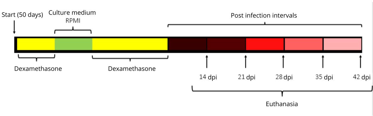
The animals were subjected to the immunodepression protocol with Dexamethasone (FARMADEX®), as performed by Chan et al. (2016), at a concentration of 50mg/kg/day. Thus, the protocol lasted twelve days, where the animals received daily doses via intraperitoneal route, starting three days before infection by the Zika virus, and ending during the nine days after infection. Immunodepression was confirmed after the global leukocyte count in the Neubauer chamber, where a low index or even absence of circulating leukocytes in the peripheral blood of the mice used in the study was observed, when compared to untreated animals. On the third day of the experiment, the animals belonging to the infected groups received, with the aid of an insulin syringe, the Zika virus (ZIKV PE243) via intraperitoneal (IP) concentration of 1x10⁴ PFU suspended in 200 µL of RPMI 1640 culture medium supplemented with 2% Fetal Bovine Serum (FBS) and 1% Antibiotic (Penicillin/Streptomycin). The control groups received, by the same route, 200 µL of the culture medium supplemented with the same components at the same concentrations, but without the virus (Figure 1). ZIKV infection was confirmed by reverse transcription quantitative polymerase chain reaction (RT-qPCR) in the serum, as described by Da Silva et al. (2025).
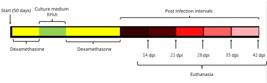
Pharmacological immunosuppression protocol with dexamethasone and ZIKV infection. Source: The author.
Study groups and crossing
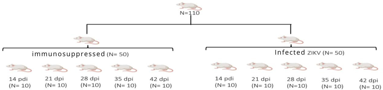
The 110 mice used in this study were distributed into 11 groups (n=10/group), including 1 water control group, 5 control groups (dexamethasone) and 5 infected groups, as illustrated below. After infection, one control group (dexa) and one infected group were euthanized each week, at 14, 21, 28, 35 and 42 days after infection (Figure 2).
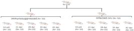
Illustration of the experimental design. Caption: N - total number of animals used in the experiment; Immunosuppressed - Animals treated with dexamethasone; ZIKV Infected - Groups of animals infected with Zika virus at a concentration of 1x10⁴ PFU. Source: The author.
Group I: Control group;
Group II: Immunocompromised control group with dexamethasone (14 days);
Group III: Immunocompromised group with dexamethasone and infected with ZIKV (14 days);
Group VI: Immunocompromised control group with dexamethasone (21 days);
Group V: Immunocompromised group with dexamethasone and infected with ZIKV (21 days);
Group VI: Immunocompromised control group with dexamethasone (28 days);
Group VII: Immunocompromised group with dexamethasone and infected with ZIKV (28 days);
Group VIII: Immunocompromised control group with dexamethasone (35 days);
Group IX: Immunocompromised group with dexamethasone and infected with ZIKV (35 days);
Group X: Immunocompromised control group with dexamethasone (42 days);
Group XI: Group immunosuppressed with dexamethasone and infected with ZIKV (42 days).
Weight of animals, submandibular-sublingual complex and Organosomatic Index (IOS)
The weight of the animals was measured daily during treatment using a precision analytical balance (BEL Mark 160/0.0001g). The weight of the submandibular-sublingual complex was measured at the time of organ collection using the same scale described above. The Organosomatic Index (IOS = {weight of the complex/body weight} x 100) was calculated from the weight obtained from the submandibular-sublingual complex and the animals.
Euthanasia and collection of the submandibular-sublingual complex
After the experimental time assigned to each group, the animals were weighed using a precision analytical balance and anesthetized intramuscularly with an anesthetic combination of xylazine (10mg/kg) and ketamine (115mg/kg), and subsequently received intraperitoneally with sodium thiopental (30mg/kg). After performing the reflex tests to stimuli and achieving the anesthetic plane, thoracotomy was performed with the animal positioned in dorsal decubitus, followed by cardiac puncture to obtain whole blood. The animal was then subjected to guillotining to certify rapid death, since after collecting a large volume of blood the individual went into hypovolemic shock. Subsequently, the submandullary-sublingual complex was collected. The complex was weighed using a precision oscillator (BEL Mark 160/0.0001 g), sectioned transversely and embedded in 10% buffered formalin (pH 7.2 and 0.01 M) for 48 hours.
Histological, histochemical and morphometric analysis
After fixation, the submandibular-sublingual complex was subjected to the paraffin embedding technique. The blocks obtained were cut in a rotary manual microtome (RM2255, Leica Biosystems) at a thickness of 3 μm. The histological sections were stained with hematoxylin and eosin (HE) and Masson’s trichrome. Digital photomicrographs were taken using an Olympus CX22 photomicroscope equipped with a “Moticam 2300 3.0M Pixel USB 2.0” digital camera capture system, and the images were used for morphometric analysis using ImageJ software.
For histopathological analysis, the HE-stained features were observed to verify: morphological aspects of the serous portion and mucosal portion according to the criteria of the national toxicology program, NIH - USA (https://ntp.niehs.nih.gov/atlas/nnl/alimentary-system/salivary-gland: presence of ductal lumen, ductal congestion, vacuolization of the ducts, vacuolization of the acini, presence of blood cells, quantity of connective and cellular tissue).
Morphometry was performed using images obtained using a 40x objective. The areas randomly selected in the histological sections (10 fields per salivary gland) were used to measure the total area of the serous acini, diameter and area of the lumen.
For the analysis of collagen fibers, Masson’s trichrome staining was performed. Ten fields were photomicrographed on each slide per animal, under 400X magnification, for measurement. After defining the color range through trial and error, the area representing collagen was selected. Then, a mask was created in the Image J program itself, providing the area and the percentage of areas that presented fibrosis in each field of the image. The Computerized Histophotometry technique was used (Dias et al. 2019). A total area of 30 × 104 μm2 was quantified for each group.
Fractal analysis
The microscopic images of the submandibular-sublingual complex were used to calculate the fractal dimension (FD) of the mucosal region using the box count method (Dbox). Briefly, the FD was calculated by covering the image with N(r) boxes, where N is the number of boxes and r is the length of one side of the box containing at least one point of the verified structure (Dos Santos et al. 2025).
The lacunarity value was described by the distribution of the lacunae in the verified structure. The image was covered by a series of classes, each class containing a number of boxes of different sizes and orientations (Dos Santos et al. 2025).
Statistical analysis
The statistical analysis was performed using the GraphPad Prism8® computer software, where the data were evaluated using nonparametric Kruskal–Wallis tests with Dunn’s post-hoc. The differences were considered statistically significant at a probability level of 5% (p<0.05).
RESULTS
Analysis of animal weight and submandibular-sublingual complex
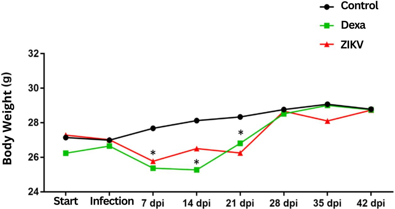
Animals in the 7 dpi Dexa and ZIKV groups showed significant weight loss compared to the control group. This weight loss can be attributed to the initial impact of ZIKV infection and dexamethasone treatment, both potentially causing metabolic stress or intense immune response. In the 14 dpi group, the weight loss trend continues for the Dexa and ZIKV groups, still showing significant differences compared to the control group. Dexamethasone, a corticosteroid, can induce muscle catabolism and weight loss, while a viral infection can reduce appetite and increase energy expenditure due to fever and immune response. In the 21 dpi group, both treated groups (Dexa and ZIKV) are still below the weight of the control group, but initially show signs of recovery. From 28 dpi onwards, a partial recovery of the animals’ weight is observed. The Dexa and ZIKV groups are close to the control group values. This indicates a recovery phase, possibly due to the organisms adapting to the treatment and/or infection.
At 35 dpi, only the ZIKV group shows a reduction in weight, which may indicate a late response or a prolonged effect of the viral infection that is still impacting the animals’ metabolism. After 42 dpi, all groups returned to body weights similar to the control group. This suggests that, by the end of the observation period, the initial adverse effects of the infection and dexamethasone treatments were overcome, allowing for a complete recovery.
Regarding the weight of the submandibular-sublingual complex, the animals in the ZIKV group at 21, 28 and 42 dpi had higher averages when compared to the control. Likewise, the Organosomatic Index of the submandibular-sublingual complex of all animals in the ZIKV groups showed higher averages when compared to the control (Table I).
Submandibular-sublingual complex weight and organosome index (OSI) of ZIKV-infected Balb/c mice at 14, 21, 28, 35, and 42 dpi.
Histopathological analysis
In the control group, the submandibular-sublingual complex presented a compound tubuloacinar gland, containing several lobules delimited by fibrous connective tissue stroma. The glandular parenchyma exhibits serous and mucous secretory units, as well as a well-developed duct system.
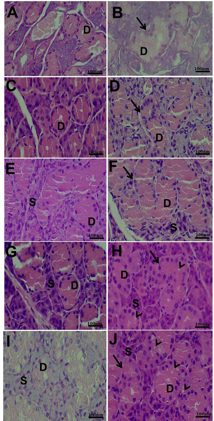
In the serous portion, more tortuous ducts and a smaller ductal lumen were observed in groups Dexa and ZIKV VII-XI (28, 35, 42 days), which may indicate the presence of congested ducts when compared to groups I-VI (control, 14, 21 days). In addition, groups X and XI (42 days) presented vacuolization of the ducts (Figure 4).
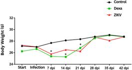
Body weight of animals during the treatment period: 7, 14, 21, 28, 35, 42 days post-infection (dpi). p<0.05.8.
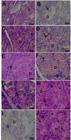
Photomicrograph of the serous portion of the submandibular-sublingual complex. a, c, e, g and i: ducts (D) and serous acini (S) of groups II-VI, respectively. 400x. b, d and f: More tortuous ducts (arrow), smaller ductal lumen of groups VII-IX, respectively. 400x. h and j: More tortuous ducts (arrow), smaller ductal lumen, and vacuolization of the ducts (arrowhead) in groups X and XI, respectively. 400x. Hematoxylin-eosin staining. Scale bar = 20μm.
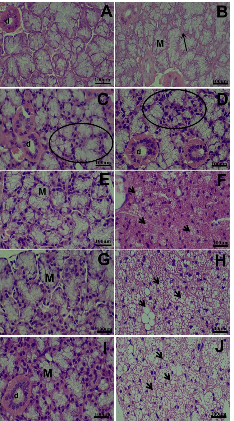
The mucosal portion of the submandibular-sublingual complex of animals in the experimental groups presented acini and intercalated, granular and striated ducts located inside the lobules. The mucous acini presented an increase in connective tissue in the interacinar space in groups II and III, an increase in the number of cells in groups IV-XI, in addition to intense vacuolization in groups VI-XI when compared to groups control, I-V (Figure 5).
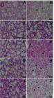
Photomicrograph of the mucosal portion of the submandibular-sublingual complex. a: acini (arrow) and ducts (d) in group II. b: increased connective tissue in the interacinar space in group III (arrow). c, d, and e: Increased cell number in groups IV, V, VI (circle). g and i: ducts (d) and mucous acini (M) in groups VIII and X. f, h and j: vacuolization in groups VII, IX, and XI. 400x. Hematoxylin-Eosin staining. Scale bar = 20μm.
Histochemical analysis
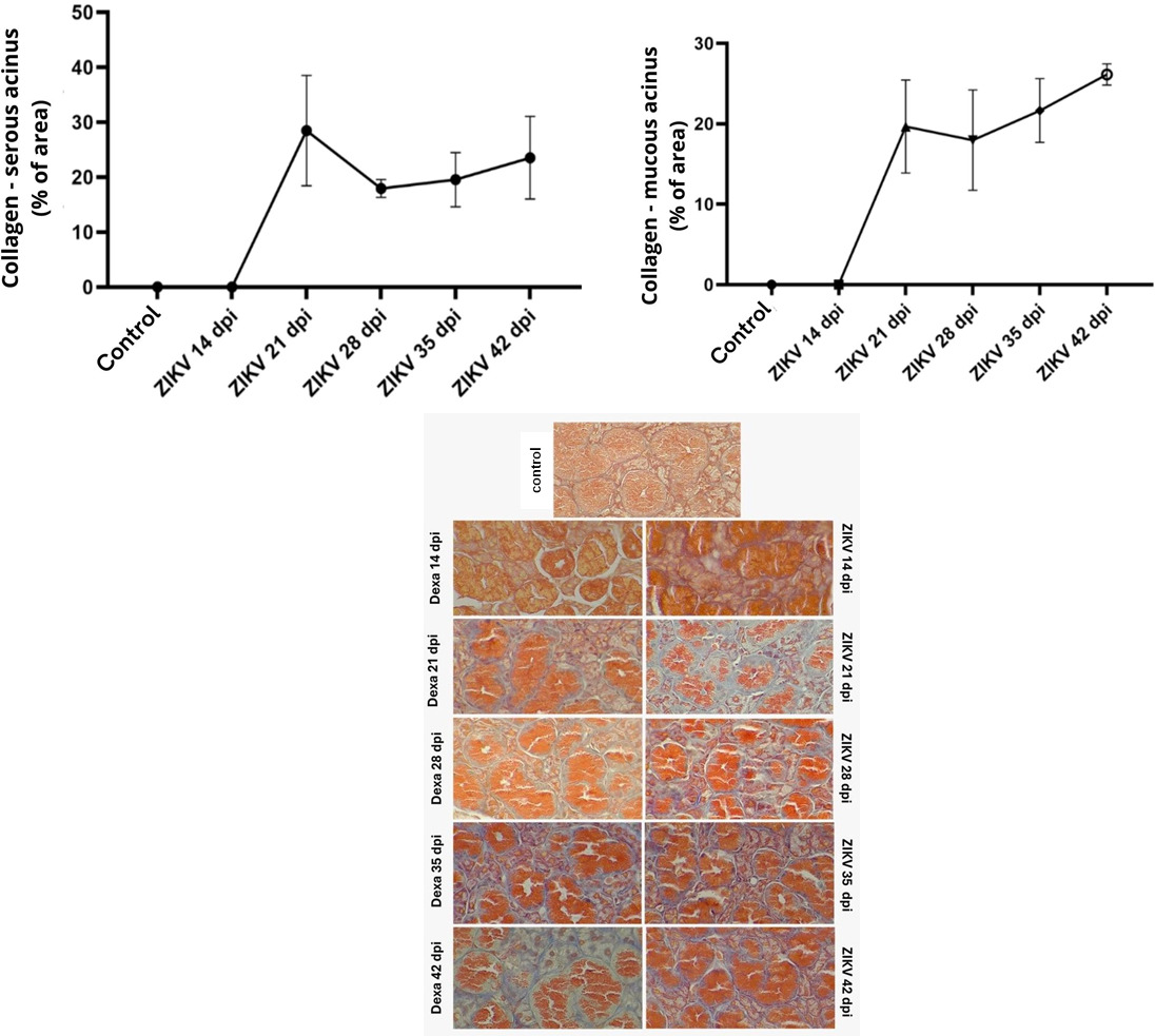
In the serous acini, the control groups and the ZIKV group at 14 dpi present minimal levels of collagen, practically zero. At 21 dpi, there is a sharp increase in the percentage of collagen, reaching approximately 30% of the area. This increase indicates a rapid onset of fibrosis in the serous acini after ZIKV infection. The percentage of collagen is reduced in the 28 dpi group, but still remains high, around 20-25% of the area. This suggests that the fibrosis process may be beginning to stabilize. In the 35 and 42 dpi groups, the amount of collagen remains relatively stable, with a tendency to increase until 42 dpi (Figure 6).
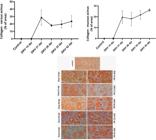
Quantification of the staining intensity of Masson’s Trichrome in the submandibular-sublingual complex of the experimental groups (p=0.001). a - Serous acini. Groups 21, 28, 35 and 42 dpi with greater staining intensity when compared to the control group and 14 dpi. b - Mucous acini. Groups 21, 28, 35 and 42 dpi with greater staining intensity when compared to the control group and 14 dpi.
In the graph of the mucous and serous acini, the control groups and the ZIKV group at 14 dpi showed the collagen levels minimal or null. There is a significant increase in the collagen percentage in the 21 dpi, rising to approximately 15-20% of the area. This demonstrates that ZIKV infection also induces fibrosis in the mucous acini, but to a lesser extent than in the serous acini initially. In the 28 dpi group, the collagen percentage remains high and stable, indicating that the fibrotic response is remaining constant after the initial peak. In the 35 and 42 dpi groups, there is a trend of continuous increase in the collagen percentage, especially at 42 dpi, where it reaches approximately 25-30% of the area. This indicates a continuous progression of the fibrotic process in the mucous acini over time (Figure 6).
Morphometric analysis
The morphometric analysis of the serous acini demonstrated a significant reduction in the lumen and an increase in the height of the epithelium in the ZIKV groups at 14, 21, 28, 35 and 42 dpi. There was no significant difference between the other groups (Table II).
Morphometric analysis of the serous acini of Balb/c mice infected with ZIKV at 14, 21, 28, 35 and 42 dpi.
The 14, 21, 28, 35 and 42 dpi groups showed an increase in the lumen of the mucous acinus and a decrease in the height of the epithelium in relation to the control group (Table III).
Morphometric analysis of the mucous acini of Balb/c mice infected with ZIKV at 14, 21, 28, 35 and 42 dpi.
Fractal Dimension (FD) analysis
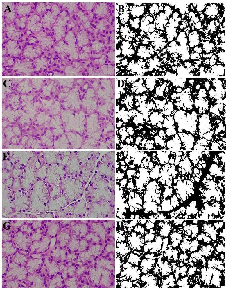
The FD of the 35 dpi and 42 dpi groups were superior to the FD of the Control group (Table IV and Figure 7). Lacunarity showed a statistically significant difference only in the 42 dpi group, demonstrating a lower value in the infected group (Table IV).
Fractal analysis and lacunarity of the mucous acini of Balb/c mice infected with ZIKV at 14, 21, 28, 35 and 42 dpi.
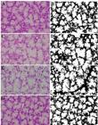
Photomicrographs and fractals of the mucosal region of the submandibular-sublingual complex of the experimental groups. a-b: Control group 35 days. c-d: Group 35 days post-infection. e-f: Control group 42 days. g-h: Group 42 days post-infection. Photomicrographs a, c, e and g decorated with HE. Images b, d, f and h processed using ImageJ software. AU 400X. Scale bar = 20μm.
DISCUSSION
In this study, we chose to use male Balb/C mice immunosuppressed with dexamethasone due to the proven efficiency of this protocol described in the study by Chan et al. (2016). Corticosteroids have been used as therapeutic agents on a large scale, as modulators or suppressors of inflammatory and immune responses, in humans and animals. Their effects on inhibiting inflammation are profound and rapid, with a decrease in the vascular phase of inflammation through vasoconstriction (Claman 1975). Mice have been the experimental model of choice for most GS studies due to their ease of acquisition and the well-established institutional protocols surrounding their use (Schapiro & Everitt 2006). The immunodeficiency induced prior to infection was implemented with the purpose of enhancing the viral cycle, allowing greater viral replication and, consequently, a higher viral load. This condition facilitates the detection and confirmation of infection, in addition to intensifying the pathological alterations associated with the infectious agent.
The data show that the animals treated with ZIKV and dexamethasone presented a significant weight loss in the first 21 days post-infection (dpi), compared to the control group. This weight loss can be attributed to the impact of ZIKV and treatment with dexamethasone, which can affect the metabolism of the animals. However, from 28 dpi, the weights of the animals became similar between the groups, with a new visualization observed only in the ZIKV group at 35 dpi. The similarity in weights was resumed at 45 dpi.
Body weight loss was also reported in Balb/c mice infected with Mayaro fever in studies by Oliveira et al. (2021) at 15 and 45 days post-infection (dpi) by ZIKV, which is another piece of data that corroborates the experimental model, since in these analyses it was also decided to infect the animals with an arbovirus and after establishing immunosuppression in this group, the peaks in body weight gain were at 28 and 42 days post-infection (dpi).
Regarding the weight of the submandibular-sublingual complex, animals infected with ZIKV presented higher average weight and organosomatic index (OSI) on days 21, 28 and 42 dpi, when compared to the control. This suggests that ZIKV infection can cause hypertrophy or inflammation in the submandibular and sublingual glands. The statistically significant data (p<0.05) reinforce the relevance of these observations.
Immunodepression by dexamethasone made mice more susceptible to ZIKV infection, where a significant increase in the relative weight of the submandibular gland was observed in infected groups. This is justified by the weight loss of the infected animal and the weight of the gland remained the same, thus having an increase in the relative weight of this organ. The increase in glandular weight in this study may also be caused by edema caused by Zika virus infection.
The submandibular-sublingual complex of Balb/C mice was found to be covered by a connective tissue capsule containing lobules and composed of their acinar and mucous portions. The results described for the rodent in question coincide with those reported for the animals studied by Frey et al. (2011) and Mohammadpour (2009). In the histopathological analysis, the control group presented a normal glandular structure, with well-defined lobules and a well-developed duct system. In the infected groups, especially at 28, 35 and 42 dpi, the ducts were more tortuous and with reduced lumen, decreasing possible congestion. In addition, vacuolization of the ducts was noted in groups X and XI (42 dpi), which may cause cellular damage or accumulation of matter in the ducts.
In the mucosal portion of the submandibular-sublingual complex, infected animals showed increased connective tissue in the interacinar space and an increase in the number of cells, in addition to intense vacuolization compared to controls. These findings indicate that ZIKV infection can induce significant structural changes in both the serous and mucosal portions of the salivary glands. Regarding the presence of vacuolization in the submandibular-sublingual complex, this data may be the result of lesions in the cell membrane (Oliveira et al. 2017), the presence of ZIKV in the organ, as has been reported in the literature in other studies. Studies show that small vacuoles can occur in acute metabolic diseases. Large vacuoles, on the other hand, are caused by slowly developing toxic or viral lesions. In Oliveira et al. (2017) the submandibular gland of mice treated with botulinum toxin (BTXA) also showed vacuolization in the cytoplasm of serous acinar cells, of varied size and poorly stained by hematoxylin and eosin.
A morphometric analysis of the serous acini demonstrated a significant reduction in the lumen and an increase in the height of the epithelium in the infected groups at 14, 21, 28, 35 and 42 dpi, indicating an adaptive response to the infection. In the mucous acini, there was a decrease in the area on days 14, 28 and 35 dpi, while on days 21 and 42 dpi there was a significant increase. The infection also caused an increase in the lumen of the mucous acini and a decrease in the height of the epithelium, altering the glandular structure due to ZIKV.
In the sublingual portion, the mucosal component showed a well-defined lumen, an increase in the area of this structure in relation to the control group, an increase in the diameter of the acini and also an increase in connective tissue in the interacinar space due to the fact that the animal is immature and therefore there is the presence of a large amount of interstitial tissue between the lobes and lobules in the first few days. This increase in the interactive space was also described by Shan et al. (2013) in their work with botulinum toxin type A.
Masson’s Trichrome staining revealed a significant increase in collagen concentration in the serous and mucous acini regions in the infected groups at 21, 28, 35, and 42 dpi, compared to the control and the 14 dpi group. This suggests that ZIKV infection can induce fibrosis in the salivary glands, evidenced by the increase in collagen deposition. Collagen fibers play a vital role in maintaining structural integrity and also in determining tissue function. In the literature, this change is related as a result of aging and tissue repair (Singh et al. 2023).
The fractal dimension (FD) of the 35 dpi and 42 dpi groups was higher than the control group, providing greater structural complexity in the mucous acini of the infected animals. Lacunarity showed a significant difference only at 42 dpi, with lower values in the infected group, indicating less uniformity or more empty spaces in the glandular structure. These changes in fractal dimension and lacunarity reinforce the idea that ZIKV infection causes significant structural changes in the salivary glands. Irregular structures, when evidenced by the fractal dimension method, are quantified in relation to their degree of complexity, so that higher fractal dimension values are related to more complex images (Dos Santos et al. 2025). Based on the data described, we suggest that ZIVK may alter the morphology of these acini, increasing the complexity and, consequently, the irregularity of these structures.
In the context, the use of saliva as a diagnostic tool offers an opportunity for simpler and more efficient molecular and proteomic analyses/diagnoses of ZIKV (Khurshid et al. 2019). Musso et al. (2015) investigated the use of saliva as an alternative sample for the routine detection of ZIKV RNA. Their results showed that ZIKV was detected more frequently in saliva than in blood: among the 182 patients analyzed, with both types of samples collected, 35 (19.2%) were positive in saliva and negative in blood, while 16 (8.8%) were positive in blood and negative in saliva. The use of saliva samples increased the molecular detection rate of ZIKV during the acute phase of the disease, but did not extend the detection window of ZIKV RNA.
Nucleases exhibit greater efficiency in degrading RNA than proteases do in degrading proteins. As a result, ZIKV proteins/peptides may persist for a longer period compared with ZIKV RNA in saliva, even during the recovery phase of the infection. This has the potential to simplify disease diagnosis. It is important to highlight that protein degradation in saliva is influenced by proteolytic processes caused by the presence of bacteria and host-derived enzymes in the oral cavity (Zuanazzi et al. 2017, Khurshid et al. 2019). Thus, the use of human glandular secretions, particularly saliva, as a diagnostic tool provides an opportunity for efficient solutions in ZIKV proteomic analysis/diagnosis.
CONCLUSIONS
Zika infection caused changes in the microscopic structure of the submandibular-sublingual salivary complex of animals in the experimental groups, providing evidence of morphological and fibrotic alterations in the glandular architecture associated with ZIKV infection. These alterations have important implications for understanding the effects of ZIKV on the salivary glands and other organs, highlighting the need for further studies to investigate the underlying mechanisms and possible therapeutic interventions.
Acknowledgements
The authors would like to express their gratitude to the Federal University of Pernambuco (UFPE), specifically the annex of the Anatomy Department and the Department of Histology and Embryology for providing the specimens and all the structure for analysis and study of the results. The authors would also like to thank the Federal Rural University of Pernambuco (UFRPE) for the professional partnership obtained in order to obtain a successful work.
References
-
CASTELLANOS JE. 2017. Could the use of saliva improve the zica diagnosis challenge? Contributions from a proteomics perspective. J Dent Res 96(10): 1076-1077. https://doi.org/10.1177/0022034517723327.
» https://doi.org/10.1177/0022034517723327 -
CHAN JF ET AL. 2016. Zika virus infection in dexamethasone-immunosuppressed mice demonstrating disseminated infection with multi-organ involvement including orchitis effectively by recombinant type I interferons. EBioMedicine 14: 112-122. DOI: 10.1016/j.ebiom.2016.11.017.
» https://doi.org/10.1016/j.ebiom.2016.11.017 -
CLAMAN HN. 1975. How corticosteroids work. J Allergy Clin Immunol 55(3): 145-151. https://doi.org/10.1016/0091-6749(75)90010-X.
» https://doi.org/10.1016/0091-6749(75)90010-X -
DA SILVA ET AL. 2025. Parental infection in males causes alterations in the locomotor system in BALB/c mice fetuses. REAS 25(8): e21307. https://doi.org/10.25248/REAS.e21307.2025.
» https://doi.org/10.25248/REAS.e21307.2025 -
DIAS FCR ET AL. 2019. Hydroalcoholic extract of Pfaffia glomerata alters the organization of the seminiferous tubules by modulating the oxidative state and the microstructural reorganization of the mice testes. J Ethnopharmacol 233: 179-189. https://doi.org/10.1016/j.jep.2018.12.047.
» https://doi.org/10.1016/j.jep.2018.12.047 -
DOS SANTOS AVF ET AL. 2025. Complex shape markers can detect alterations in the spatial distribution of cell nuclei in human lung squamous cell carcinoma: a useful tool for automatic analysis? Histochem Cell Biol 163(1): DOI: 10.1007/s00418-024-02336-3.
» https://doi.org/10.1007/s00418-024-02336-3 -
DUFFY MR ET AL. 2009. Zika Virus Outbreak on Yap Island, Federated States of Micronesia. NEJM 360: 2536-2543. https://doi.org/10.1056/NEJMoa0805715.
» https://doi.org/10.1056/NEJMoa0805715 -
ENFISSI A, CODRINGTON J, ROOSBLAD J, KAZANJI M & ROUSSET D. 2016. Zika virus genome from the Americas. Lancet 387(10015): 227-228. https://doi.org/10.1016/S0140-6736(16)00003-9.
» https://doi.org/10.1016/S0140-6736(16)00003-9 -
FREY R, MARKGRAF U & HOFMANN RR. 2011. Evolutionary morphology of the zygomatic gland and lacrimal bulla in roe deer Capreolus capreolus Linnaeus, 1758 (Mammalia, Cervidae). Zoologischer Anzeiger 240: 181-195. https://doi.org/10.1078/0044-5231-00020.
» https://doi.org/10.1078/0044-5231-00020 -
KHURSHID Z, ZAFAR M, KHAN E, MALI M & LATIF M. 2019. Human saliva can be a diagnostic tool for Zika virus detection. J Infect Public Health 12(5): 601-604. https://doi.org/10.1016/j.jiph.2019.05.004.
» https://doi.org/10.1016/j.jiph.2019.05.004 -
LETA S ET AL. 2018. Global risk mapping for major diseases transmitted by Aedes aegypti and Aedes albopictus. Int J Infect Dis 67: 25-35. https://doi.org/10.1016/j.ijid.2017.11.026.
» https://doi.org/10.1016/j.ijid.2017.11.026 -
LOPES N, NOZAWA C & LINHARES REC. 2014. Características gerais e epidemiologia dos arbovírus emergentes no Brasil. RPAS 5(3): 55-64. https://doi.org/10.5123/S2176-62232014000300007.
» https://doi.org/10.5123/S2176-62232014000300007 - MOHAMMADPOUR AA. 2009. Investigations on the shape and size of molar and zygomatic salivary glands in shorthair domestic cats. Bulgarian J Vet Med 12(4): 221-225. Available at: https://profdoc.um.ac.ir/paper-abstract-1011783.html.
-
MUSSO D, ROCHE C, NHAN TX, ROBIN E, TEISSIER A & CAO-LORMEAU VM. 2015. Detection of Zika virus in saliva. J Clin Virol 68(29): 5-53. https://doi.org/10.1016/j.jcv.2015.04.021.
» https://doi.org/10.1016/j.jcv.2015.04.021 -
OLIVEIRA AA ET AL. 2021. Manifestações orais de arboviroses com ênfase em dengue, zica e chikungunya: revisão de literatura. Arch Health Invest 10(2): 323-328. https://doi.org/10.21270/archi.v10i2.4677.
» https://doi.org/10.21270/archi.v10i2.4677 -
OLIVEIRA JB, EVÊNCIO-NETO J & BARATELLA-EVÊNCIO L. 2017. Histological and immunohistochemical findings of the action of botulinum toxin in salivary gland: systematic review. Braz J Biol 77(2): 251-259. https://doi:10.1590/1519-6984.11115.
» https://doi.org/10.1590/1519-6984.11115 -
SCHAPIRO SJ & EVERITT JI. 2006. Preparation of Animals for Use in the Laboratory: Issues and Challenges for the Institutional Animal Care and Use Committee (IACUC). Diário Ilar 47(4): 370-375. https://doi.org/10.1093/ilar.47.4.370.
» https://doi.org/10.1093/ilar.47.4.370 -
SHAN XF ET AL. 2013. Botulinum toxin A inhibits salivary secretion of rabbit submandibular gland. Int J Oral Sci 5: 217-223. https://doi.org/10.1038/ijos.2013.82.
» https://doi.org/10.1038/ijos.2013.82 -
SINGH D, RAI V & AGRAWAL DK. 2023. Regulation of Collagen I and Collagen III in Tissue Injury and Regeneration. Cardiol Cardiovasc Med 7(1): 5-16. https://doi.org/10.26502/fccm.92920302.
» https://doi.org/10.26502/fccm.92920302 -
SIQUEIRA WL, SALIH E, WAN DL, HELMERHORST EJ & OPPENHEIM FG. 2008. Proteome ofhuman minor salivary gland secretion. J Dent Res 87: 445-450. https://doi.org/10.1177/154405910808700508.
» https://doi.org/10.1177/154405910808700508 -
ZUANAZZI D, ARTS EJ, JORGE PK, MULYAR Y, GIBSON R, XIAO Y, BRINGEL DOS SANTOS M, MACHADO MAAM & SIQUEIRA WL. 2017. Postnatal Identification of Zika Virus Peptides from Saliva. J Dent Res 96: 1078-1084. https://doi.org/10.1177/0022034517723325.
» https://doi.org/10.1177/0022034517723325
Edited by
-
Handling editor
Guilherme Baldo
The authors confirm that the data supporting the findings of this study are available within the article.