In vitro evaluation of Zymomonas mobilis UFPEDA 363 as a probiotic: safety profile, antagonism, and simulated gastrointestinal resistance
Abstract
The growing interest in non-conventional probiotics has led to investigations of new candidates with health promoting properties. This study aimed to evaluate the in vitro probiotic potential of Zymomonas mobilis UFPEDA 363. The strain exhibited exponential growth, reaching 8.8 log CFU/mL within 24 h, and demonstrated strong acid tolerance with survival rates of 54% at pH 3.0, 99% at pH 3.5, and 100% at pH 7. It exhibited notable surface properties, including auto-aggregation (64%), co-aggregation with Staphylococcus aureus and high hydrophobicity in the presence of organic solvents. The strain also tolerated bile salts, maintaining 96-100% survival and viability above log 8 CFU/mL under simulated gastrointestinal conditions. Antagonistic activity assays revealed effective inhibition of Salmonella enteritidis and Escherichia coli. Regarding safety, the strain was susceptible to most clinically relevant antibiotics, such as tetracycline and ampicillin, and it did not exhibit hemolytic or gelatinase activity. These results indicate the acid and bile salt resistance, antimicrobial potential and safety profile of Zymomonas mobilis UFPEDA 363, supporting its candidacy as a probiotic microorganism. Further in vivo studies are needed to confirm its efficacy and explore applications in functional foods or therapeutic formulations aimed at promoting human health.
Key words
Antimicrobial activity; Bacterial adhesion; Gastrointestinal survival; Gut microbiota
INTRODUCTION
Probiotics are live microorganisms that, when administered in adequate quantities, play a significant role in maintaining the host’s health, being able to reestablish and preserve the microbial balance in the gastrointestinal tract. By reducing pathogen populations and promoting the growth of beneficial bacteria, probiotics strengthen intestinal mucosal defenses, thereby diminishing susceptibility to both intestinal and systemic infections (FAO 2001).
The demand for probiotic supplements has increased significantly as a natural alternative to antibiotics. The most widely used probiotic microorganisms include lactic acid bacteria (LAB) such as Lactobacillus spp., Enterococcus spp., and Pediococcus spp., as well as Bifidobacterium spp., Bacillus spp., and yeast species like Saccharomyces boulardii (Zaghari et al. 2020, Ramlucken et al. 2020).
Although there are characterized and commercially available probiotic strains, the search for new probiotic strains is justified by the potential to discover promising candidates that offer health benefits for humans. Furthermore, from a technological point of view, these strains should exhibit resistance to the conditions during product formulation, ensuring good viability. Consequently, the application of probiotics as constituents of a less aggressive alternative therapy becomes increasingly appealing, considering their importance in promoting a healthy and balanced lifestyle.
Among the potential candidates, Zymomonas mobilis stands out as a Gram-negative bacterium known for unique metabolic properties and efficient fermentative capacity. It is generally recognized as safe (GRAS) by the Food and Drug Administration (FDA) and is endorsed by Bulletin 495 of the International Dairy Federation, issued in 2018, affirming its use for human consumption.
Studies suggest that Z. mobilis strains may exhibit both probiotic and prebiotic characteristics and are capable of stimulating the immune system (Azerêdo et al. 2010, Almo et al. 2023). This bacterium demonstrated inhibitory action against various pathogenic microorganisms, which may be attributed to its production of bacteriocins (Lima et al. 2011) or the generation of acidic metabolites such as propionate and acetate (Rutkis et al. 2022).
In this context, Z. mobilis has emerged as a promising alternative due to its unique physiological traits and demonstrated benefits in various studies. Additionally, Z. mobilis is capable of producing bacteriocin’s proteinaceous substances with antimicrobial properties that specifically target pathogens, including Escherichia coli and Salmonella spp. (Lima et al. 2011). The dual role of organic acid production and bacteriocin synthesis enhances this strain’s potential as an effective probiotic capable of promoting gut health and protecting against infections. Beyond its antimicrobial properties, other studies have reported that Zymomonas mobilis may influence cholesterol metabolism, potentially aiding in cholesterol reduction.
One critical challenge for any probiotic strain is its ability to survive transit through the gastrointestinal tract (Ayyash et al. 2021). The acidic conditions of the stomach and the presence of bile in the intestines can be lethal to many bacteria. Therefore, a potential probiotic strain must withstand the adversities of the digestive tract and be capable of colonizing the intestine to ensure beneficial health effects (Saad 2006). To achieve this, the bacteria must demonstrate resistance to acidic pH, bile, and pancreatic juice. ANVISA recommends that the characteristics of probiotic microorganisms in preparations should be resistant to storage conditions, maintain an adequate concentration of cells (108 CFU/g) and ensure the viability of the microorganism so that it can reach the target and confer benefits to animal health (Clavijo & Flórez 2018).
Thus, studies involving probiotic strains of Z. mobilis can provide valuable insights for developing new products that offer additional benefits to human health. The aim of the present study was to investigate the probiotic potential of Z. mobilis UFPEDA 363 in vitro, encompassing antimicrobial activity, cell surface characteristics, and the evaluation of tolerance to simulated gastrointestinal conditions.
MATERIALS AND METHODS
Bacterial strain and growth conditions
Z. mobilis UFPEDA 363, previously isolated from sugarcane juice in Pernambuco and deposited in the culture collection of the Department of Antibiotics of Federal University of Pernambuco (UFPEDA), was grown in SSDL broth (20 g/L glucose, 5 g/L yeast extract, pH 6.8) at 30°C for 48 h. For antimicrobial activity assays, the following pathogenic bacteria were used: Staphylococcus aureus UFPEDA 02, Escherichia coli UFPEDA 224, Pseudomonas aeruginosa UFPEDA 416 and Salmonella enteritidis UFPEDA 414. The pathogenic strains were cultured on nutrient agar at 37°C for 24 h.
Microbial growth kinetics
Z. mobilis UFPEDA 363, freshly grown, was inoculated into a flask containing 250 mL of SSDL broth at 30°C and samples were taken at 3h intervals during 48 h. The bacterial count was given as CFU/mL and the assay was performed in triplicate.
Evaluation of probiotic properties in vitro
Acid tolerance
Z. mobilis UFPEDA 363 was inoculated in SSDL medium with pH adjusted to 2.0; 2.5; 3.0; 3.5 and 7.0 incubated at 30°C. One hundred microliter samples were taken following different exposure time intervals (1, 2, 3 and 4 h). Serial dilutions were performed and the number of viable cells was estimated as CFU/mL. The methodology used was that of Śliżewska & Chlebicz-Wójcik (2020) with modifications.
Tolerance to bile
Bile tolerance was assessed according to the method described by Berebon et al. (2019) with some modifications. Z. mobilis UFPEDA 363 was inoculated in SSDL medium at 30°C for 48 h, after inoculated in SSDL with addition of 0.1%, 0.3%, 0.5% and 1.0 % (w/v) of bile salt and incubated at 30°C. One milliliter samples were taken following different exposure time intervals (1, 2, 3 and 4 h), serial dilutions were performed and the number of viable cells was estimated as CFU/mL.
Aggregation capacity
Zymomonas mobilis UFPEDA 363 cells were harvested from a 24 h SSDL broth culture, washed with PBS (pH 7.2), and resuspended to measure initial absorbance (At0) at 625 nm. After incubation at 30°C for 3.5 h and 24 h, supernatants were removed to measure absorbance (ATs).
Autoaggregation (A%) was calculated as: A% = 1 – (ATs / At0) × 100, with ≥ 40%, considered high, 10–40%, moderate, and ≤ 10%, weak (Wang et al. 2010). For co-aggregation, Z. mobilis UFPEDA 363 and pathogen suspensions were mixed, incubated for 24 h, and absorbance measured at 3 h, 5 h, and 24 h. Co-aggregation (CoA%) was calculated as: CoA% = ODTOT – ODs / ODTOT × 100, where ODTOT is the initial absorbance and ODs is the absorbance at specific times (Scillato et al. 2021).
Hydrophobicity
The hydrophobicity of Z. mobilis UFPEDA 363 strain was assessed using xylene (nonpolar), chloroform (acidic), and ethyl acetate (basic) solvents. Cells from a fresh culture were centrifuged, washed with PBS, and resuspended to measure initial absorbance (A0) at 625 nm. Equal volumes of cell suspension and solvent were mixed, vortexed for 2 min, and left for 3 h to separate into organic and aqueous phases. Absorbance of the aqueous phase (AT) was measured, and hydrophobicity (H%) was calculated as: H% = (1 – AT / A0) × 100 (Lee et al. 2017).
Survival of Z. mobilis UFPEDA 363 when exposed to in vitro simulated gastrointestinal conditions
The simulated gastric fluid (SGF) and intestinal fluid (SIF) were prepared according to the method described by Farias et al. (2019). One milliliter of Z. mobilis UFPEDA 363 suspension containing approximately 1 × 10¹³ CFU/mL was mixed with 9 mL of SGF (NaCl: 0.73 g/L; KCl: 0.05 g/L; pepsin: 0.3 g/L pH 3.0) and incubated at 30 °C in the shaker at 150 rpm during 120 min. After 2 h, the intestinal fluid (6.0 g /L NaCL, 0.22 g/L CaCl2, 1.0386 g/L NaHCO3 0.22 g/L KCl, 1.0 g/L pancreatin and 3.0 g/L bile salts, pH 6.8) was added and incubated at 30 °C in the shaker at 150 rpm during 180 min. Then, viability was determined by the plate counting method and survival rate was calculated.
Antimicrobial activity against pathogens
The antagonistic activity of the Z. mobilis UFPEDA 363 strain was evaluated using the agar spot method (Lima et al. 2011). A 10 µL aliquot of strain (108 CFU/mL), freshly grown, was inoculated into four spots in the Petri dish containing SSDL agar and incubated at 30°C for 48 h. Next, 10 mL of semi-solid Müeller-Hinton agar medium containing 107 CFU/mL of each test microorganism: Salmonella enteritidis UFPEDA 414, Escherichia coli UFPEDA 224, Staphylococcus aureus UFPEDA 02 and Pseudomonas aeruginosa UFPEDA 416, were poured onto the plate. The plates were incubated at 37°C for 24 h. Antagonistic action was assessed by the formation of a protective halo with a diameter > 10 mm.
Safety aspects
Antibiogram
Z. mobilis UFPEDA 363 strain was tested for antibiotic susceptibility profile with a standard disk diffusion assay according to the CLSI (2019) protocol. Different antibiotics were used as followed: Amikacin (30 µg), Ampicillin (10 µg), Azithromycin (15 µg), Aztreonam (30 µg), Cefuroxime (30 µg), Ciprofloxacin (5 µg), Penicillin (10 µg), Polymyxin (300 µg) and Tetracycline (30 µg). The antibiotic susceptibility of Z. mobilis UFPEDA 363 was evaluated by measuring the diameter of growth inhibition zones and classified as susceptible (S) or resistant (R).
Hemolysis test
The hemolysis test was performed by initially cultivating Zymomonas mobilis UFPEDA 363 in liquid SSDL medium for 18 h at 30°C. The microorganism was then inoculated onto Petri dishes containing 10 mL of Blood Agar medium and incubated at 30°C for 48 h. Hemolysis was assessed by observing the presence or absence of a color change in the medium. Staphylococcus aureus UFPEDA 02 was used as a positive control (Süle et al. 2022).
Gelatinase test
The gelatinase detection test was conducted in a modified SSDL medium, supplemented with gelatin (15%), glucose (12%), and yeast extract (5%). Z. mobilis UFPEDA 363 was inoculated into a gelatin tube and incubated at 30°C for five days and subsequently refrigerated for 20 min. Staphylococcus aureus was used as a positive control, while Escherichia coli was employed as a negative control. Tubes that remained liquid after refrigeration were considered positive for gelatinase production (Pereira et al. 2009).
Statistical analysis
The data were analyzed using statistical parameters, including standard deviation and confidence intervals, analysis of variance (ANOVA), and Tukey’s test for mean comparison at a 5% significance level. Statistical analysis was performed using Origin for Windows 11, Paired Comparison Plot version 3.7, and Past version 4.03.
RESULTS
Growth kinetics
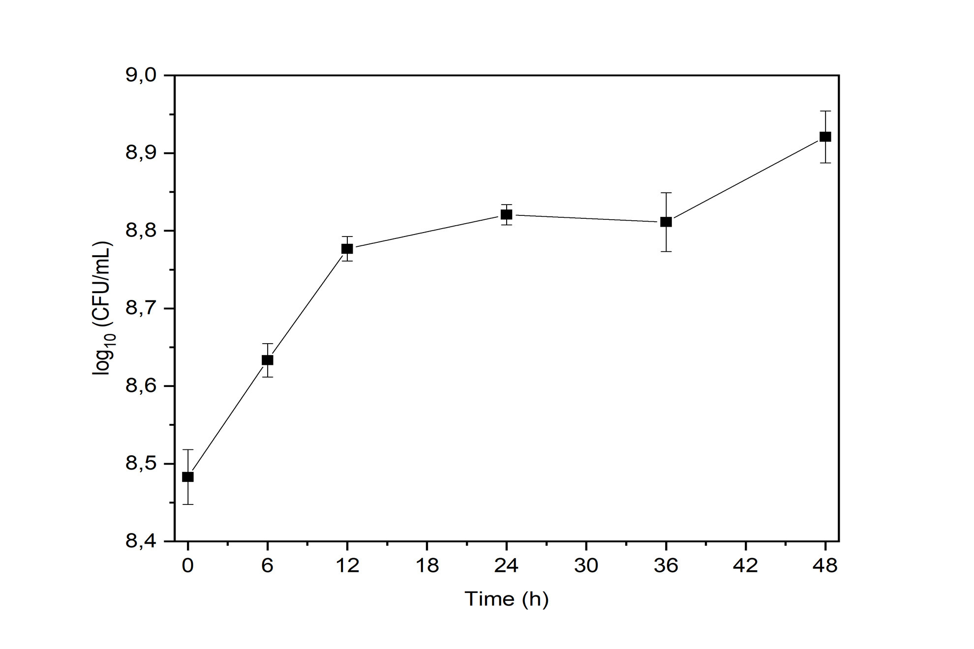
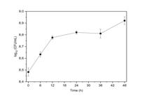
The Zymomonas mobilis UFPEDA 363 strain showed exponential growth, reaching 8.8 ± 0.01 log CFU/mL after 24 h. This strain remained in the stationary phase from 24 h to 42 h, remaining at 8.7 ± 0.01 log CFU/mL, not reaching its death phase during 48 h of testing (Figure 1).
In vitro probiotic properties
Tolerance to acids and bile salts
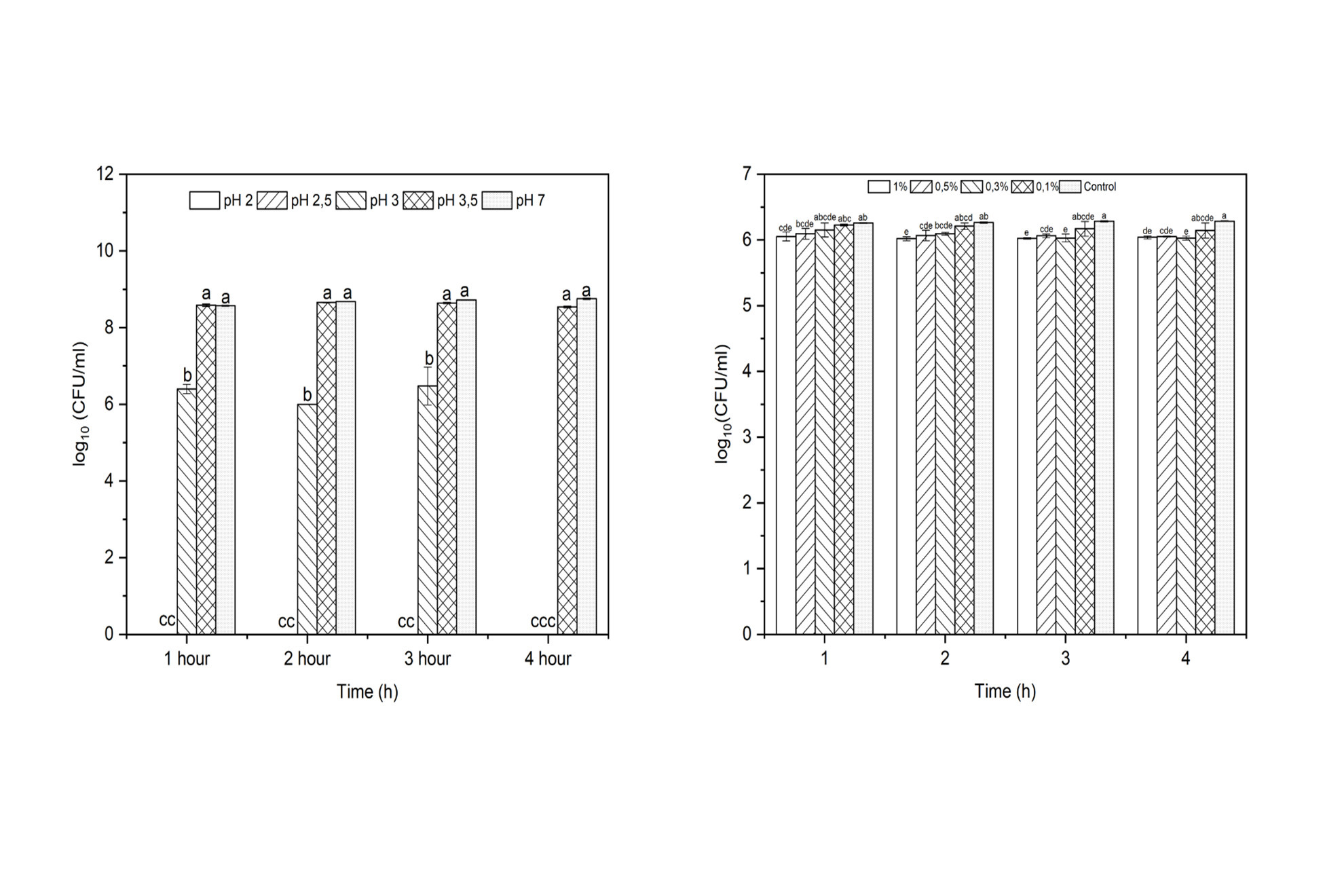
The acid tolerance of Z. mobilis UFPEDA 363 was assessed by exposing the strain to pH levels ranging from 2.0 to 7.0. No growth was observed at pH 2.0 and 2.5, whereas at pH 3.0, the strain retained 54% viability after 3 h of exposure. At pH 3.5 and 7.0 (control condition), survival rates were 99% and 100%, respectively (Figure 2a). These results indicate that Z. mobilis UFPEDA 363 can withstand moderately acidic conditions, an essential feature for probiotics intended for gastrointestinal applications.
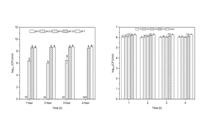
Tolerance of Z. mobilis UFPEDA 363 during exposition of different pH (a) and bile salt (b). (a) Means labeled with “a” within the same row are not significantly different (P > 0.05), while means labeled with “b” and “c” across different groups indicate a significant difference (P < 0.05). (b) Statistical analysis was performed using one-way ANOVA. Measures with “a, b, c, d, e” with the same letters on the same line show no significant difference (P > 0.05); e, c, d, e: averages for different groups show a significant difference (P > 0.05).
The strain exhibited high survival rates, ranging from 96% to 100% after 4 h of exposure (Figure 2b). Statistical analysis revealed a significant difference only when the 1.0% bile salt concentration was compared to the control group. However, this difference was minimal, with a reduction of less than 0.28 log CFU/mL, indicating that the strain maintains its viability even under higher bile salt concentrations typically encountered in the human intestinal tract.
Aggregation
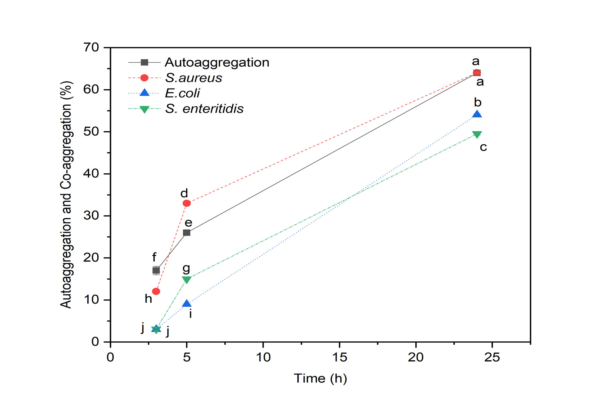
The ability of Zymomonas mobilis UFPEDA 363 to auto-aggregate reached 64% after 24 h, indicating strong intercellular adhesion (Figure 3). Additionally, co-aggregation assays revealed that the strain effectively interacted with Staphylococcus aureus (64%), Escherichia coli (53%), and Salmonella enteritidis (49%).
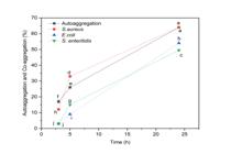
Percentage of auto-aggregation and co-aggregation of Zymomonas mobilis UFPEDA 363 in interaction with pathogenic bacteria. Comparison of the auto aggregation capacity of Zymomonas mobilis UFPEDA 363 (■) and its co-aggregation with (●) Staphylococcus aureus, (▲) Escherichia coli, and (▼) Salmonella enteritidis. Error bars represent the standard deviations of the mean values from three independent experiments.
Hydrophobicity
The hydrophobicity assessment of Zymomonas mobilis UFPEDA 363 revealed significant differences in adhesion capacity depending on the solvent used. The strain exhibited 93.6% hydrophobicity with chloroform, 85.2% with ethyl acetate, and 50.79% with xylene (Table I).
Survival of Zymomonas mobilis under simulated gastrointestinal conditions
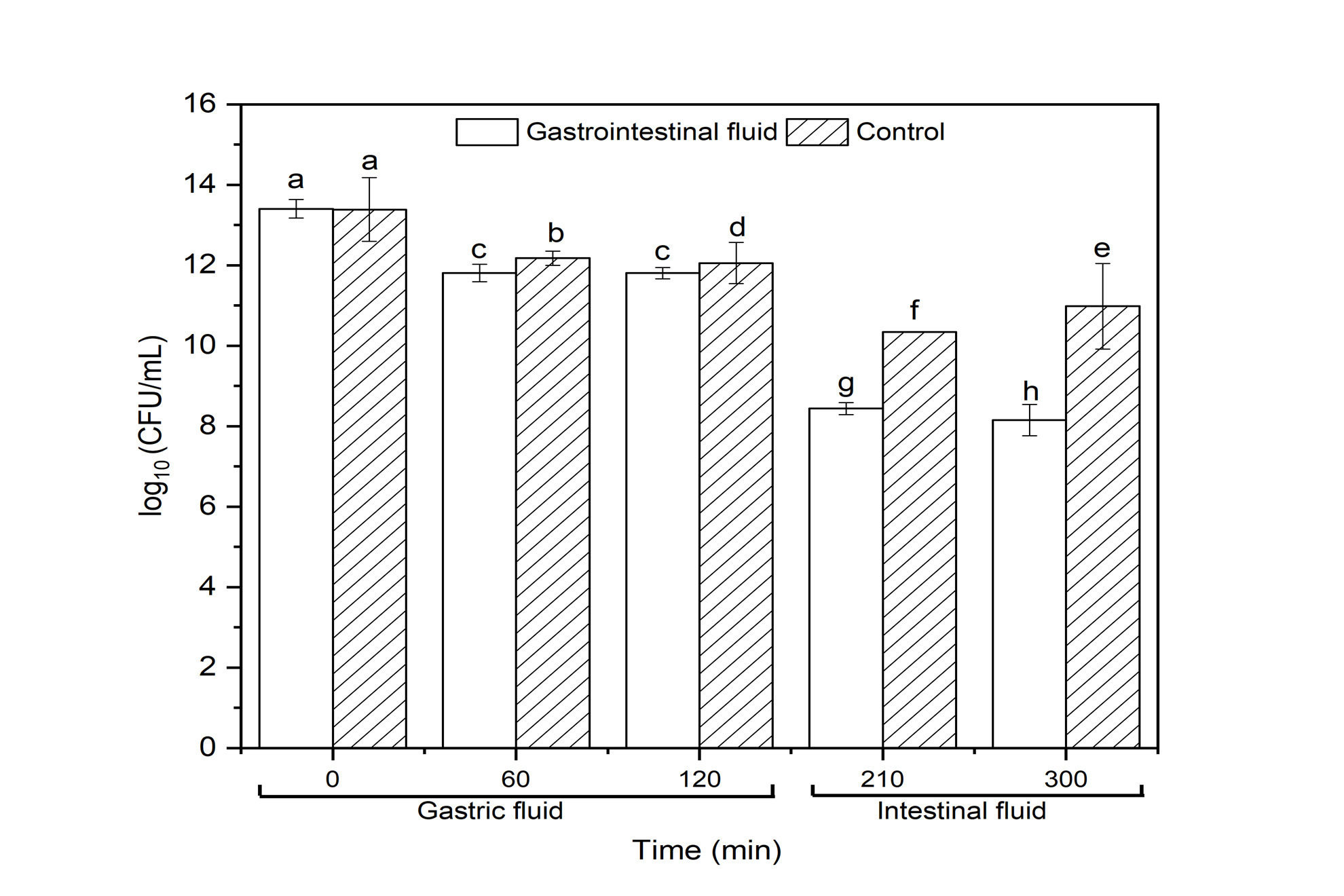
The in vitro evaluation of Zymomonas mobilis UFPEDA 363 in simulated gastrointestinal fluid demonstrated its ability to survive under conditions representative of the human digestive system. At time zero, immediately after inoculation into gastric initial bacterial count was 13.4 log CFU/mL. Over the first 120 min of incubation, the population decreased to 11.8 log CFU/mL, indicating that the strain remained viable despite exposure to acidic gastric conditions.
Following an additional 90 min in simulated intestinal fluid (totaling 210 min of incubation), the count further declined to 8.44 log CFU/mL, reflecting the progressive yet controlled reduction in viability. After another 180 min in simulated intestinal conditions (totaling 300 min of incubation), the bacterial count stabilized at 8.15 log CFU/mL, as shown in Figure 4.
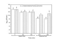
Survival rate of Zymomonas mobilis UFPEDA 363 under simulated gastrointestinal conditions. Means with the same letters (a, c) in the same row are not significantly different (P > 0.05), while different letters (b, d, f, h, e) indicate a significant difference (P < 0.05). Calculated using one-way ANOVA. FG = Simulated gastric juice; FI = Simulated intestinal juice.
Antimicrobial activity against pathogenic bacteria
Zymomonas mobilis UFPEDA 363 exhibited antimicrobial activity against several pathogenic bacteria. The inhibition zone diameters ranged from 19 mm to 22 mm, demonstrating effectiveness against Staphylococcus aureus, Escherichia coli, Salmonella enteritidis, and Pseudomonas aeruginosa (Table II). These results suggest that Z. mobilis UFPEDA 363 possesses antagonistic properties that may contribute to its probiotic potential by inhibiting the growth of common intestinal pathogens.
Antibiotic susceptibility test
The antibiogram analysis revealed that Zymomonas mobilis UFPEDA 363 was susceptible to eight out of nine tested antibiotics, with inhibition zone diameters ranging from 10.5 mm to 43.5 mm. As shown in Table III, the strain exhibited resistance only to ciprofloxacin, while maintaining susceptibility to a broad spectrum of antibiotics.
Hemolytic Activity
Z. mobilis UFPEDA 363 did not exhibit hemolytic activity, confirming that the studied strain is not capable of causing hemolysis of blood, thus not inducing the rupture of the erythrocyte membrane and therefore posing no health risk. The importance of using probiotics without hemolytic activity lies in the assurance of safety and efficacy.
Gelatinase test
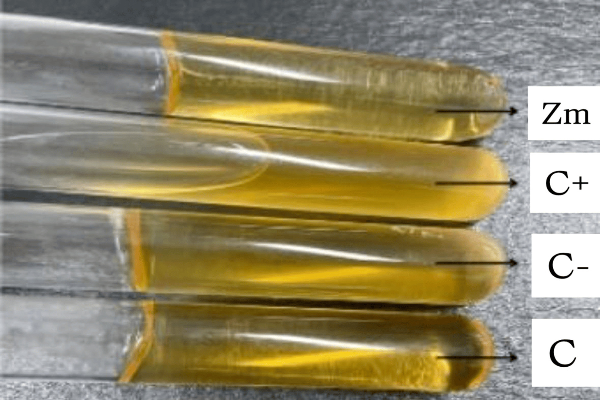
The gelatinase assay demonstrated that Z. mobilis UFPEDA 363 did not produce gelatinase (Figure 5), confirming the absence of this enzyme, which is responsible for the hydrolysis of gelatin, collagen, casein, hemoglobin, and other bioactive peptides.
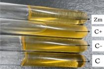
Gelatinase assay of Zymomonas mobilis UFPEDA 363. Zm = Zymomonas mobilis UFPEDA 363; C+ = Staphylococcus aureus; C- = Escherichia coli; C = SSDL+GELATIN culture media.
DISCUSSION
To be effective, probiotic bacteria must ensure cell viability at the time of consumption. Essential traits of effective probiotics include their capacity to survive processing, storage, and passage through the gastrointestinal tract. In this regard, Zymomonas mobilis stands out due to its unique growth pattern, reaching its peak density at around 24 h. This extended stationary phase is associated with high viability over time (Dai et al. 2022), which is particularly beneficial for probiotic strains, as it suggests their ability to maintain viability in various conditions. Moreover, research has shown that Lactobacillus acidophilus exhibits similar growth behavior, reaching peak density around 18 h before stabilizing (Lee et al. 2017). This study demonstrates that the ability of Zymomonas mobilis to survive at pH 3.0 positions it as a strong candidate for human probiotic applications, demonstrating persistence during gastric transit, where pH levels typically range from 2.0 to 3.5 (Campos et al. 2013). Comparatively, Lactobacillus rhamnosus GG, a well-established human probiotic, shows similar survival rates under acidic conditions, suggesting that Z. mobilis has comparable acid tolerance (Lee et al. 2017). Acid resistance is a crucial trait for any probiotic strain intended for oral consumption, as it ensures bacterial survival long enough to reach the intestines.
The high bile salt tolerance of strain Z. mobilis UFPEDA 363, ranging from 94% to 100%, is comparable to that reported for probiotic strains such as Lactobacillus acidophilus and Nitrobacterium (Ruiz et al. 2013). This level of bile salt resistance is characteristic of probiotics that perform well in vivo, and Z. mobilis exhibits performance comparable to commonly used strains such as Lactiplantibacillus plantarum (Zhu et al. 2020).
The strong auto-aggregation ability of Zymomonas mobilis UFPEDA 363 is comparable to other probiotic strains, such as Enterococcus faecium CRL 183 and Lactobacillus helveticus ssp. jugurti 416, which is known for biofilm formation that enhances probiotic efficacy (Kós et al. 2003). The auto-aggregation of Z.mobilis UFPEDA 363 reached 17% at 3 h, 25% at 5 h, and 65% at 24 h. This aggregation is influenced by various factors, including cellular strategies that enhance survival and replication, as well as interactions between adhesion molecules and cellular receptors that facilitate environmental adaptation, reinforcing its potential to compete with and limit pathogen colonization in the human gut. Both auto-aggregation and hydrophobicity are crucial for probiotic functionality. These processes play a key role in preventing pathogen colonization by forming physical barriers, occupying ecological niches, producing antimicrobial compounds, and creating an unfavorable environment for pathogens (Campana et al. 2017).
Co-aggregation refers to the ability to form associations with other bacterial species, as observed by Scillato et al. (2021). When bacterial cells cluster spontaneously, they form more resilient microbial communities capable of secreting antagonistic substances and inhibiting pathogens. This process offers key advantages, such as greater defense against environmental stresses, increased pathogen resistance, and improved adhesion to intestinal surfaces (Collado et al. 2007). The co-aggregation of Z. mobilis UFPEDA 363 with pathogenic test bacteria ranged from 49% to 65% at 24 h, demonstrating characteristics directly related to probiotic properties, as probiotics are live microorganisms that, in adequate doses, provide health benefits to the host.
The high hydrophobicity levels observed suggest a significant presence of hydrophobic groups on the surface of Z.mobilis UFPEDA 363, indicating strong bacterial adhesion potential (Alander et al. 1997, Fuller & Freter 1992, Pedersen & Tannock 1989). Higher affinity for chloroform and ethyl acetate indicates a greater interaction with apolar compounds, which may contribute to enhanced adhesion to intestinal epithelial cells and subsequent colonization. Compared to the Bacillus MKSK-J1 strain reported by Lee et al. (2017), Zymomonas mobilis Z.UFPEDA 363 exhibited higher hydrophobicity, particularly in interaction with chloroform (88.2% for MKSK-J1), highlighting its superior ability to interact with hydrocarbons. The ability of Z.mobilis to function as a more efficient electron donor and acceptor is significant, as it enhances adhesion to intestinal epithelial cells. Additionally, the strain’s high hydrophobicity may interfere with pathogen attachment by blocking receptors or through steric interactions, providing a competitive advantage in the gastrointestinal environment (Otero et al. 2004).
This study also evaluated the effects of simulated gastric and intestinal fluids on the viability of the strain Zymomonas mobilis UFPEDA 363 under conditions that replicate the human digestive process. The gastric environment typically maintains a pH range of 2.5 to 3.5; accordingly, this work simulated gastric conditions by maintaining the culture at pH 3.0 for the initial 120 min. Z. mobilis UFPEDA 363 demonstrated significant resistance. These results align with those reported by Li et al. (2023), who observed a survival rate of Lacticaseibacillus chiayiensis AACE3 exceeding 80% in gastrointestinal fluid tests. Moreover, the authors emphasized the antioxidant properties of these bacteria, comparable to those of lactic acid bacteria (LAB), which play a pivotal role in mitigating oxidative damage and preventing conditions such as diabetes, cardiovascular diseases, and intestinal inflammation (Kaushik et al. 2009, Wang et al. 2017).
The inhibitory effect, ranging from 19 to 22.5 mm against Staphylococcus aureus, Escherichia coli, Salmonella enteritidis, and Pseudomonas aeruginosa, may be attributed to the production of organic acids and bacteriocins, which acidify the environment and inhibit pathogen growth (Lima et al. 2011, Rutkis et al. 2022). The broad-spectrum antagonistic activity exhibited by Z. mobilis UFPEDA 363 underscores its potential for controlling gastrointestinal pathogens in humans. Its inhibition of Escherichia coli and Staphylococcus aureus is particularly significant, as these bacteria are commonly associated with gastrointestinal infections and foodborne illnesses. Previous studies on Lactobacillus acidophilus have reported similar antagonistic activity, primarily attributed to organic acid production (Hauka et al. 2014, Gilliland & Speck 1977).
Probiotics can influence health by modulating inflammatory responses, altering gut microbiota composition, and carrying antibiotic resistance genes. These factors highlight the importance of critically reassessing their use and regulatory frameworks (Mousa et al. 2023). The antibiogram analysis confirmed that Z. mobilis UFPEDA 363 is susceptible to most tested antibiotics, ensuring its safety profile.
One of the key tests in this context is the hemolysis assay, which plays an important role in determining microbial pathogenicity, classifying bacteria, and guiding the development of effective treatments (Guastalli et al. 2010). Solanki et al. (2023) examined four potential probiotic strains, Lactobacillus fermentum (BM1), Lactobacillus crispatus (H39), Enterococcus faecium (H31), and Lactobacillus helveticus (IF1), all of which exhibited no hemolytic activity. Similarly, Zymomonas mobilis UFPEDA 363 showed no signs of hemolysis, further reinforcing its probiotic potential.
Another critical safety criterion is the absence of gelatinase production, as this enzyme is associated with pathogenicity and is undesirable in probiotic strains intended for human consumption. In this regard, Mahasneh et al. (2015) analyzed 17 Lactobacillus isolates with probiotic potential and found no gelatinolytic activity. Likewise, the present study confirms that Z. mobilis UFPEDA 363 does not produce gelatinase. In summary, Zymomonas mobilis UFPEDA 363 demonstrated probiotic properties across all tests performed, yielding results comparable to or superior to the strains currently utilized in probiotic production. Additionally, it proved to be safe for human use in the conducted safety assessments, highlighting its potential applications in antimicrobial therapy and the production of bioactive compounds to maintain gut microbiota balance.
CONCLUSIONS
The findings of this study demonstrate that Zymomonas mobilis UFPEDA 363 exhibits robust growth under adverse conditions, including acidic pH and the presence of bile salts. The strain showed promising antimicrobial activity, producing compounds that effectively inhibit the growth of Salmonella enteritidis UFPEDA 414, Escherichia coli UFPEDA 224, Staphylococcus aureus UFPEDA 02, and Pseudomonas aeruginosa UFPEDA 416. A high aggregation potential was observed in both auto-aggregation and co-aggregation assays, along with significant hydrophobicity, indicating a strong capacity for cell adhesion. The strain is considered safe, as it does not produce hemolytic or gelatinolytic enzymes, and it exhibits high susceptibility to various antimicrobials. However, further studies are necessary to confirm its probiotic properties in vivo models.
Acknowledgements
The authors would like to thank Fundação de Amparo à Ciência e Tecnologia de Pernambuco (FACEPE) and Coordenação de Aperfeiçoamento de Pessoal de Nível Superior (CAPES).
References
-
ALANDER M, KORPELA R, SAXELIN M, VILPPONEN-SALMELA T, MATTILA-SANDHOLM T & VON WRIGHT A. 1997. Recovery of Lactobacillus rhamnosus GG from human colonic biopsies. Lett Appl Microbiol 24: 361-364. doi: https://doi.org/10.1046/j.1472-765X.1997.00140.x.
» https://doi.org/10.1046/j.1472-765X.1997.00140.x -
ALMO MMD, SOUSA IG, OLINTO VG, PINHATE SB, JIVAGO JLP, SOUSA DER, CASTRO MB, RUBINI MR, MARANHÃO AQ & BRIGIDO MM. 2023. Therapeutic effects of Zymomonas mobilis on experimental DSS-induced colitis mouse model. Microorganisms 11: 2793. doi: https://doi.org/10.3390/microorganisms11112793.
» https://doi.org/10.3390/microorganisms11112793 -
AYYASH MM, ABDALLA AK, ALKALBANI NS, BAIG MA, TURNER MS, LIU S Q & SHAH NP. 2021. Characterization of new probiotics from dairy and nondairy products - Insights into acid tolerance, bile metabolism and tolerance, and adhesion capability. J Dairy Sci 104: 8363-8379. doi: https://doi.org/10.3168/jds.2021-20398.
» https://doi.org/10.3168/jds.2021-20398 -
AZERÊDO GA, STAMFORD TLM, SOUZA EL, VERAS FF, ALMEIDA ER & ARAÚJO JM. 2010. In vivo assessment of possible probiotic properties of Zymomonas mobilis in a Wistar rat model. Electron J Biotechnol 13: 3-4. doi: 10.2225/vol13-issue2-fulltext-4.
» https://doi.org/10.2225/vol13-issue2-fulltext-4 -
BEREBON D, OFOKANSI K, ATTAMA A, EZE C, ONWUSOBA R & UGWOKE I. 2019. Preliminary studies on isolation, bile tolerance and antibiogram of potential probiotics (probionts) from locally fermented food products at Beach Market, Nsukka Metropolis, Enugu State, Nigeria. Biotechnol J Int 22: 1-10. doi: https://doi.org/10.9734/bji/2018/v22i330060.
» https://doi.org/10.9734/bji/2018/v22i330060 -
CAMPANA R, HEMERT S & BAFFONE W. 2017. Strain-specific probiotic properties of lactic acid bacteria and their interference with human intestinal pathogens invasion. Gut Pathog 9: 1-12. doi: https://doi.org/10.1186/s13099-017-0162-4.
» https://doi.org/10.1186/s13099-017-0162-4 -
CAMPOS IA, XIMENES EA, JÚNIOR CHRC, MESQUITA ARC, SILVA JBN, MAIA MBS, FRANCO ES, MEDEIROS PL, PEIXOTO CA & SILVA TG. 2013. Zymomonas mobilis culture protects against sepsis by modulating the inflammatory response, alleviating bacterial burden and suppressing splenocyte apoptosis. Eur J Pharm Sci 48: 1-8. doi: https://doi.org/10.1016/j.ejps.2012.10.011.
» https://doi.org/10.1016/j.ejps.2012.10.011 -
CLAVIJO V & FLOREZ MJV. 2018. The gastrointestinal microbiome and its association with the control of pathogens in broiler chicken production: A review. Poult Sci 97: 1006-1021. doi: https://doi.org/10.3382/ps/pex359.
» https://doi.org/10.3382/ps/pex359 -
CLSI - CLINICAL AND LABORATORY STANDARDS INSTITUTE. 2019. Performance standards for antimicrobial susceptibility testing. 29th ed. CLSI Supplement M100, Clinical and Laboratory Standards Institute, Wayne, PA. Available at: https://www.standards-global.com/wp-content/uploads/pdfs/preview/2031512
» https://www.standards-global.com/wp-content/uploads/pdfs/preview/2031512 -
COLLADO MC, MERILUOTO J & SALMINEN S. 2007. Measurement of aggregation properties between probiotics and pathogens: in vitro evaluation of different methods. J Microbiol Methods 71(1): 71-74. doi: https://doi.org/10.1016/J.MIMET.2007.07.005.
» https://doi.org/10.1016/J.MIMET.2007.07.005 -
DAI J, FANG L, ZHANG M, DENG H, CHENG X, YAO M & HUANG L. 2022. Isolation and identification of new source of bacteriocin-producing Lactobacillus plantarum C010 and growth kinetics of its batch fermentation. World J Microbiol Biotechnol 38: 4-67. doi: https://doi.org/10.1007/s11274-022-03244-1.
» https://doi.org/10.1007/s11274-022-03244-1 -
FAO – FOOD & AGRICULTURE ORGANIZATION. 2001. Manual on the Application of the HACCP System in Mycotoxin Prevention and Control. FAO Food and Nutrition Paper. Available at: https://www.fao.org/4/Y1390E/y1390e00.htm
» https://www.fao.org/4/Y1390E/y1390e00.htm -
FARIAS TGS, DE LADISLAU HFL, STAMFORD TCM, MEDEIROS JAC, SOARES BLM, ARNAUD TMS & STAMFORD TLM. 2019. Viabilities of Lactobacillus rhamnosus ASCC 290 and Lactobacillus casei ATCC 334 (in free form or encapsulated with calcium alginate-chitosan) in yellow mombin ice cream. LWT 100: 391-396. doi: https://doi.org/10.1016/j.lwt.2018.10.084.
» https://doi.org/10.1016/j.lwt.2018.10.084 -
FULLER R & FRETER R. 1992. Factors affecting the microecology of the gut. Probiotics, Springer, p. 111-144. doi: https://doi.org/10.1007/978-94-011- 2364-8_6.
» https://doi.org/10.1007/978-94-011-2364-8_6 -
GILLILAND S & SPECK M. 1977. Antagonistic Action of Lactobacillus acidophilus Toward Intestinal and Foodborne Pathogens in Associative Cultures. J Food Prot 40(12): 820-823. doi: https://doi.org/10.4315/0362-028X-40.12.820.
» https://doi.org/10.4315/0362-028X-40.12.820 - GUASTALLI E, GAMA NMSQ, BUIM MR, OLIVEIRA R, FERREIRA AJP & LEITE D. 2010. Índice de patogenicidade, produção de hemolisina e sorogrupo de amostras de Escherichia coli isoladas de aves de postura comercial. Arq Inst Biol 77(1): 153-157.
-
HAUKA F, SELIM A, EL-SAWAH M & RASHAD EM. 2014. Organic acids production and antagonistic effect of some strains of probiotics. J Agr Chem & Biotechnol 5(4): 101-112. doi: https://doi.org/10.21608/jacb.2014.49886.
» https://doi.org/10.21608/jacb.2014.49886 -
KAUSHIK JK, KUMAR A, DUARY RK, MOHANTY AK, GROVER S & BATISH VK. 2009. Functional and probiotic attributes of an indigenous isolate of Lactobacillus plantarum PloS One 4(12): 8099. doi: https://doi.org/10.1371/journal.pone.0008099.
» https://doi.org/10.1371/journal.pone.0008099 -
KOS B, ŠUŠKOVIĆ J, VUKOVIĆ S, ŠIMPRAGA M, FRECE J & MATOŠIĆ S. 2003. Adhesion and aggregation ability of probiotic strain Lactobacillus acidophilus M92. J Appl Microbiol 94: 981-987. doi: https://doi.org/10.1046/j.1365-2672.2003.01915.x.
» https://doi.org/10.1046/j.1365-2672.2003.01915.x -
LEE S, LEE J, JIN Y-I, JEONG J-C, CHANG YH, LEE Y, JEONG Y & KIM M. 2017. Probiotic characteristics of Bacillus strains isolated from Korean traditional soy sauce. LWT 79: 518-524. doi: https://doi.org/10.1016/j.lwt.2016.08.040.
» https://doi.org/10.1016/j.lwt.2016.08.040 -
LI X-D, LIN Y-C, YANG R-S, KANG X, XIN W-G, WANG F, ZHANG Q-L, ZHANG W-P & LIN L-B. 2023. Genomic and in-vitro characteristics of a novel strain Lacticaseibacillus chiayiensis AACE3 isolated from fermented blueberry. Front Microbiol 14: 1168378. doi: https://doi.org/10.3389/fmicb.2023.1168378.
» https://doi.org/10.3389/fmicb.2023.1168378 -
LIMA GMS, SOUZA LIO, ROSATO YB & ARAÚJO MJ. 2011. Detection of bacteriocins in Zymomonas mobilis and RAPD fingerprinting of the producer strains. Afr J Pharm Pharmacol 5(19): 2132-2139. doi: https://doi.org/10.5897/AJPP11.328.
» https://doi.org/10.5897/AJPP11.328 -
MAHASNEH AM, HAMDAN S & MAHASNEH SA. 2015. Probiotic Properties of Lactobacillus Species Isolated from Local Traditional Fermented Products. Jordan J Biol Sci 8: 81-87. DOI: 10.12816/0027552.
» https://doi.org/10.12816/0027552 -
MOUSA WK, MOUSA S, GHEMRAWI R, OBAID D, SARFRAZ M, CHEHADEH F & HUSBAND S. 2023. Probiotics modulate host immune response and interact with the gut microbiota: shaping their composition and mediating antibiotic resistance. Int J Mol Sci 24(18): 13783. doi: https://doi.org/10.3390/ijms241813783.
» https://doi.org/10.3390/ijms241813783 -
OTERO MC, OCAÑA VS & NADER-MACÍAS ME. 2004. Bacterial Surface Characteristics Applied to Selection of Probiotic Microorganisms. In: Spencer JFT & Ragout de Spencer AL (Eds), Public Health Microbiology. Methods Mol Biol, Humana Press, p. 268. https://doi.org/10.1385/1-59259-766-1:435.
» https://doi.org/10.1385/1-59259-766-1:435 -
PEDERSEN K & TANNOCK G. 1989. Colonization of the porcine gastrointestinal tract by lactobacilli. AEM 55(2): 279-283. doi: https://doi.org/10.1128/aem.55.2.279-283.1989.
» https://doi.org/10.1128/aem.55.2.279-283.1989 -
PEREIRA V, LOPES C, CASTRO A, SILVA J, GIBBS P & TEIXEIRA P. 2009. Characterization for enterotoxin production, virulence factors, and antibiotic susceptibility of Staphylococcus aureus isolates from various foods in Portugal. Food Microbiol 26(3): 278-282. doi: https://doi.org/10.1016/j.fm.2008.12.008.
» https://doi.org/10.1016/j.fm.2008.12.008 -
RAMLUCKEN U, RAMCHURAN SO, MOONSAMY G, LALLOO R, THANTSHA MS & RENSBURG CJV. 2020. A novel Bacillus based multi-strain probiotic improves growth performance and intestinal properties of Clostridium perfringens challenged broilers. Poult Sci 99(1): 331-341. doi: https://doi.org/10.3382/ps/pez496.
» https://doi.org/10.3382/ps/pez496 -
RUIZ L, MARGOLLES A & SANCHEZ B. 2013. Bile resistance mechanisms in Lactobacillus and Bifidobacterium Front Microbiol 4: 396. doi: https://doi.org/10.3389/fmicb.2013.00396.
» https://doi.org/10.3389/fmicb.2013.00396 -
RUTKIS R, ĻAŠA Z, RUBINA M, ŠČERBAKA R, KALNIŅŠ G, BOGANS J & KALNENIEKS U. 2022. Antimicrobial Activity of Zymomonas mobilis Is Related to Its Aerobic Catabolism and Acid Resistance. Fermentation 8(2): 77. doi: https://doi.org/10.3390/fermentation8020077.
» https://doi.org/10.3390/fermentation8020077 -
SAAD SMI. 2006. Probióticos e prebióticos: o estado da arte. Rev Bras Cienc Farm 42(1): 1-6. Available at: https://doi.org/10.1590/S1516-93322006000100002.
» https://doi.org/10.1590/S1516-93322006000100002 -
SCILLATO M, SPITALE A, MONGELLI G, PRIVITERA GF, MANGANO K, CIANCI A, STEFANI S & SANTAGATI M. 2021. Antimicrobial properties of Lactobacillus cell free supernatants against multidrug-resistant urogenital pathogens. Microbiology Open 10(2): 1173. doi: https://doi.org/10.1002/mbo3.1173.
» https://doi.org/10.1002/mbo3.1173 -
ŚLIŻEWSKA K & CHLEBICZ-WÓJCIK A. 2020. Growth kinetics of probiotic Lactobacillus strains in the alternative, cost-efficient semi-solid fermentation medium. Biology 9(12): 423. doi: https://doi.org/10.3390/biology9120423.
» https://doi.org/10.3390/biology9120423 -
SOLANKI R, BHATT A, PATEL J & KOTHARI R. 2023. Evaluation of Prebiotic Utilization and Probiotic Properties of Potential Probiotic Bacteria of Food and Human Origin. Food Sci Nutr 8(2): 1-12. doi: https://doi.org/10.23880/fsnt-16000297.
» https://doi.org/10.23880/fsnt-16000297 -
SÜLE J, VARGA L, HATVAN Z & ZOLTÁN K. 2022. Application of an in vitro test system for the selection of probiotic bacterial strains. J Food Inv 68(2): 3916-3927. doi: https://doi.org/10.52091/EVIK-2022/2-4-ENG.
» https://doi.org/10.52091/EVIK-2022/2-4-ENG -
WANG L-Q, MENG X-C, ZHANG B-R, WANG Y & SHANG Y-L. 2010. Influence of cell surface properties on adhesion ability of bifidobacteria. World J Microbiol Biotechnol 26: 1999-2007. doi: https://doi.org/10.1007/s11274-010-0384-9.
» https://doi.org/10.1007/s11274-010-0384-9 -
WANG M, WU Y, LI Y-D & ZENG J-B. 2017. Progress in toughening poly (lactic acid) with renewable polymers. Polym Rev 57(4): 557-593. doi: https://doi.org/10.1080/15583724.2017.1287726.
» https://doi.org/10.1080/15583724.2017.1287726 -
ZAGHARI M, SARANI P & HAJATI H. 2020. Comparison of two probiotic preparations on growth performance, intestinal microbiota, nutrient digestibility and cytokine gene expression in broiler chickens. J Appl Anim Res 48(1): 166-175. doi: https://doi.org/10.1080/09712119.2020.1754218.
» https://doi.org/10.1080/09712119.2020.1754218 -
ZHU W, LYU F, NAUMOVSKI N, AJLOUNI S & RANADHEERA CS. 2020. Functional efficacy of probiotic Lactobacillus sanfranciscensis in apple, orange and tomato juices with special reference to storage stability and in vitro gastrointestinal survival. Beverages 6(1): 13. doi: https://doi.org/10.3390/beverages6010013.
» https://doi.org/10.3390/beverages6010013
Edited by
-
Handling editor
Vasco Azevedo
The data supporting the findings of this study are available from the corresponding author upon reasonable request.