Growth kinetics, secondary metabolite profiles and antioxidant activity in cell cultures for bioprospecting of a native sweet potato variety from La Guajira, Colombia
Abstract
Sweet potato (Ipomoea batatas) is a versatile crop with considerable nutritional, medicinal, and industrial potential. In field-grown plants, caffeic acid, chlorogenic acid, and their derivatives are among the most abundant secondary metabolites and are closely associated with the biological activity of sweet potato extracts. Although tissue and organ culture techniques have been explored for decades, their application beyond plant regeneration remains limited. Optimizing callus proliferation is essential for biotechnological applications, particularly when aiming to utilize native genotypes. This study aimed to develop an in vitro protocol to enhance biomass production from a sweet potato variety traditionally grown in La Guajira and explore its potential for bioactive compound production. The application of 1.5 μM 2,4-dichlorophenoxyacetic acid (2,4-D) effectively induced high callus formation from leaf explants, while a pre-adaptation period in basal medium improved cell proliferation in both in gelled and liquid media. Thin-layer chromatography and LC-QTOF-MS confirmed the presence of secondary metabolites, with variations observed in total phenolic content and antioxidant activity during cell culture growth. These findings represent an advancement in biotechnological processes related to sweet potato callus proliferation. The developed protocol provides a foundation for future research on the production of novel bioactive compounds from this traditional crop.
Key words
In vitro culture; Cell suspensions; Biotechnology; Bioactivity; Ipomoea batatas
INTRODUCTION
Ipomoea batatas (L.) Lam. (sweet potato) is a plant species that has gained importance across various sectors due to its high capacity to store starch and essential nutrients for human health. As a result, it has been labeled a “superfood” or a health-promoting food (Alam 2021, Behera et al. 2022, Wang 2024) and is considered vital for food security in several Asian and African countries (Daurov et al. 2018, Dery et al. 2020, Echodu et al. 2019). This species has also been reported to contain significant amounts of phenols, anthocyanins, tocopherol, vitamin C, and β-carotene (Chen et al. 2012, Daurov et al. 2018), contributing to its medicinal and industrial potential, particularly due to its high antioxidant activity (Salgado et al. 2023).
The genetic diversity of sweet potato is vast, which can be appreciated in its extensive morphological variability (Jackson et al. 2020, Zhang et al. 2018). Accordingly, the composition of secondary metabolites varies depending on this biodiversity (Sun et al. 2019). Colombia is a center of origin for sweet potato and in La Guajira (northern Colombia) several types of sweet potatoes are cultivated. While some have been introduced by organizations engaged in genetic improvement of this species, others are considered traditional due to their generational cultivation. The bioactivity of these traditionally cultivated varieties must be evaluated, especially because the high light intensity and the saline soils in La Guajira force plants to adjust their metabolome as an adaptation strategy.
Despite the evident bioprospecting potential of sweet potato, its use as a raw material in the cosmetics and pharmaceutical industries faces challenges in meeting anticipated demand (Behera et al. 2024). Sweet potato field cultivation requires extensive land, continuous care, and is exposed to environmental conditions that cannot be controlled (Katayama et al. 2017). Field cultivation presents several limitations that hinder the optimal utilization of sweet potato, including: 1) vegetative propagation – sweet potato is primarily propagated vegetatively through cuttings or tuberous roots, which increases labor and production costs (Akomeah et al. 2019, Denham et al. 2020); 2) susceptibility to diseases – sweet potato is highly vulnerable to a wide range of pathogens, including nematodes, insects, microorganisms, and viruses, which significantly impact its health and productivity (Behera et al. 2022, Daurov et al. 2018); 3) variability in chemical composition – sweet potato exhibits substantial variation in nutrient content and bioactive compounds due to its extensive genetic diversity (Das et al. 2019, Jackson et al. 2020, Luo et al. 2021, Zhang et al. 2018). This variability is further influenced by environmental conditions during cultivation and the harvest season (Kobayashi et al. 2019, Suárez et al. 2020).
Micropropagation allows to overcome the limitations described above (Fadaladeen et al. 2022) and is a fundamental stage for carrying out other activities that involve the use and manipulation of in vitro plant genetic resources (Vollmer et al. 2023). Plants established in vitro can be used to induce the formation of undifferentiated tissue with high proliferation capacity, called callus (Hasnain et al. 2022). In this regard, cultivating them in gelled or liquid medium (cell suspensions) permits the generation of large amounts of new tissue for an indefinite period from minimal initial plant material (Efferth 2019, Mohaddab et al. 2022), with potential use in various biotechnological applications. The in vitro biomass produced can be used in large-scale plant regeneration (organogenesis, somatic embryogenesis), genetic transformation, somaclonal variation induction, synthetic seed development, haploid production (anther and ovary culture), somatic hybridization (protoplast fusion), or the sustained production of bioactive substances (Behera et al. 2022, Chandran et al. 2020, Kaur et al. 2022, Masekesa et al. 2021, Mohaddab et al. 2022, Ozyigit et al. 2023).
Sweet potato callus induction has been a research topic of interest for several decades, aiming to establish efficient methods for vegetative propagation and plant regeneration. In this context, the combined use of auxins and cytokinins has proven effective, enabling the formation of both embryogenic and non-embryogenic callus with high multiplication rates from various explant types, with leaf explants showing particularly favorable responses (Liu & Cantliffe 1984, Ochoa & López 1987). Similarly, the type of callus obtained may vary depending on the auxin used for induction; in this regard, 2,4-D has been shown to induce friable callus, an essential prerequisite for the establishment of continuous cell suspension cultures aimed at secondary metabolite production (Salah et al. 2024).
Since sweet potato responses under in vitro conditions, as well as the production of bioactive compounds, largely depend on genotype (Otani & Shimada 1996, Suhendy et al. 2023, Salah et al. 2024), it is essential to determine the culture conditions required for the successful establishment of each variety, evaluate their multiplication potential, and assess their suitability for industrial applications. Despite the callus morphogenetic and biosynthetic plasticity, it is necessary to develop efficient establishment and multiplication protocols for these cell cultures (Liu & Cantliffe 1984), along with strategies that can direct metabolic pathways towards the production of bioactive substances. However, the production of specific compounds is still a challenge due to the complexity of metabolic pathways and extraction methods. Additionally, culture age and cell differentiation processes can alter the phytochemical profile of the extracts (Dias et al. 2016, Wang et al. 2017). Therefore, it is crucial to identify the metabolites naturally produced in cell cultures in order to enhance the production of those with potential for use.
Several studies have reported significant anthocyanin production in sweet potato cell cultures from purple varieties, which produce these pigments in high concentrations (Konczak et al. 2003, 2005). On the other hand, the sweet potato genotype used in this investigation corresponds to the white type. White sweet potatoes are not characterized by their high pigment production; however, their phenolic content and bioactivity are comparable to those of the yellow, purple, and orange sweet potatoes (Šlosár et al. 2020, Sun et al. 2014). The establishment of cell cultures from unimproved sweet potato varieties offers a unique opportunity to preserve the genetic heritage of this species while also exploiting its biochemical potential. Based on the statements presented, this research aimed to determine the conditions for the induction and multiplication of calluses from a sweet potato variety traditionally grown in La Guajira and to evaluate its potential for obtaining secondary metabolites, particularly those with antioxidant activity.
MATERIALS AND METHODS
Obtaining the initial plant material
Sampling site for plant material was located in the municipality of Dibulla (La Guajira) at coordinates 4955729.822 East - 2794867.654 North (Magna Sirgas System). The collection was covered by Amendment No. 3 to the Access to Genetic Resources Contract No. 237, RGE-308-03, processed by the Technological University of Pereira (Risaralda, Colombia) with the Ministry of Environment and Sustainable Development of the Republic of Colombia.
In vitro plants establishment and multiplication
A population of sweet potato plants grown in pots was maintained for six months; subsequently, a single mother plant was selected to ensure genetic homogeneity of the plant material used in the in vitro experiments. Leaves were removed, and their nodal segments (explants) were isolated and disinfected following the protocol described by Mengs et al. (2018) with some modifications. Explants were washed with soapy water + Tween 20® (4 drops per 100 mL) for 30 min, then rinsed with distilled water and placed in a 4.0 gL-1 copper oxychloride solution for 2 h. They were then transferred into a laminar flow chamber, where they were immersed in 70% ethanol for 30 s and rinsed with sterile distilled water. Finally, they were disinfected with NaOCl (2%) for 10 min and rinsed again with sterile distilled water.
In vitro establishment was performed in glass flasks containing MS culture medium (Murashige & Skoog 1962) without plant growth regulators (PGRs), supplemented with 3% of sucrose and 8.0 gL-1 of agar-agar, with lighting provided by white LED lamps, at a temperature of 22 ± 2 °C and a photoperiod of 12 h, until new stems developed from the explants. The obtained stems were multiplied in gelled MS medium under the same culture conditions, with subcultures performed every eight weeks.
Callus induction and multiplication
Leaves were collected from sweet potato plants established in vitro, and the methodology described by Guevara et al. (2012) was followed. Three types of leaf explants were evaluated (complete leaf, leaf blade segments, and petiole segments), which were placed in gelled MS culture medium (3% of sucrose; 8.0 gL-1 of agar-agar), supplemented (independently) with three concentrations of 2,4-dichlorophenoxyacetic acid (2,4-D) (1.5, 2.5 and 3.5 µM), in addition to a control without PGRs. All experimental units were kept in dark conditions for eight weeks, with weekly evaluations.
Induced callus were subcultured several times and subsequently used in multiplication experiments to establish the best proliferation conditions in gelled and liquid culture media. Two initial conditions were evaluated: callus without pre-adaptation in basal culture medium and callus with pre-adaptation for 15 d in basal culture medium (medium without 2,4-D) before being transferred to fresh media supplemented with 2,4-D. All experimental units were kept in dark conditions, and biomass gain was recorded over 10 weeks, with fresh weight data taken every 2–3 d. Five repetitions were used per treatment (2,4-D concentration or control), and 300 ± 0.0047 mg of callus was used as inoculum.
Establishment of cell suspensions
Cell suspensions were established in Erlenmeyer flasks with a capacity of 100 mL, containing 40 mL of MS culture medium supplemented with sucrose (3%), 1.5 µM 2,4-D and 4.0 ± 0.01 g of callus as inoculum. A growth curve was constructed by independently quantifying the fresh and dry biomass produced every 5 d in three Erlenmeyer flasks. Prior to the establishment of suspensions, callus pre-adaptation was carried out for 15 d in liquid basal MS medium. Experimental units were maintained under constant agitation at 120 rpm in an orbital shaker and kept in darkness. In parallel with the suspension growth curve, preliminary monitoring of total phenolic content (TPC) and antioxidant activity was conducted to identify the growth stage at which these parameters reached their maximum values.
Kinetic parameters; including the specific growth rate (μ), doubling time (td), and growth index (GI), were calculated using both fresh weight (FW) and dry weight (DW) as biomass variables. Subsequently, new cell suspension cultures were established, and the resulting biomass was harvested after 19 days of culture. The biomass from each suspension was washed with distilled water to remove any remaining culture medium and placed in sterile, properly labeled plastic jars with lids and stored at -80 °C until use. Subsequently, the biomass in each jar was freeze-dried using a Labconco FreeZone® 4.5 freeze-dryer and then pulverized.
Obtaining crude extracts
Extraction was performed using HPLC-grade methanol at a 1:10 ratio. The extraction process lasted 15 min, utilizing ultrasound (HF-Freq. 35 kHz) at a temperature of 40 °C. Each extract was obtained by combining three successive extractions from the same plant material and were subsequently dried under reduced pressure using a rotary evaporator.
Total phenol content (TPC)
Total phenols quantification was performed using the Folin-Ciocalteu method in 96-well microplates. Prior to the tests, extracts were diluted to an appropriate concentration; in all cases, deionized water was used as the diluent. Subsequently, 50 µL of extract was placed into the wells, followed by the addition of 50 µL of Folin-Ciocalteu reagent diluted with deionized water at a 1:50 ratio. Then, 100 µL of a 0.35 M NaOH solution was added. The microplate was incubated in the dark at room temperature for 3 min, and absorbance was measured at 760 nm using a microplate reader (Thermo Scientific, Multiskan GO).
Absorbance values were used to determine the TPC using calibration curves and were expressed as μg chlorogenic acid equivalents per mg of dry extract (μg CAE·mg⁻¹ DE)
Antioxidant potential of extracts
Antioxidant activity of the extracts was determined using the DPPH• assay, the oxygen radical absorbance capacity (ORAC) assay, and the total antioxidant capacity (CAT) method. In all cases, extracts were diluted at the same ratio used for total phenols quantification.
For DPPH• assay, aliquots (25 µL) of each extract were mixed with 100 µL of a freshly prepared 20 mg·L⁻¹ 1,1-diphenyl-2-picrylhydrazyl (DPPH•) solution. The reaction mixture was incubated at room temperature for 30 min in the dark, and absorbance was measured at 517 nm. A hydroquinone solution (1,000 ppm) was used as a positive control, while methanol served as a negative control. The antioxidant activity was expressed as μg Trolox equivalents per mg of dry extract (µg TE·mg⁻¹ DE).
The ORAC assay was performed using aliquots (20 µL) of each extract mixed with 120 µL of a freshly prepared fluorescein solution (120 nM) in 75 mM phosphate buffer (pH 7.4) and incubated at 37 °C for 15 min. After which 60 µL of 2,2’-Azobis (2-methylpropionamide) dihydrochloride (AAPH) were added. Fluorescence was recorded for 60 min at 37 °C using a fluorimeter (excitation 485 nm; emission 538 nm). Antioxidant activity was expressed as µg TE·mg⁻¹ DE.
Total antioxidant capacity was determined using the phosphomolybdate method. Aliquots (100 µL) of each extract were mixed with 1,000 µL of phosphomolybdate reagent (0.6 M H₂SO₄, 4 mM (NH₄)₂MoO₄, 28 mM Na₂HPO₄). The mixture was incubated in a water bath at 95 °C for 90 min and then allowed to cool to room temperature. Aliquots (200 µL) were transferred to a 96-well microplate, and absorbance was measured at 695 nm using a microplate reader. Results were expressed as μg ascorbic acid equivalents per mg of dry extract (µg AAE·mg⁻¹ DE).
Chromatographic analysis
The phytochemical profile of crude extracts was established using thin-layer chromatography (TLC) and LC-QTOF-MS. For the characterization of chemical nuclei by TLC, a mobile phase composed of ethyl acetate: formic acid: acetic acid: water (100:11:11:26) was used, with silica gel chromatoplates (60 F254; Merk Millipore) of 8.0 cm as the stationary phase. For LC-QTOF-MS analysis (Agilent Technologies), 2 μL of extract was injected onto a C18 column (InfinityLab Poroshell 120 EC-C18, 100 × 2.1 mm, 1.9 μm) at 30 °C, using a gradient elution composed of 0.1% (v/v) formic acid in Milli-Q water (Phase A) and 0.1% (v/v) formic acid in acetonitrile (Phase B) at a constant flow rate of 0.4 mLmin-1. Mass spectrometry detection was performed in negative ESI mode with a full scan from 50 to 1100 m/z and GNPS (autoMSMS) at 20 eV.
The detected compounds were analyzed and manually inspected using Agilent MassHunter Profinder 10.0 software with the Recursive Molecular Extraction algorithm. Metabolite annotation was performed using MS-DIAL software.
Data processing and information analysis
All experiments followed a completely randomized design, and assays were performed in triplicate. Data were tabulated, graphed, and subjected to normality and homogeneity of variance tests; subsequently, they were analyzed using ANOVA. When statistically significant differences were observed in response to any of the analyzed variables, means were separated using Duncan’s test. Additionally, the relationship between TPC and the antioxidant activity of the extracts was determined using Pearson correlation analysis. In all cases, statistical analyses were performed using SAS® OnDemand for Academics (SAS® Institute 2021).
RESULTS AND DISCUSSION
In vitro plants establishment and multiplication
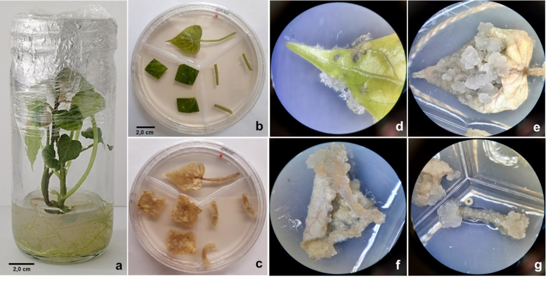
The disinfection treatment resulted in 22% axenic explants, which were used for in vitro plant establishment. During in vitro establishment and subsequent plant multiplication, the culture medium was not supplemented with PGRs. Consequently, only 50% of the explants regenerated shoots, with an average of 1.23 ± 0.43 shoots per explant, indicating low morphogenetic stimulation but confirming the regenerative capacity of the buds despite the absence of PGRs in the culture medium. Root formation occurred spontaneously in the basal region of the explants from the third day of culture, with roots elongating and branching in subsequent days. In all cases, plants exhibited normal development (Figure 1a).
In vitro establishment of sweet potato plants and callus induction. a) Plant established in vitro (six weeks of culture); b) Explants on callus induction medium (1 d); c) Explants established on culture medium with 3.5 μM of 2,4-D (six weeks of culture); d) Explants on control treatment (week 1) (15x). Callus induction by explant (eight weeks of culture): e) Complete leaf (8x); f) Leaf blade fragment (8x); g) Fragment of petiole (8x).
Callus induction
During the second week of culture, swelling was observed in explants cultured in medium supplemented with 2,4-D, while callus formation became evident from the fourth week. In some control treatment explants (“complete leaf” and “leaf blade fragments”), cell proliferation occurred at the wound sites and explant edges from the first week of culture. However, this proliferation was not abundant, had a translucent color, and persisted until the sixth week (Figure 1d), after which it underwent oxidation and died.
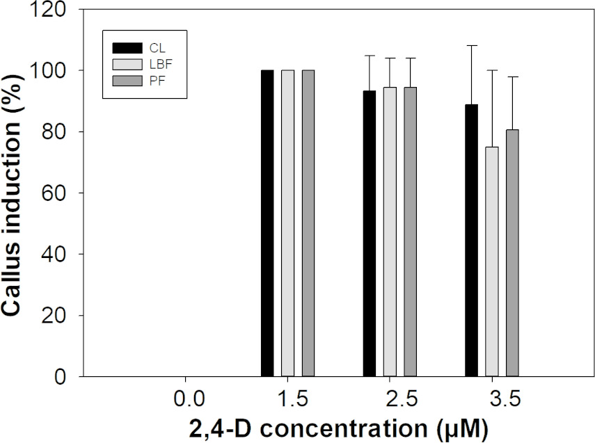
Callus induction evaluation at the eighth week of culture showed that callus formation occurred in all explants across treatments containing any concentration of 2,4-D, whereas no callus formation was observed in explants subjected to the control treatment (Figure 2).
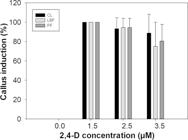
Percentage of sweet potato explants induced towards callus formation, eighth week of cultivation. CL: complete leaves; LBF: leaf blade fragments; PF: petiole fragments. Percentages correspond to the average of three replicates, with each replicate consisting of six repetitions per treatment and explant.
Some studies, such as those conducted by Masekesa et al. (2021) and Mohanraj & Subha (2015), indicate an early response of explants to callus formation in sweet potato, characterized by explant swelling and cell proliferation at wound sites from the first week of culture, as well as callus formation itself from the second or third week when using 2,4-D or an auxin + cytokinin mixture in ratios favoring auxin. Based on this and previous research findings (Guevara et al. 2012, Masekesa et al. 2021), it is considered that the response observed in explants before the third week of induction does not correspond to callus formation. Instead, this event indicates the dedifferentiation of specialized tissues in preparation for callus proliferation.
Friable callus proliferation was evident in all explants and treatments where 2,4-D was used. Statistical analysis indicated the presence of significant differences in response to 2,4-D concentrations (p < 0.0001), with the best response observed at a concentration of 1.5 μM (Figure 2). On the other hand, no significant differences in callus formation were found among the explants evaluated (p = 0.0964); therefore, subsequent experiments were conducted using calli obtained from leaf blade explants.
Ochoa & López (1987) reported callus induction from petiole segments by combining 10 μM of 2,4-D + 0.01 μM of 6-benzylaminopurine (BAP) under dark conditions. The calluses obtained were pale yellow, a coloration attributed to light deprivation during the experiment. However, the authors stated that their growth did not differ from that of calluses induced under light conditions. Chen et al. (2012) obtained morphogenetic callus from tuberous roots of sweet potato using 2.26 μM of 2,4-D + 0.46 μM of kinetin (KIN), while Masekesa et al. (2021) reported embryogenic callus formation using this same combination of PGRs.
Auxins and cytokinins induce cell division, and evidence suggests that the combination of these substances is more effective in promoting callus formation (Cambaz & Çördük 2023). In sweet potato, the use of a single PGR for the induction of this morphogenetic event is less common, especially at low concentrations. Mohanraj & Subha (2015) reported low callus induction rates (30.6–32.8%) when using root explants and 2,4-D concentrations equal to or greater than 9.0 μM. In contrast, they achieved induction rates above 87% with different concentrations of a combination of naphthalene-acetic acid (NAA) and KIN.
Similarly, Rahman & Sultana (2017) reported callus induction from sweet potato petiole segments using 10 μM of 2,4-D, which was superior to other concentrations of this PGR (even in combination with KIN); however, the induction percentage was only 55%. Guevara et al. (2012) achieved 100% friable and pale-yellow callus formation in sweet potato leaf explants using only 2,4-D at a minimum concentration of 2.26 μM. However, in the present study, the same result was obtained with a lower concentration of this PGR (1.5 μM).
Michael (2019) obtained white and yellow calluses on explants consisting of internodes, leaf blade fragments, and petiole fragments by supplementing the culture medium with 0.1% (w/v) picloram. Regarding this, Fehér (2019) stated that auxin-induced callus exhibits a well-defined gene expression pattern, regardless of the explant type. Furthermore, Ikeuchi et al. (2018), Xin et al. (2022) and Yu et al. (2020) concur that there is a convergence in the gene regulation pathways of auxin and wound response, which could explain the cell proliferation observed in some explants of the control treatment.
Callus proliferation experiments
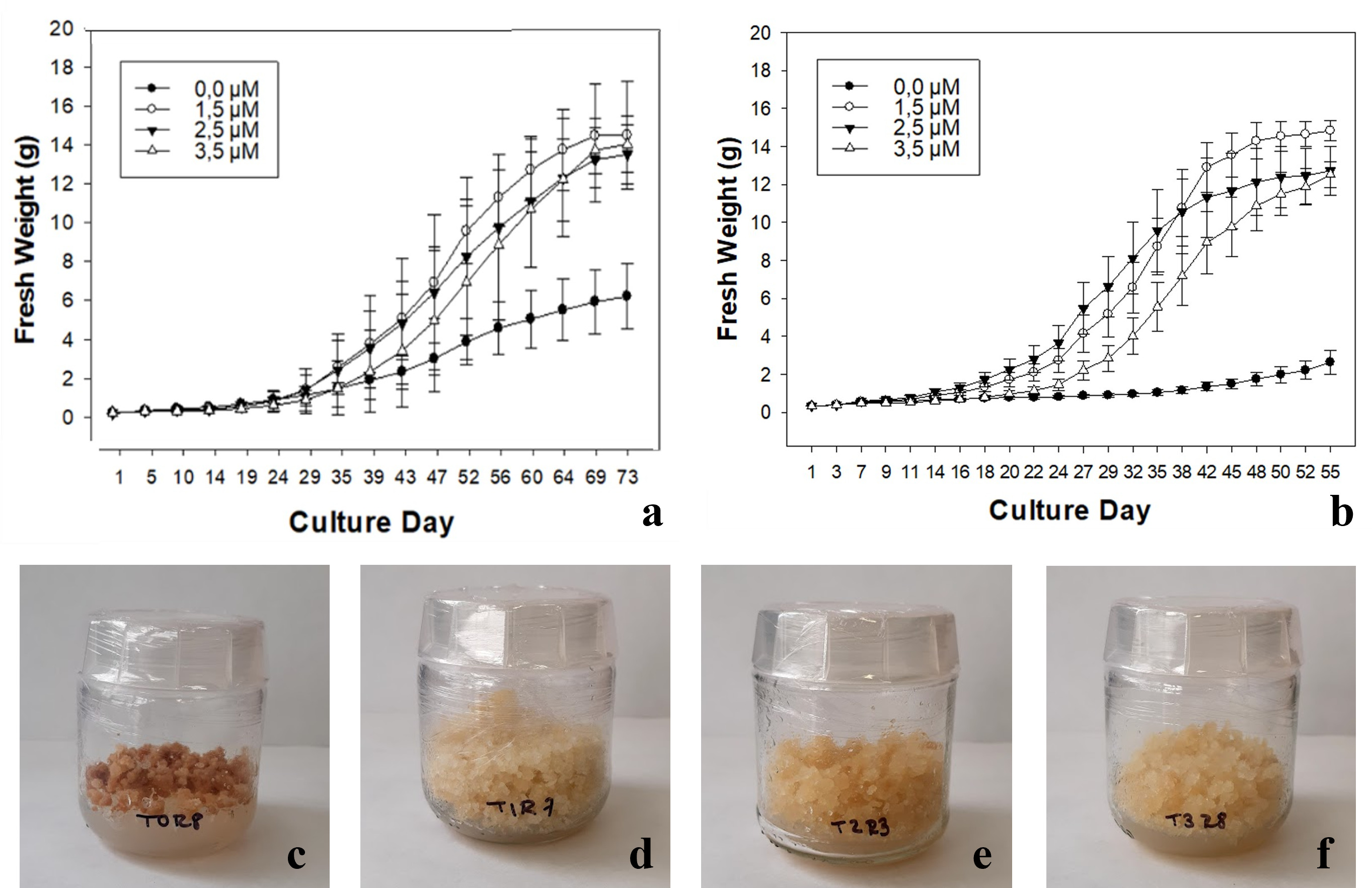
The callus biomass obtained in the presence of 2,4-D significantly exceeded that reported in previous studies (González et al. 2011, Guevara et al. 2012) under similar cultivation conditions, demonstrating the potential of this PGR to optimize the process. Growth analyses revealed typical patterns in the form of sigmoidal curves, characterized by an initial lag phase, followed by an accelerated growth phase, and finally a stationary phase. The duration of the dormancy phase was notably prolonged in callus that were not pre-adapted in basal medium (24 d), while in pre-adapted callus, it was reduced to 14 d. The growth phase was mostly linear in cultures without pre-adaptation until 69 d, when the stationary phase was reached. In contrast, pre-adapted cultures exhibited a greater degree of exponential growth, which lasted until 50 d of culture (Figure 3).
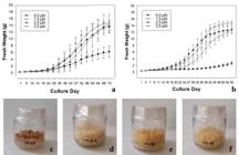
Growth curves of callus proliferation experiments, each point represents the average of eight repetitions a) Callus without pre-adaptation in basal MS culture medium. b) Pre-adapted callus in basal MS culture medium. Calluses with eight weeks of culture: c) 0.0 μM 2,4-D; d) 1.5 μM 2,4-D; e) 2.5 μM 2,4-D; f) 3.5 μM 2,4-D.
González et al. (2011) reported linear growth of sweet potato cell suspensions until 65 d of culture (with a lag phase of 7 d), using MS medium supplemented with 2.26 μM of 2,4-D and 1.1 μM BAP. However, the biomass obtained was less than 3.5 g of fresh mass. In contrast, Guevara et al. (2012) achieved a maximum production of 1.7 g of fresh mass in sweet potato callus cultured in MS medium supplemented with 2.26 μM of 2,4-D. In the latter case, linear growth was evident until 15 d of culture (without a lag phase), followed by an exponential growth phase until 25 d.
In pre-adapted cultures, the control treatment showed a lower fresh weight gain compared to the cultures without pre-adaptation (Figure 3). This difference suggests that cell proliferation in sweet potato callus is induced by 2,4-D and that a pre-adaptation period in basal medium reduces the effects of this PGR on fresh weight gain. Furthermore, in the control treatment with pre-adapted callus, the stationary phase was reached after 55 d of culture, which implies a shorter evaluation time compared to cultures without pre-adaptation. Statistical analysis revealed significant differences (p < 0.0001) in fresh weight gain between callus exposed to 2,4-D and the control (Figures 3a, b). However, no significant differences were observed in the effect of different concentrations of 2,4-D evaluated in relation to this variable in any of the experiments.
Maximum biomass production in pre-adapted cultures was achieved in gelled medium supplemented with 1.5 μM of 2,4-D (14.82 ± 1.45 g), compared to the control treatment, which reached a maximum fresh weight of 2.63 ± 1.73 g at the end of the experiment. In contrast, in the control treatment of cultures without pre-adaptation, a relatively high biomass gain was obtained (5.69 ± 1.66 g). This can be explained as the residual effect of 2,4-D, as the callus used originated from a culture medium supplemented with this PGR, in addition to the apparent high biomass production potential of the sweet potato variety used. Konczak et al. (2005) indicate that PGRs can accumulate intracellularly, which may prevent growth suppression in the absence of exogenous PGRs.
Oggema et al. (2007) emphasized the importance of determining the optimal concentration of this PGR for plant material multiplication, especially in local cultivars. The results presented here complement existing knowledge on the in vitro cultivation of sweet potatoes and represent an advancement in the in vitro manipulation of indigenous sweet potato genotypes from Colombia. Moreover, it was demonstrated that, although the in vitro response of sweet potato tissues is highly dependent on genotype (Otani & Shimada 1996), 2,4-D is one of the most effective auxins for callus induction in this species.
Growth dynamics of suspension cultures
The cell suspension cultures exhibited an exponential growth phase between days 5 and 20 of culture. Afterward, a decline in biomass production and crop deterioration became evident (Table I). Rahman & Sultana (2017) reported the establishment of sweet potato cell suspensions with a lag phase of two weeks, followed by an exponential growth phase between the third and seventh week, culminating in a stationary phase at the eighth week. In our suspensions, no lag phase was observed, which was attributed to pre-adaptation in liquid basal medium, making the effect of 2,4-D more pronounced once the cultures were transferred to media containing this PGR.
Growth dynamics and variation in total phenolic content and antioxidant activity (DPPH method) in sweet potato cell suspensions.
During the exponential phase, the specific growth rate (μ) calculated based on FW was 0.108 d⁻¹, with a doubling time (td) of 6.43 d. Similarly, analysis based on DW yielded a μ value of 0.112 day⁻¹ and a td of 6.21 d. The close agreement between these values indicates that biomass accumulation resulted from both effective organic matter accumulation and active cell division. The GI, calculated between the onset of cultivation and the point of maximum growth (day 20), was higher when FW (5.93) was used than when DW (4.86) was considered, confirming the high efficiency of the system in converting nutrients into structural biomass.
There is evidence that culture media supplemented with PGRs other than 2,4-D induces lower proliferation rates in sweet potato cell suspensions. For instance, Mohanraj & Subha (2017) reported a maximum biomass gain (measured as fresh weight) of only 4 g using a combination of 11 µM NAA and 1.0 µM KIN. These results are significant because some biotechnological processes depend on available biomass.
Konczak et al. (2003) calculated GI (final fresh weight/initial fresh weight) of sweet potato cell suspensions in modified MS culture medium, without PGRs, and with 1.0 g of inoculum. In their study, a maximum GI of 3.5 to 4.0 was recorded after 18 d of cultivation. In contrast, in the present study, this index reached a value of 5.93 after 20 days of culture, indicating greater biomass production, which was enhanced by the presence of 2,4-D regardless of the inoculum size. Considering the differences in GI values, it is worth noting that the callus used to establish cell suspension cultures in Konczak et al. (2003) study originated from a medium supplemented with 4.52 µM of 2,4-D. Therefore, the observed biomass increase may be partly explained by a residual response to 2,4-D, which, through intracellular accumulation (Konczak et al. 2005), promoted sustained cell growth. Furthermore, exogenously applied PGRs can alter the endogenous levels of other regulators, thereby affecting the physiology of cultured cells (Yu et al. 2020).
Quantification of total phenolic content (TPC) revealed a temporal pattern partially decoupled from growth. During the initial stages of culture (days 1–15), TPC values remained relatively low, coinciding with a period of high proliferative activity. This pattern suggests that, during early phases, cellular metabolism was predominantly oriented toward primary metabolic processes and cell expansion rather than secondary metabolite synthesis. From day 20 onward, when FW reached its maximum and the growth rate began to decline, a progressive increase in total phenolic accumulation was observed, reaching a maximum on day 25 (4.65 µg CAE mg⁻¹) (Table I). The stabilization of FW and its subsequent decrease after maximum growth suggest favorable conditions for the induction or accumulation of secondary metabolites. Accordingly, the period immediately following peak biomass accumulation (around day 20) may be considered strategic for elicitor application, controlled stress induction, or biomass harvesting for biotechnological purposes.
Antioxidant activity displayed a less variable pattern throughout the culture period, with relatively high values during the exponential phase and a tendency to decline toward the end of the experiment. Maximum antioxidant activity was recorded on day 15 (7,48 ± 0,06 µg TE.mg-1), coinciding with active growth and a rapid increase in DW (Table I). These results suggest that antioxidant capacity is not determined exclusively by total phenolic concentration but also depends on the qualitative composition of the metabolites produced and the physiological state of the tissue. The partial decoupling between TPC and antioxidant activity, particularly evident between days 20 and 25, indicates that increased phenolic accumulation in sweet potato cell cultures does not necessarily result in a proportional increase in antioxidant activity. This effect may be associated with changes in phenolic composition, including the accumulation of compounds with lower reducing capacity, or with the contribution of non-phenolic antioxidant systems.
Solís et al. (2013) support not only the superiority of cell suspensions for biomass production but also for secondary metabolite production, compared to more complex systems such as complete plants. This approach provides an unlimited supply of undifferentiated, uniform cells with short life cycles. Combined with controlled culture conditions, this reduces variability in experimental results when the production of substances of interest is induced (Babich et al. 2020, Cambaz & Çördük 2023, Mohaddab et al. 2022, Salgado et al. 2023, Vasyutkina et al. 2022).
The death phase, evident after 25 d of culture, indicates the accumulation of toxic substances in the culture medium and low nutrient availability (Mahendran et al. 2021), as a consequence of a relatively high inoculum amount. Mohanraj & Subha (2017) reported an increase in the biomass of sweet potato cell suspensions six times greater than the initial inoculum, with cessation of growth from 15 d of culture, using 20 mL of MS medium supplemented with NAA + KIN. This shorter growth period could have been due to the use of a smaller medium volume.
Total phenol content (TPC) and antioxidant activity
The TPC calculated in the biomass from cell suspensions after 19 d of cultivation was 11.96 ± 0.47 μg CAE·mg⁻¹ DW, while the antioxidant activity yielded values of 2.08 ± 0.06 (DPPH), 244.25 ± 12.07 (ORAC) μg TE·mg⁻¹ DW, and 34.48 ± 0.98 μg AAE·mg⁻¹ DW (CAT). Correlation analysis showed a moderate to low association between TPC and antioxidant activity. The CAT method produced a correlation of 0.81 (p = 0.1871), the ORAC method a correlation of 0.69 (p = 0.3089), and the DPPH method a correlation of 0.35 (p = 0.5544).
Results show that sweet potato callus produces low concentrations of phenolic compounds, which in turn affects the antioxidant activity of its extracts. However, published studies suggest that this could be improved by manipulating biosynthetic pathways (Mohaddab et al. 2022, Solís et al. 2013, Vasyutkina et al. 2022). Elicitation has been recognized as the most effective strategy for inducing the production of secondary metabolites (Isah 2019).
Phenolic compound production in sweet potato has been achieved through elicitation, stimulating callus with precursors, RCVs, and modifying nutrient concentrations in the culture medium (Mohanraj & Subha 2017, Salgado et al. 2023). Additionally, Ghasemzadeh et al. (2016) found that certain RCVs, such as salicylic acid (SA), methyl jasmonate (MeJA), and abscisic acid (ABA), affect phenolic content in sweet potato plants by acting as elicitors, modulating the activity of phenylalanine ammonia-lyase (PAL). Their research demonstrated that the application of elicitors also enhances the bioactivity of the extracts (antioxidant/antiproliferative).
Mendoza et al. (2018) significantly increased the biosynthesis of phenols in cell suspensions of Thevetia periviana using SA, MeJA, and a mixture of both, particularly after 24 h of exposure. In all cases, the CFT was significantly higher compared to the control; however, treatment with MeJA was superior, establishing the following hierarchy: MeJA (3.0 mM) > MeJA (3.0 mM)/SA (300 mM) > SA (300 mM) > control. It should be noted that, in this case, previous tests to establish the growth curve of the suspensions showed that the maximum phenol production occurred at the beginning of the exponential phase, rather than at the end of it as observed with the suspensions used in this research.
Plant cells express metabolic pathways depending on their genotype and physiological state (Ho et al. 2020, Isah 2019). In this regard, Adil et al. (2019) recorded greater biomass gain in Cnidium officinale callus maintained in dark conditions compared to callus exposed to white light. Similarly, they found that both TPC and total flavonoid content (TFC), as well as antioxidant activity (DPPH• method) of extracts obtained, were higher under light conditions, although they recognize that this response may vary between plant species. In contrast, Ali et al. (2018) reported that both callus grown in gelled medium and cell suspensions of Ajuga bracteosa maintained in dark conditions exhibited the highest biomass gain, as well as the highest TPC, TFC, and antioxidant enzyme activity (superoxide dismutase, peroxidase) when exposed to 2,23 µM of MeJA.
Chromatographic analysis
Cell suspensions exhibited a variety of phytochemical compounds, which varied as their age increased (Table II). The majority of the identified metabolites showed low intensity, which is consistent with the findings of Cambaz & Çördük (2023), who reported that some secondary metabolites of Verbascum scamandri Murb. are produced at lower concentrations in callus than in the plants from which they were induced. However, Mahendran et al. (2021) emphasize the importance of cell suspensions in enhancing the accumulation of bioactive substances.
Tan et al. (2024) reported organic acids, flavonoids, carbohydrates, phenolic acids, coumarins, terpenes, and alkaloids as major compound classes present in 32 sweet potato varieties. In the present study, Flavonoids were not detected. Although phenolic acids were present, band intensities were weak, indicating low concentrations of these compounds. This contrasts with reports for field-grown sweet potato plants, in which phenolic compounds occur at substantially higher levels (Luo et al. 2021, Salgado et al. 2023, Zhao et al. 2022). Therefore, the absence or very low concentration of certain chemical nuclei in the extracts may be attributed to the influence of in vitro conditions. Alkaloids were detected throughout most of the suspension culture period, although at low intensity. These findings support further research on the production of these metabolites under in vitro conditions, given their reported potential in cancer therapy and other biomedical applications due to their antimicrobial, antiviral, and anti-inflammatory activities (Yan et al. 2021, Srivastava & Tiwari 2022).
Coumarins were clearly detected in the extracts, exhibiting relatively high band intensities, whereas terpene presence was moderate throughout most of the culture period and declined toward the end. Quinone production was variable, with the highest levels observed between days 5 and 15 of culture. Coumarins are found throughout the plant kingdom and are attributed with various biological and pharmacological activities, including antimicrobial, antimutagenic, and anti-inflammatory effects (Alagesan et al. 2019, Sulaiman et al. 2019), all of which have been reported in sweet potato. Batiga et al. (2019) highlight coumarins, along with alkaloids, terpenes, and phenolic compounds, as important bioactive compounds in the genus Ipomoea, therefore, their presence was expected in the cell cultures evaluated in this study.
Carbohydrate accumulation up to the final growth stage of the suspensions is consistent with expectations, as in vitro cultured tissues receive a carbon supplement (in this case, sucrose) that allows for cell proliferation. It has been shown that mitotically active cells increase their internal glucose concentration; furthermore, sugars are necessary to maintain osmotic pressure in cells, and their presence in high concentrations has been linked to defense mechanisms against stress (Gangola & Ramadoss 2018, Sharma et al. 2023, Siddiqui et al. 2020). The decrease in carbohydrates concentration at the end of the culture may be associated with the utilization of both the external carbon source and internal cell reserves in stress-induced cellular survival mechanisms, as well as marked cell death at this stage of suspension development. Regarding this latter aspect, Malerba & Cerana (2021) indicate that factors such as darkness and depletion of nutrients like phosphates and carbon sources induce apoptosis in plant cell suspensions. The drop in the concentration of different groups of secondary metabolites on 30 d of cultivation is associated with cell death.
Table III shows the compounds identified by LC-QTOF-MS. The presence of compounds associated with the metabolism of dividing cells is evident, including amino acids, peptides, fatty acids, vitamins (pantothenic acid) and intermediates of primary metabolism. Although caffeic acid, chlorogenic acid, and derivatives such as 3,4,5-tricaffeoylquinic acid are commonly reported in sweet potato, these compounds were absent in the extracts obtained from the cell suspensions. Additionally, the proportion of caffeic glucoside and dicaffeoylquinic acids is lower compared to reports on extracts obtained from the leaves and roots of field-grown plants, which may be linked to the dedifferentiation of cells in the suspension.
Compounds identified by LC-QTOF-MS present in extracts of cell suspensions from sweet potato.
The presence of vanilloloside, skimmin, dihydroxy-valeric acid, lariciresinol glucoside and narcissin stands out, as these are not frequently reported in sweet potato extracts. Due to their proven bioactivity, their elicitation could be considered.
Vanilloloside has demonstrated reversible acetylcholinesterase inhibitory activity, making it a promising drug candidate for the treatment of Alzheimer’s disease (Avetyan et al. 2022). Skimmin, on the other hand, decreases the levels of several factors associated with inflammation, including TNF-alpha, IL-1 beta, and IL-6 (Su et al. 2023). It also has a gastroprotective effect (Razuvaeva et al. 2023), could be effective in treating type 2 diabetes (Zhang et al. 2020), and has been considered a potential agent for the treatment of postpartum stroke (Sun et al. 2023). Compounds such as narcissin, caffeic glucoside, and dicaffeoylquinic acids exhibit significant antioxidant activity, and their concentrations in cell suspension extracts can be increased by elicitors (Da Silva et al. 2022).
Lariciresinol glucoside was found among the major constituents of a methanolic extract of Aconitum heterophyllum, exhibiting antiproliferative, anti-inflammatory, and antinociceptive activity (Ilyas et al. 2024). It also has potential in reducing the risk of certain types of cancer, particularly breast cancer (Anjum et al. 2017). Furthermore, lariciresinol glucoside has antioxidant activity, and in silico studies have demonstrated its potential for developing antimicrobial drugs (Singh et al. 2023). Additionally, Chen et al. (2024) reported its antiviral and anti-hepatitis effects.
CONCLUSIONS
Supplementation of the culture medium with 1.5 μM of 2,4-D is both necessary and sufficient to induce callus formation and high multiplication in sweet potato. Furthermore, a 15 d preadaptation period of the callus in basal culture medium improves cell proliferation, resulting in a 46.3-fold increase in biomass in gelled medium and a 5.93-fold increase in liquid medium. The established protocol allows for the continuous generation of biomass from minimal plant material, providing a scalable platform for biotechnological applications such as genetic transformation, somaclonal variation, and bioactive compound production.
Although the white sweet potato variety is not known for high pigment production, callus cultures of the tested genotype exhibited diverse bioactive metabolites, some previously unreported in similar studies. The identification of various secondary metabolites through TLC and LC-QTOF-MS, along with the observed variations in the TPC and antioxidant activity, highlights the bioactive potential of the callus generated from this traditional sweet potato variety cultivated in La Guajira. These findings provide a solid foundation for future research aimed at the sustained production of bioactive compounds on a large scale, which could contribute to strengthening the sweet potato value chain in sectors such as pharmaceuticals, cosmetics, and food. Furthermore, the establishment of in vitro cell cultures represents a key tool for preserving the genetic heritage of traditional varieties and enhancing their use in bioprospecting contexts.
It is crucial to evaluate the elicitation of phenolic compounds from different sweet potato genotypes at the in vitro level. Future research should explore the influence of different growth regulators, carbon sources, and environmental conditions (light, temperature) to maximize the production of specific metabolites of interest. Additionally, it is recommended to conduct more detailed metabolomic studies to identify and quantify the bioactive compounds synthesized during callus development, as well as evaluate their stability and functionality.
The developed protocol can be adapted for large-scale production of plant biomass intended for the extraction of compounds with antioxidant potential. Furthermore, it is necessary to assess the economic viability and environmental impact of these processes for industrial implementation. Likewise, since the composition of secondary metabolites depends on the genotype, it would be relevant to replicate this study with other traditional sweet potato varieties grown in different regions of Colombia to identify those with greater biosynthetic capacity.
Acknowledgements
The authors express their gratitude to the University of La Guajira, Technological University of Pereira (TUP), and the Bicentennial Doctoral Excellence Scholarship Program of the Ministry of Science, Technology, and Innovation (Minciencias), Colombia. Likewise, the authors thank the Ministry of Environment and Sustainable Development (Colombia) for allowing the use of the plant material required in this research. This document is the result of the research project “Evaluation of Antioxidant and Antitumor Potential of Extracts and Fractions Obtained from Plants and Cell Suspensions of Ipomoea batatas (L.) Lam. (Convolvulaceae).” This project was supported by the Biotechnology research group (University of La Guajira - Riohacha, Colombia) and the Biotechnology - Natural Products research group (TUP - Pereira, Colombia).
-
Data availability
The datasets generated and analyzed during the current study are available from the corresponding author upon reasonable request. Data may be shared for academic and research purposes, subject to appropriate justification and compliance with applicable ethical and institutional regulations.
References
-
ADIL M, REN X & JEONG B. 2019. Light elicited growth, antioxidant enzymes activities and production of medicinal compounds in callus culture of Cnidium officinale Makino. J Photochem Photobiol B 196: 111509. https://doi.org/10.1016/j.jphotobiol.2019.05.006.
» https://doi.org/10.1016/j.jphotobiol.2019.05.006 -
AKOMEAH B, QUAIN M, RAMESH S, ANAND L & RODRÍGUEZ C. 2019. Common garden experiment reveals altered nutritional values and DNA methylation profiles in micropropagated three elite Ghanaian sweet potato genotypes. PLoS One 14(4): e0208214. https://doi.org/10.1371/journal.pone.0208214.
» https://doi.org/10.1371/journal.pone.0208214 -
ALAGESAN E, RAMALINGAM S, KIM M & VENUGOPAL S. 2019. Antioxidant activity guided isolation of a coumarin compound from Ipomoea pes-caprea (Convolvulaceae) leaves acetone extract and its biological and molecular docking studies. Eur J Integr Med 32: 100984. https://doi.org/10.1016/j.eujim.2019.100984.
» https://doi.org/10.1016/j.eujim.2019.100984 -
ALAM M. 2021. A comprehensive review of sweet potato (Ipomoea batatas [L.] Lam): Revisiting the associated health benefits. Trends Food Sci Technol 115: 512-529. https://doi.org/10.1016/j.tifs.2021.07.001.
» https://doi.org/10.1016/j.tifs.2021.07.001 -
ALI H, KHAN MA, ULLAH N & KHAN RS. 2018. Impacts of hormonal elicitors and photoperiod regimes on elicitation of bioactive secondary volatiles in cell cultures of Ajuga bracteosa J Photochem Photobiol B 183: 242-250. https://doi.org/10.1016/j.jphotobiol.2018.04.044.
» https://doi.org/10.1016/j.jphotobiol.2018.04.044 -
ANJUM S, ABBASI BH, DOUSSOT J, FAVRE-RÉGUILLON A & HANO C. 2017. Effects of photoperiod regimes and ultraviolet-C radiations on biosynthesis of industrially important lignans and neolignans in cell cultures of Linum usitatissimum L. (Flax). J Photochem Photobiol B 167: 216-227. https://doi.org/10.1016/j.jphotobiol.2017.01.006.
» https://doi.org/10.1016/j.jphotobiol.2017.01.006 -
AVETYAN DL, SHATSKIY A, KÄRKÄS MD & STEPANOVA EV. 2022. Scalable total synthesis of natural vanillin-derived glucoside ω-esters. Carbohydr Res 522: 108683. https://doi.org/10.2139/ssrn.4191727.
» https://doi.org/10.2139/ssrn.4191727 -
BABICH O, SUKHIKH S, PUNGIN A, IVANOVA S, ASYAKINA L & PROSEKOV A. 2020. Modern trends in the in vitro production and use of callus, suspension cells and root cultures of medicinal plants. Molecules 25(24): 5805. https://doi.org/10.3390/molecules25245805.
» https://doi.org/10.3390/molecules25245805 -
BATIGA S, VALLI M, ZERAIK M, FRAIGE K, LEME G, PITANGUI N, ALMEIDA A, MICHEL S, YOUNG M & BOLZANI V. 2019. Chemical composition and biological properties of Ipomoea procumbens Rev Bras Farmacogn 29(2): 191-197. https://doi.org/10.1016/j.bjp.2018.08.010.
» https://doi.org/10.1016/j.bjp.2018.08.010 -
BEHERA S, CHAUHAN VBS, PATI K, BANSODE V, NEDUNCHEZHIYAN M, VERMA AK, MONALISA K, NAIK PK & NAIK SK. 2022. Biology and biotechnological aspect of sweet potato (Ipomoea batatas L.): a commercially important tuber crop. Planta 256: 40. https://doi.org/10.1007/s00425-022-03938-8.
» https://doi.org/10.1007/s00425-022-03938-8 -
BEHERA S ET AL. 2024. In vitro plant regeneration, genetic fidelity, biochemical analysis and anticancer activity of anthocyanin-rich purple flesh sweet potato var. ‘Bhu Krishna’. S Afr J Bot 166: 332-343. https://doi.org/10.1016/j.sajb.2024.01.041.
» https://doi.org/10.1016/j.sajb.2024.01.041 -
CAMBAZ E & ÇÖRDÜK N. 2023. Secondary metabolite production in callus culture of Verbascum scamandri Murb. Acta Soc Bot Pol 92(1): 1-11. https://doi.org/10.5586/asbp/165894.
» https://doi.org/10.5586/asbp/165894 -
CHANDRAN H, MEENA M, BARUPAL T & SHARMA K. 2020. Plant tissue culture as a perpetual source for production of industrially important bioactive compounds. Biotechnol Rep 26: e00450. https://doi.org/10.1016/j.btre.2020.e00450.
» https://doi.org/10.1016/j.btre.2020.e00450 -
CHEN R ET AL. 2024. The ERF transcription factor LTF1 activates DIR1 to control stereoselective synthesis of antiviral lignans and stress defense in Isatis indigotica roots. Acta Pharm Sin B 14(1): 405-420. https://doi.org/10.1016/j.apsb.2023.08.011.
» https://doi.org/10.1016/j.apsb.2023.08.011 - CHEN SY, YAP WS & YONG WTL. 2012. Comparison of anthocyanin and phenolic contents between tuber and callus of Ipomoea batatas (L.). Pertanika J Trop Agric Sci 35(1): 9-14.
-
DA SILVA SANTOS É ET AL. 2022. Low-cost alternative for the bioproduction of bioactive phenolic compounds of callus cultures from Cereus hildmannianus (K.) Schum. J Biotech 356: 8-18. https://doi.org/10.1016/j.jbiotec.2022.07.001.
» https://doi.org/10.1016/j.jbiotec.2022.07.001 -
DAS G, PATRA J, BASAVEGOWDA N, VISHNUPRASAD C & SHIN H. 2019. Comparative study on antidiabetic, cytotoxicity, antioxidant and antibacterial properties of biosynthesized silver nanoparticles using outer peels of two varieties of Ipomoea batatas (L.) Lam. Int J Nanomed 14: 4741-4754. https://doi.org/10.2147/ijn.s210517.
» https://doi.org/10.2147/ijn.s210517 -
DAUROV D, ZHAPAR K, DAUROVA A, VOLKOV D, BAKBERGENOVA M, TOLEGENOVA D, SHAMEKOVA M & ZHAMBAKIN K. 2018. Production of virus-free sweet potato planting material for the southeast of Kazakhstan. Int J Agric Biol 20(4): 851-856. https://doi.org/10.17957/IJAB/15.0576.
» https://doi.org/10.17957/IJAB/15.0576 -
DENHAM T, BARTON H, CASTILLO C, CROWTHER A, DOTTE-SAROUT E, FLORIN A, PRITCHARD J, BARRON A, ZHANG Y & FULLER D. 2020. The domestication syndrome in vegetatively-propagated field crops. Ann Bot 125(4): 581-597. https://doi.org/10.1093/aob/mcz212.
» https://doi.org/10.1093/aob/mcz212 -
DERY E, CAREY E, SSALI R, LOW J, JOHANNINGSMEIER S, ODURO I BOAKYE A, OMODAMIRO R & YUSUF H. 2020. Sensory characteristics and consumer segmentation of fried sweetpotato for expanded markets in Africa. Int J Food Sci Technol 56(3): 1419-1431. https://doi.org/10.1111/ijfs.14847.
» https://doi.org/10.1111/ijfs.14847 -
DIAS M, SOUSA M, ALVES R & FERREIRA I. 2016. Exploring plant tissue culture to improve the production of phenolic compounds: A review. Ind Crop Prod 82: 9-22. https://doi.org/10.1016/j.indcrop.2015.12.016.
» https://doi.org/10.1016/j.indcrop.2015.12.016 -
ECHODU R, EDEMA H, WOKORACH G, ZAWEDDE C, OTIM G, LUAMBANO N, ATEKA E & ASIIMWE T. 2019. Farmers’ practices and their knowledge of biotic constraints to sweetpotato production in East Africa. Physiol Mol Plant Pathol 105: 3-16. https://doi.org/10.1016/j.pmpp.2018.07.004.
» https://doi.org/10.1016/j.pmpp.2018.07.004 -
EFFERTH T. 2019. Biotechnology applications of plant callus cultures. Eng 5: 50-59. https://doi.org/10.1016/j.eng.2018.11.006.
» https://doi.org/10.1016/j.eng.2018.11.006 -
FADALADEEN L, TOMA R, SAHEEN A & AHMED H. 2022. A rapid micropropagation protocol for sweet potato (Ipomoea batatas L.) via tissue culture technique. DASJ 14(1): 31-39. https://doi.org/10.52951/dasj.22140104.
» https://doi.org/10.52951/dasj.22140104 -
FEHÉR A. 2019. Callus, dedifferentiation, totipotency, somatic embryogenesis: What these terms mean in the era of molecular plant biology? Front Plant Sci 10: 536. https://doi.org/10.3389/fpls.2019.00536.
» https://doi.org/10.3389/fpls.2019.00536 - GANGOLA M & RAMADOSS B. 2018. Chapter 2 - Sugars play a critical role in abiotic stress tolerance in plants. In: Hussain S (Ed), Biochemical, physiological and molecular avenues for combating abiotic stress tolerance in plants. Academic Press, p. 17-38. https://doi.org/10.1016/B978-0-12-813066-7.00002-4.
-
GHASEMZADEH A, TALEI D, JAAFAR H, JURAIMI A, MOHAMED M, PUTEH A & HALIM M. 2016. Plant-growth regulators alter phytochemical constituents and pharmaceutical quality in Sweet potato (Ipomoea batatas L.). BMC Complement Altern Med 16: 152. https://doi.org/10.1186/s12906-016-1113-1.
» https://doi.org/10.1186/s12906-016-1113-1 - GONZÁLEZ O, HERNÁNDEZ M, SILVA J & ESPINOSA A. 2011. Evaluación de la dinámica del crecimiento in vitro en callos de Ipomoea batatas Rev Colomb Biotecnol 13(1): 148-155. Available at: http://www.scielo.org.co/scielo.php?script=sci_arttext&pid=S0123-34752011000100020&lng=en&tlng=es.
-
GUEVARA Y, SUÁREZ I & SALGADO J. 2012. Inducción y proliferación in vitro de tejidos celulares de batata (Ipomoea batatas (L.) Lam.) en medio con 2,4-D. Temas Agrar 17(2): 9-17. DOI: 10.21897/rta.v17i2.698.
» https://doi.org/10.21897/rta.v17i2.698 -
HASNAIN A ET AL. 2022. Plants in vitro propagation with its applications in food, pharmaceuticals and cosmetic industries; current scenario and future approaches. Front Plant Sci 13: 1009395. https://doi.org/10.3389/fpls.2022.1009395.
» https://doi.org/10.3389/fpls.2022.1009395 -
HO TT, MURTHY HN & PARK SY. 2020. Methyl jasmonate induced oxidative stress and accumulation of secondary metabolites in plant cell and organ cultures. Int J Mol Sci 21: 716. https://doi.org/10.3390/ijms21030716.
» https://doi.org/10.3390/ijms21030716 -
IKEUCHI M ET AL. 2018. A Gene regulatory network for cellular reprogramming in plant regeneration. Plant Cell Physiol 59(4): 770-782. https://doi.org/10.1093/pcp/pcy013.
» https://doi.org/10.1093/pcp/pcy013 -
ILYAS M, SHAD A, BAKHT J, VILLALTA P & SHIER WT. 2024. Insights into metabolites profiling and pharmacological investigation of Aconitum heterophyllum wall ex. royle stem through experimental and bioinformatics techniques. ACS Omega 9(25): 26922-26940. https://doi.org/10.1021/acsomega.3c09668.
» https://doi.org/10.1021/acsomega.3c09668 -
ISAH T. 2019. Stress and defense responses in plant secondary metabolites production. Biol Res 52: 39. https://doi.org/10.1186/s40659-019-0246-3.
» https://doi.org/10.1186/s40659-019-0246-3 -
JACKSON D, HARRISON H, JARRET R & WALD P. 2020. Phenotypic variation in leaf morphology of the USDA, ARS sweetpotato (Ipomoea batatas) germplasm collection. HortScience 55(4): 465-475. https://doi.org/10.21273/HORTSCI14703-19.
» https://doi.org/10.21273/HORTSCI14703-19 -
KATAYAMA K, KOBAYASHI A, SAKAI T, KURANOUCHI T & KAI Y. 2017. Recent progress in sweetpotato breeding and cultivars for diverse applications in Japan. Breed Sci 67(1): 3-14. https://doi.org/10.1270/jsbbs.16129.
» https://doi.org/10.1270/jsbbs.16129 -
KAUR K, DOLKER D, BEHERA S & PATI P. 2022. Critical factors influencing in vitro propagation and modulation of important secondary metabolites in Withania somnifera Dunal. PCTOC 149: 41-60. https://doi.org/10.1007/s11240-021-02225-w.
» https://doi.org/10.1007/s11240-021-02225-w -
KOBAYASHI T, KURATA R & KAI Y. 2019. Seasonal variation in the yield and polyphenol content of sweet potato (Ipomoea batatas L.) foliage. Hort J 88(2): 270-275. https://doi.org/10.2503/hortj.utd-025.
» https://doi.org/10.2503/hortj.utd-025 -
KONCZAK I, OKUNO S, YOSHIMOTO M & YAMAKAWA O. 2003. Composition of phenolics and anthocyanins in a sweet potato cell suspension culture. Biochem Eng J 14: 155-161. https://doi.org/10.1016/s1369-703x(02)00216-4.
» https://doi.org/10.1016/s1369-703x(02)00216-4 -
KONCZAK I, TERAHARA N, YOSHIMOTO M, NAKATANI M, YOSHINAGA M & YAMAKAWA O. 2005. Regulating the composition of anthocyanins and phenolic acids in a sweetpotato cell culture towards production of polyphenolic complex with enhanced physiological activity. Trends Food Sci Technol 16: 377-388. https://doi.org/10.1016/j.tifs.2005.02.007.
» https://doi.org/10.1016/j.tifs.2005.02.007 -
LIU J & CANTLIFFE D. 1984. Somatic embryogenesis and plant regeneration in tissue cultures of sweet potato (Ipomoea batatas Poir.). Plant Cell Rep 3(3): 112-115. https://doi.org/10.1007/BF02441013.
» https://doi.org/10.1007/BF02441013 -
LUO D, MU T & SUN H. 2021. Profiling of phenolic acids and flavonoids in sweet potato (Ipomoea batatas L.) leaves and evaluation of their anti-oxidant and hypoglycemic activities. Food Biosci 39: 100801. https://doi.org/10.1016/j.fbio.2020.100801.
» https://doi.org/10.1016/j.fbio.2020.100801 -
MAHENDRAN G, IQBAL Z, KUMAR D, VERMA S, ROUT P & RAHMAN L. 2021. Enhanced gymnemic acids production in cell suspension cultures of Gymnema sylvestre (Retz.) R.Br. ex Sm. through elicitation. Ind Crops Prod 162: 113234. https://doi.org/10.1016/j.indcrop.2020.113234.
» https://doi.org/10.1016/j.indcrop.2020.113234 -
MALERBA M & CERANA R. 2021. Plant cell cultures as a tool to study programmed cell death. Int J Mol Sci 22: 2166. https://doi.org/10.3390/ijms22042166.
» https://doi.org/10.3390/ijms22042166 -
MASEKESA R, GASURA E, KUJEKE G, NGADZE E & CHIDZWONDO F. 2021. Induction of somatic embryogenesis and organogenesis in zimbabwean sweet potato (cv Brondal). Adv Agric 9976800: 8. https://doi.org/10.1155/2021/9976800.
» https://doi.org/10.1155/2021/9976800 -
MENDOZA D, CUASPUD O, ARIAS J, RUIZ O & ARIAS M. 2018. Effect of salicylic acid and methyl jasmonate in the production of phenolic compounds in plant cell suspension cultures of Thevetia peruviana Biotechnol Rep 19: e00273. https://doi.org/10.1016/j.btre.2018.e00273.
» https://doi.org/10.1016/j.btre.2018.e00273 -
MENGS B, CHIMDESSA M & ABRAHA E. 2018. In vitro propagation of sweet potato (Ipomoea batatas (L.) Lam.) through lateral bud culture. IJIPSR 6(07): 1-12. DOI: 10.21276/IJIPSR.2018.06.07.721.
» https://doi.org/10.21276/IJIPSR.2018.06.07.721 -
MICHAEL P. 2019. Responses of different explants of sweet potato on modified MS and LS based nutrient media in vitro Asian J Adv Agric Res 11(4): 1-7. https://doi.org/10.9734/AJAAR/2019/v11i430064.
» https://doi.org/10.9734/AJAAR/2019/v11i430064 -
MOHADDAB M, EL GOUMI Y, GALLO M, MONTESANO D, ZENGIN G, BOUYAHYA A & FAKIRI M. 2022. Biotechnology and in vitro culture as an alternative system for secondary metabolite production. Molecules 27: 8093. https:// doi.org/10.3390/molecules27228093.
» https://doi.org/10.3390/molecules27228093 - MOHANRAJ R & SUBHA S. 2015. Production of 4-ipomeanol, an anticancer agent from the root tubers and rhizogenic callus of Ipomoea batatas Lam.: a comparative study. Indian J Exp Biol 53(5): 297-304.
- MOHANRAJ R & SUBHA S. 2017. Optimization of process parameters for bioproduction, isolation and purification of 4-Ipomeanol from cell suspension cultures of Ipomoea batatas (L.) Lam. Indian J Exp Biol 55(3): 191-196.
-
MURASHIGE T & SKOOG F. 1962. A Revised medium for rapid growth and bio assays with tobacco tissue cultures. Physiol Plant 15(3): 473-497. https://doi.org/10.1111/j.1399-3054.1962.tb08052.x.
» https://doi.org/10.1111/j.1399-3054.1962.tb08052.x -
OCHOA N & LÓPEZ F. 1987. Effect of light and NaCI salinity on the growth of callus cultures of Ipomoea pes-caprae (L.) R. Brown and Ipomoea batatas (L.) Lam. Ann Bot 59(5): 495-497. https://doi.org/10.1093/oxfordjournals.aob.a087342.
» https://doi.org/10.1093/oxfordjournals.aob.a087342 -
OGGEMA J, KINYUA M & OUMA J. 2007. Optimum 2,4-D concentration suitable for embryogenic callus induction in local Kenyan sweet potato cultivars. Asian J Plant Sci 6(3): 484-489. https://doi.org/10.3923/ajps.2007.484.489.
» https://doi.org/10.3923/ajps.2007.484.489 -
OTANI M & SHIMADA T. 1996. Efficient embryogenic callus formation in sweet potato (Ipomoea batatas (L.) Lam.). Breed Sci 46(3): 257-260. https://doi.org/10.1270/jsbbs1951.46.257.
» https://doi.org/10.1270/jsbbs1951.46.257 -
OZYIGIT I, DOGAN I, HOCAOGLU-OZYIGIT A, YALCIN B, ERDOGAN A, YALCIN I, CABI E & KAYA Y. 2023. Production of secondary metabolites using tissue culture-based biotechnological applications. Front Plant Sci 14: 1132555. https://doi.org/10.3389/fpls.2023.1132555.
» https://doi.org/10.3389/fpls.2023.1132555 - RAHMAN M & SULTANA R. 2017. Sweet potato (Ipomoea batatas L.): suspension culture for cell aggregation. J Plant Sci Res 4(2): 171.
-
RAZUVAEVA YG, TOROPOVA AA, SALCHAK SM & OLENNIKOV DN. 2023. Coumarins of Ferulopsis hystrix: LC–MS profiling and gastroprotective and antioxidant activities of skimmin and peucenidin. Appl Sci 13(17): 9653. https://doi.org/10.3390/app13179653.
» https://doi.org/10.3390/app13179653 -
SALAH E, EISSA M & EL-FEKY F. 2024. In vitro calli developing and characterizing from sweet potato (Ipomoea batatas) to maximize secondary metabolites production. Al-Azhar J Agric Res 49(1): 149-155. https://doi.org/10.21608/ajar.2024.263165.1324.
» https://doi.org/10.21608/ajar.2024.263165.1324 -
SALGADO J, RAMÍREZ L & MOSQUERA Ó. 2023. Ipomoea batatas (L.) Lam. (Convolvulaceae) as a source of polyphenols with antitumor activity and prospects for in vitro production using chemical elicitors-A Review. Bol Latinoam Caribe Plant Med Aromat 22(2): 156-179. https://doi.org/10.37360/blacpma.23.22.2.12.
» https://doi.org/10.37360/blacpma.23.22.2.12 -
SAS® INSTITUTE. 2021. Software de Analítica & Soluciones (versión OnDemand for Academics). Windows. Estados Unidos: SAS Institute Inc. Available at: https://www.sas.com/es_mx/software/on-demand-for-academics.html Accessed on: January 31, 2024.
» https://www.sas.com/es_mx/software/on-demand-for-academics.html - SHARMA P, KAPOOR N, DHIMAN S, KOUR J, SINGH A, SHARMA A & BHARDWAJ R. 2023. Chapter 16 - Role of sugars in regulating physiological and molecular aspects of plants under abiotic stress. In: Sharma A, Pandey S, Bhardwaj R, Zheng B & Kumar D (Eds), The Role of Growth Regulators and Phytohormones in Overcoming Environmental Stress. Academic Press, p. 355-374. https://doi.org/10.1016/B978-0-323-98332-7.00010-X.
-
SIDDIQUI H, SAMI F & HAYAT S. 2020. Glucose: Sweet or bitter effects in plants - A review on current and future perspective. Carbohydr Res 487: 107884. https://doi.org/10.1016/j.carres.2019.107884.
» https://doi.org/10.1016/j.carres.2019.107884 -
SINGH U, SINGH P, SINGH AK, SINGH S, KUMAR D, SHRIVASTAVA S & ASTHANA RK. 2023. In silico and in vitro evaluation of extract derived from Dunaliella salina, a halotolerant microalga for its antifungal and antibacterial activity. J Biomol Struct Dyn 41(15): 7069-7083. https://doi.org/10.1080/07391102.2022.2115556.
» https://doi.org/10.1080/07391102.2022.2115556 -
ŠLOSÁR M, HEGEDŰSOVÁ A, HEGEDŰS O, MEZEYOVÁ I & TIMORACKÁ M. 2020. The effect of cultivar on selected quantitative and qualitative parameters of sweet potatoes (Ipomoea batatas L.) grown in Slovak republic. J Cent Eur Agric 21(2): 344-353. https://doi.org/10.5513/JCEA01/21.2.2684.
» https://doi.org/10.5513/JCEA01/21.2.2684 - SOLÍS L, CARBALLO L & VALDEZ M. 2013. Establishment of cell suspension cultures of two Costa Rican Jatropha species (Euphorbiaceae). Rev Biol Trop 61(3): 1095-1107. Available at: https://www.scielo.sa.cr/scielo.php?script=sci_arttext&pid=S0034-77442013000400009.
-
SRIVASTAVA R & TIWARI P. 2022. Medicinal plant used against cancer: A review. Asian J Pharm Res Dev 10(4): 1-10. https://doi.org/https://doi.org/10.22270/ajprd.v10i4.1150.
» https://doi.org/10.22270/ajprd.v10i4.1150 -
SU M, CUI J, ZHAO J & FU X. 2023. Skimmin ameliorates cardiac function via the regulation of M2 macrophages in a myocardial infarction mouse model. Perfusion 38(6): 1298-1307. https://doi.org/10.1177/02676591221100742.
» https://doi.org/10.1177/02676591221100742 -
SUÁREZ S, MU T, SUN H & AÑÓN M. 2020. Antioxidant activity, nutritional, and phenolic composition of sweet potato leaves as affected by harvesting period. Int J Food Prop 23(1): 178-188. https://doi.org/10.1080/10942912.2020.1716796.
» https://doi.org/10.1080/10942912.2020.1716796 -
SUHENDY H, FIDRIANNY I & INSANU M. 2023. Phytochemical compounds and pharmacological activities of Ipomoea batatas L.: An updated review. Pharmacia 70(4): 1283-1294. https://doi.org/10.3897/pharmacia.70.e108922.
» https://doi.org/10.3897/pharmacia.70.e108922 -
SULAIMAN C, DEEPAK M, SUNIL A, LIJINI K & BALACHANDRAN I. 2019. Characterization of coumarins from Ipomoea mauritiana Jacq by LC-APCI-MS/MS analysis and evaluation of its anti-amnesic activity. Beni-Suef Univ J Basic Appl Sci 8(1): 24. https://doi.org/10.1186/s43088-019-0022-z.
» https://doi.org/10.1186/s43088-019-0022-z -
SUN H, MU T, XI L, ZHANG M & CHEN J. 2014. Sweet potato (Ipomoea batatas L.) leaves as nutritional and functional foods. Food Chem 156: 380-389. https://doi.org/10.1016/j.foodchem.2014.01.079.
» https://doi.org/10.1016/j.foodchem.2014.01.079 -
SUN Y, JIANG Y, ZHANG R & WANG J. 2023. A sensitive LC-MS/MS method for the simultaneous determination of skimmin, a potential agent for treating postpartum stroke, and its metabolite umbelliferone in rat plasma. J AOAC Int 106(4): 1010-1016. https://doi.org/10.1093/jaoacint/qsad012.
» https://doi.org/10.1093/jaoacint/qsad012 -
SUN Y, PAN Z, YANG C, JIA Z & GUO X. 2019. Comparative assessment of phenolic profiles, cellular antioxidant and antiproliferative activities in ten varieties of sweet potato (Ipomoea batatas) storage roots. Molecules 24: 4476. https://doi.org/10.3390/molecules24244476.
» https://doi.org/10.3390/molecules24244476 -
TAN W, GUO X, WANG Z, ZHANG R, TANG C, JIANG B, JIA R, DENG Y, YANG S & CHEN J. 2024. Metabolic profiles and morphological characteristics of leaf tips among different sweet potato (Ipomoea batatas Lam.) varieties. J Integr Agric 23(2): 494-510. https://doi.org/10.1016/j.jia.2023.04.029.
» https://doi.org/10.1016/j.jia.2023.04.029 -
VASYUTKINA E, YUGAY Y, GRIGORCHUK V, GRISHCHENKO O, SOROKINA M, YAROSHENKO Y, KUDINOVA O, STEPOCHKINA V, BULGAKOV V & SHKRYL Y. 2022. Effect of stress signals and ib-rolb/c overexpression on secondary metabolite biosynthesis in cell cultures of Ipomoea batatas Int J Mol Sci 23(23): 15100. http://dx.doi.org/10.3390/ijms232315100.
» https://doi.org/10.3390/ijms232315100 -
VOLLMER R, ESPIRILLA J, SÁNCHEZ J, ARROYO L, ACOSTA M, FLORES G, ROJAS A, ELLIS D & AZEVEDO V. 2023. Thiamine improves in vitro propagation of sweetpotato [Ipomoea batatas (L.) Lam.] – confirmed with a wide range of genotypes. PCTOC 152: 253-266. https://doi.org/10.1007/s11240-022-02400-7.
» https://doi.org/10.1007/s11240-022-02400-7 -
WANG J, LI J, LI J, LI J, LIU S, HUANG L & GAO W. 2017. Production of active compounds in medicinal plants: from plant tissue culture to biosynthesis. Chin Herb Med 9(2): 115-125. https://doi.org/10.1016/s1674-6384(17)60085-6.
» https://doi.org/10.1016/s1674-6384(17)60085-6 -
WANG S. 2024. Anthocyanin biosynthesis in sweetpotato: Current status and future perspectives. J Food Compos Anal 132: 106353. https://doi.org/10.1016/j.jfca.2024.106353.
» https://doi.org/10.1016/j.jfca.2024.106353 -
XIN Q, LIU B, SUN J, FAN X, LI X, JIANG L, HAO G, PEI H & ZHOU X. 2022. Heat shock treatment promoted callus formation on postharvest sweet potato by adjusting active oxygen and phenylpropanoid metabolism. Agriculture 12: 1351. https://doi.org/10.3390/agriculture12091351.
» https://doi.org/10.3390/agriculture12091351 -
YAN Y, LI X, ZHANG C, LV L, GAO B & LI M. 2021. Research progress on antibacterial activities and mechanisms of natural alkaloids: A review. Antibiotics (Basel, Switzerland) 10(3): 318. https://doi.org/10.3390/antibiotics10030318.
» https://doi.org/10.3390/antibiotics10030318 -
YU L, LI X, TIAN H, LIU H, XIAO Y, LIANG N, ZHAO X & ZHAN Y. 2020. Effects of hormones and epigenetic regulation on the callus and adventitious bud induction of Fraxinus mandshurica Rupr. Forests 11(5): 590. https://doi.org/10.3390/f11050590.
» https://doi.org/10.3390/f11050590 -
ZHANG G, CAI X, HE L, QIN D, LI H & FAN X. 2020. Skimmin improves insulin resistance via regulating the metabolism of glucose: in vitro and in vivo models. Front Pharmacol 11: 540. https://doi.org/10.3389/fphar.2020.00540.
» https://doi.org/10.3389/fphar.2020.00540 -
ZHANG L, ZHAO L, BIAN X, GUO K, ZHOU L & WEI C. 2018. Characterization and comparative study of starches from seven purple sweet potatoes. Food Hydrocoll 80: 168-176. https://doi.org/10.1016/j.foodhyd.2018.02.006.
» https://doi.org/10.1016/j.foodhyd.2018.02.006 -
ZHAO D, ZHAO L, LIU Y, ZHANG A, XIAO S, DAI X, YUAN R, ZHOU Z & CAO Q. 2022. Metabolomic and transcriptomic analyses of the flavonoid biosynthetic pathway for the accumulation of anthocyanins and other flavonoids in sweetpotato root skin and leaf vein base. J Agric Food Chem 70(8): 2574-2588. https://doi.org/10.1021/acs.jafc.1c05388.
» https://doi.org/10.1021/acs.jafc.1c05388
Edited by
-
Handling editor
Marilia Goulart
The datasets generated and analyzed during the current study are available from the corresponding author upon reasonable request. Data may be shared for academic and research purposes, subject to appropriate justification and compliance with applicable ethical and institutional regulations.