Comparison of the effects of Prunella Vulgaris, Symphytum officinale, and Plantago major on wound healing in rats with an excisional wound model
ABSTRACT
This study investigated the effects of Prunella vulgaris, Plantago major, and Symphytum officinale on wound healing. Forty-two rats were divided into six groups of seven: Plantago major (PM), Prunella vulgaris (PV), Symphytum officinale (SO), olive oil-beeswax-vitamin E mixture (OBEG), control group (CG), and healthy control group (HCG). A 6 mm excisional wound was created in all groups except HCG using a biopsy punch. Herbal ointments were applied topically three times daily to the treatment groups. The OBEG group received an olive oil-beeswax-vitamin E mixture, while the wounds in the CG group were cleaned with isotonic solution three times daily. No treatment was administered to the HCG group. Macroscopic findings showed significantly better wound healing rate, closure percentage, and perimeter reduction in the PM, PV, and SO groups compared to the CG group. Histopathological evaluation revealed vascular granulation tissue composed of fibroblasts and dense collagen fibers, partially or fully covered by an epithelial layer in the PM, PV, and SO groups. In contrast, the control group exhibited a higher density of inflammatory cells and only early epithelialization. In conclusion, these herbal extracts positively affected wound healing and may have potential for clinical use.
Keywords:
Plantago major; Prunella vulgaris; Symphytum officinale; wound healing; rat
RESUMO
Este estudo investigou os efeitos de Prunella vulgaris, Plantago major e Symphytum officinale na cicatrização de feridas. Quarenta e dois ratos foram divididos em seis grupos de sete: Plantago major (PM), Prunella vulgaris (PV), Symphytum officinale (SO), mistura de azeite de oliva, cera de abelha e vitamina E (OBEG), grupo controle (CG) e grupo controle saudável (HCG). Uma ferida excisional de 6mm foi criada em todos os grupos, exceto no HCG, utilizando um punch para biópsia. Pomadas herbais foram aplicadas topicamente três vezes ao dia nos grupos de tratamento. O grupo OBEG recebeu a mistura de azeite de oliva, cera de abelha e vitamina E, enquanto as feridas do grupo CG foram limpas com solução isotônica três vezes ao dia. Nenhum tratamento foi administrado ao grupo HCG. Os achados macroscópicos mostraram taxas de cicatrização, percentuais de fechamento e reduções de perímetro significativamente melhores nos grupos PM, PV e SO em comparação com o grupo CG. A avaliação histopatológica revelou tecido de granulação vascular composto por fibroblastos e fibras densas de colágeno, parcialmente ou totalmente coberto por uma camada epitelial nos grupos PM, PV e SO. Em contraste, o grupo controle apresentou maior densidade de células inflamatórias e apenas epitelização inicial. Conclui-se que esses extratos herbais tiveram efeito positivo na cicatrização de feridas e podem ter potencial para uso clínico.
Palavras-chave:
Plantago major; Prunella vulgaris; Symphytum officinale; cicatrização de feridas; rato
INTRODUCTION
A wound is a condition in which the functions of soft tissues are disrupted by causing changes in their normal anatomical condition. Skin wounds are mostly caused by trauma resulting from sharp or piercing objects (Karasu and Bakir, 2008; Baktir, 2019). Wound healing is a tissue repair process involving a sequence of cellular, biochemical, and systemic activities mediated by local and systemic factors that begin immediately after wound formation (Behm et al., 2012; Polat, 2014; Polat et al., 2025).
It is known that various practices aimed at wound healing have been used throughout human history, starting from prehistoric times, and that this is supported by cave paintings and documents that appeared after the invention of writing (e.g., the Ebers Papyrus). The Ebers Papyrus mentions around 900 medicines, many of which are plant-based, used for wound healing. Again, in the course of history, it is known that many organic and inorganic substances were also applied to ensure healthy and faster healing of wounds (Akdogan, 2019; Polat et al., 2025).
According to research by the World Health Organization (WHO), there are around 20,000 medicinal plants used for therapeutic purposes and have been used since ancient times to add flavor, aroma, and color to foods. Natural plants and their essential oils, which are used as raw materials in many industries such as medicine, food, perfume, and cosmetics, have been the subject of numerous studies since 1940, particularly in terms of their antimicrobial effects, and important results have been obtained (Faydaoglu and Surucuoglu, 2013).
Biological activity studies have shown that Prunella species have anti-inflammatory, antiviral, anticarcinogenic, antioxidant, antihypertensive, and antihyperglycemic effects (Ahmed and Ezer, 2008). The medicinal benefits of Plantago major have been recognized worldwide for hundreds of years. This plant contains biologically diverse active components such as flavonoids, alkaloids, terpenoids, phenolic acid derivatives, iridoid glycosides, fatty acids, polysaccharides, and vitamins, and these components are known to contribute to its therapeutic effects. Studies have shown that Plantago major is not only effective in wound healing, but also effective as an antiulcerative, antidiabetic, antidiarrheal, anti-inflammatory, antinociceptive, antibacterial, and antiviral agent (Adom et al., 2017). Especially in the Black Sea Region (in Turkey), the Plantago major plant is widely used among the people for wound healing, and positive results have been obtained from this application (Calapoglu et al., 2003). The extract obtained from the leaves of Symphytum officinale has been reported to exhibit measurable antibacterial activity against all gram-positive and gram-negative bacteria (Sumathi et al., 2011).
This study examined the effects of ointments obtained from Prunella vulgaris, Plantago major, and Symphytum officinale plants on wound healing. Based on the clinical, histopathological, and biochemical data obtained during the research process, the therapeutic potential of these herbal ointments was evaluated and their suitability for clinical use was investigated within the framework of a translational medicine approach.
ETHICAL ASPECTS
This study was approved by the Local Ethics Committee on Animal Experiments at Fırat University (dated 05.04.2024, Session Number 23604).
MATERIALS AND METHOD
Forty-two Sprague Dawley rats were used in this study. The experimental phase of the study was conducted between October and November 2024. Eight-week-old female rats weighing an average of 250-350 grams were randomly divided into six groups. The groups, each containing seven rats, were as follows: Plantago major group (PM), Prunella vulgaris group (PV), Symphytum officinale group (SO), olive oil + beeswax + E vitamin group (OBEG), control group (CG), and healthy control group (HCG). The animals were housed in cages containing 3-4 rats each, under standard laboratory conditions with a temperature of 22±2 °C, 50-60% humidity, and a 12-hour light/dark cycle. The rats were fed standard pellet feed and had ad libitum access to water. The experimental phase of the study began after a 7-day adaptation period. The formation of groups, the procedures to be implemented, and the duration of the study are presented in detail in Table 1.
In the study, an excision wound model was created on the back regions of all rats except those in the HCG group using a 6 mm diameter biopsy punch.
The relevant parts of Plantago major (leaves), Prunella vulgaris (herb), and Symphytum officinale (root) plants grown in the plots of Balikesir Metropolitan Municipality Farmer Training Center (BAÇEM) were harvested in July-August 2024 and left to dry.
Plants were extracted at the BAÇEM Medical Aromatic Plants Ar-Ge Laboratory, and ointments were prepared from the plant extracts obtained. Plant explants were subjected to Soxhlet extraction (Büchi Labortechnik AG) for 2 hours using methanol (Sigma-Aldrich) with a purity of 99.7% at a ratio of 5g dry plant/150mL solvent. Then, methanol solvent was removed from the obtained extracts using a rotary evaporator (Büchi R-300) device.
The ointments are made using 79% olive oil, 1% vitamin E, 10% beeswax, and 10% plant extracts. First, beeswax was melted, and olive oil and vitamin E were added to the mixture at the same temperature to create a homogeneous mixture. When the temperature of the mixture dropped below 30°C, three different plant extracts were added separately to the olive oil-beeswax-vitamin E mixture, and ointments containing each of the different plant extracts were prepared. For the control group, ointment was produced using only olive oil, beeswax, and vitamin E, without any plant extracts. The prepared ointments were applied to the wound line and surrounding area three times a day.
The total phenolic and flavonoid amounts of the plant extracts in this study were determined by the Fitoterapi Education, Research, and Application Center at Bezmiâlem Vakıf University. The Folin Ciocalteu method was used to determine the amounts of phenolic compounds, while the Aluminum Chloride Colorimetric analysis method was used to determine the amounts of flavonoids. In the study, a ultraviolet-visible (UV-VIS) spectrophotometer device was used for this purpose.
As part of the bacteriological examination conducted for sterility control, ointment samples were inoculated onto blood agar (Blood Agar Base Oxoid) medium containing 5% defibrinated sheep blood. The samples were incubated at 37°C for 24-48 hours in an aerobic environment.
During the clinical evaluation of the study, photographs taken of the wound area of the subjects on days 0, 3, 7, and 10 were examined using the Image J program. As a result of these examinations, the wound area and perimeter were measured and recorded. Using the data obtained, wound healing rate coefficient (WHRC), wound closure percentage (WCP), and wound perimeter change percentage (WPCP) were calculated according to the formulas in Table 2 (Polat et al., 2026).
On the last day of the study, rats were euthanized by decapitation under anesthesia.
Skin tissue samples were fixed with 10% neutral formalin, washed in water for 8 hours, and then dehydrated with increasing concentrations of alcohol (50-100%). It was embedded in paraffin blocks after being clarified with xylol. 5 μm sections were taken from the tissues embedded in paraffin at 60°C using a microtome. The sections were rehydrated through alcohols after deparaffinization with xylene and then stained with hematoxylin. The sections differentiated with 1% acid alcohol were stained with eosin, then dehydrated again, cleared with xylene, and mounted under a coverslip using Entellan. The preparations were examined and photographed under a light microscope. When performing histopathological scoring, a modified version of the method developed by Greenhalgh et al. (1990) was used (Kant et al., 2014). A score between 1 and 15 was assigned to 10 randomly selected areas of the wound line under a 40x objective lens. Scoring was based on criteria such as inflammatory cells, fibroblast density, collagen deposition, vascularization, and epithelialization. Regions containing a small number of inflammatory cells and granulation tissue were scored between 1 and 3, while areas consisting of immature connective tissue characterized by a high density of inflammatory cells and a low number of fibroblasts, blood vessels, and collagen fibers were given scores between 4 and 6. Scores between 7 and 9 were given to areas where fibroblasts and collagen fibers were densely present, thick connective tissue occurred, blood vessels increased, epithelial formation began, and a small number of inflammatory cells were present. Areas with fibroblasts and dense collagen fibers, vascular granulation tissue, and a partially and/or completely covering epithelial layer were given a score of 10-12. Scores between 13 and 15 were given to areas with well-developed epithelial tissue covering the mature connective tissue layer and a reduced number of fibroblasts and blood vessels. Low-scoring areas exhibited inflammatory characteristics, while high-scoring areas showed features indicative of tissue nearing healing.
After the experiment, blood samples collected into tubes (BD Vacutainer) containing Ethylenediaminetetraacetic acid (EDTA). Blood samples collected in EDTA were centrifuged using a refrigerated centrifuge (NF NUVE NF800R, Turkey) at 3000 rpm for 10 minutes to obtain plasma. Plasma was then used for the determination of malondialdehyde (MDA), an indicator of lipid peroxidation. Glutathione (GSH) and glutathione peroxidase (GSH-Px) were determined in whole blood. Erythrocytes were washed three times with 0.9% saline (at 3000 rpm for 10 minutes). Catalase (CAT) and superoxide dismutase (SOD) activities were determined in this washed blood sample.
Malondialdehyde (MDA) levels were measured spectrophotometrically based on the method described by Placer et al. (1966), which relies on the reaction between thiobarbituric acid (TBA) and MDA, a by-product of lipid peroxidation. Glutathione (GSH) levels were determined according to the method of Ellman et al. (1961), which is based on the spectrophotometric detection of a yellow-colored compound formed when sulfhydryl groups react with 5,5’-dithiobis-2-nitrobenzoic acid (DTNB). Catalase (CAT) activity was evaluated using the method of Aebi (1984), which measures the decomposition rate of hydrogen peroxide (H₂O₂) by catalase at 240 nm. Glutathione peroxidase (GSH-Px) activity was assessed based on the method of Beutler (1984), involving the oxidation of GSH to glutathione disulfide (GSSG) in the presence of H₂O₂, catalyzed by GSH-Px. The rate of GSSG formation was measured via the glutathione reductase (GR)-coupled reaction. Superoxide dismutase (SOD) activity was determined using the method described by Sun et al. (1988), which is based on the inhibition of nitroblue tetrazolium (NBT) reduction by superoxide anions generated through the xanthine-xanthine oxidase system.
All statistical analyses were performed using SPSS 22 software. Prior to analysis, data normality was checked using the Shapiro-Wilk test and Q-Q plots, while sphericity was checked using the Mauchly test. Numerical variables are presented as mean ± standard error. Statistical differences in the biochemical parameters and wound healing outcomes among the experimental groups were analyzed using one-way analysis of variance (ANOVA) for data that met the assumption of normality. When significant differences were observed, the Tukey HSD test was applied for datasets with homogeneous variances, while the Tamhane's T2 test was used when variances were not homogeneous. The Kruskal Wallis test was used to determine whether there were intergroup differences in data that did not show a normal distribution, and the Mann Whitney U test was used to determine the group that constituted the significance. Changes in clinical findings of wound healing parameters on days 0, 3, 7, and 10 were analyzed using repeated measures analysis of variance and the Bonferroni multiple comparison test when the sphericity assumption is met; when the sphericity assumption is not met, repeated measures analysis of variance with the Greenhouse-Geisser correction and the Bonferroni test were used for evaluation (Sumbuloglu and Sumbuloglu, 2019).
RESULTS
Content Analysis of Plant Extracts. The content analyses of the P. major, P. vulgaris, and S. officinale plant extracts used in the study are presented in Table 3.
As a result of the microbiological examination of the ointments used in the study, no microbial growth was observed in any of them until the end of the incubation period.
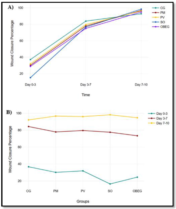
The statistical analysis results of intra-group (time-dependent) and inter-group (same time period) differences in WHRC, WCR (Figure 1), and WPCP are presented in Table 4.
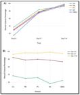
Changes in wound closure percentage over time for three different groups (A); Changes in wound closure percentage for three different groups (B).
When evaluating the differences between groups in WHRC data within the same time period, no statistical difference was found in any group between days 3 and 7. It was determined that rats in the SO group between days 0-3 and rats in the CG group between days 7-10 had statistically significantly lower WHRC than other groups. According to the data obtained, at the end of the 10th day, it was determined that the averages of the groups to which ointments prepared from all three plant extracts were applied were statistically significantly higher than those of the CG group in terms of WCP and WPCP. It was determined that the SO group, which had the lowest WCP and WPCP values between days 0 and 3, reached the highest level at the end of day 10 when compared to other groups. Although the average WCP and WPCP values of rats in the OBEG group were higher than those in the CG group, they were lower than those in the other groups (PM, PV, and SO). (Figures 2, 3, 4, and 5).
When the intra-group time-dependent relationship of WHRC data was evaluated, no statistical difference was found between the PM and OBEG groups. It was determined that WHRC was statistically significantly higher in the PV and SO groups between days 3-7, and in the CG group between days 0-3 and 3-7, compared to other days. When evaluating the intra-group time-dependent relationship of WCP data, statistically significant differences were observed on days 3, 7, and 10 in all groups. When evaluating the intra-group time-dependent relationship of WPCP data, no statistical difference was found between days 3-7 and 7-10 in all groups, but both periods showed a statistically significant increase compared to days 0-3.

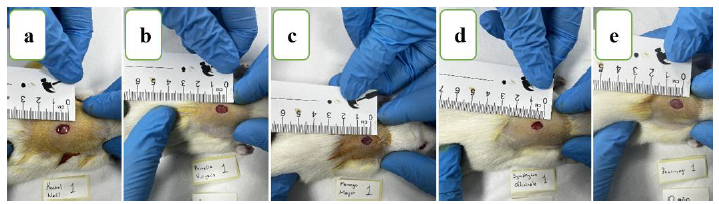
Macroscopic view of wounds in rats in all groups on day 0 of the study: CG group (a), PV group (b), PM group (c), SO group (d), OBEG group (e)
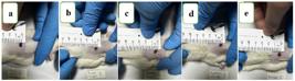
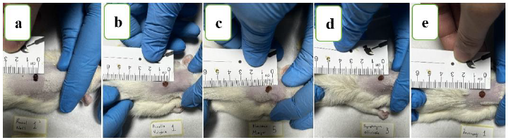
Macroscopic view of wounds in rats in all groups on the 3rd day of the study. CG group (a), PV group (b), PM group (c), SO group (d), OBEG group (e)
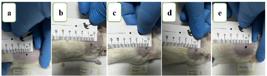
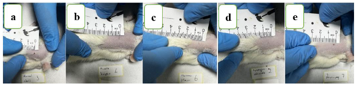
Macroscopic view of the wounds of rats in all groups on the 7th day of the study. CG group (a), PV group (b), PM group (c), SO group (d), OBEG group (e)

Macroscopic view of the wounds of rats in all groups on the 10th day of the study. CG group (a), PV group (b), PM group (c), SO group (d), OBEG group (e)
In the histopathological evaluation of the study, vascular granulation tissue composed of fibroblasts and dense collagen fibers was observed in the wound areas of rats from the PM, PV, and SO groups, with an epithelial layer partially or completely covering this granulation tissue. Histopathological examination of the wound areas of rats in the CG group revealed a higher concentration of inflammatory cells and the onset of epithelialization. When evaluated based on histopathological scores, rats in the PM, PV, and SO groups showed statistically significant improvement compared to those in the CG and OBEG groups. Although there was no statistical difference between the PM, PV, and SO groups, histopathological improvement was found to be better in the PV group (Table 5 and Figure 6).
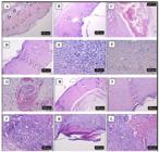
A) Normal histological appearance of the skin, (HxE staining, HCG group, scale bar: 500 µm), B) Epidermis not completely closed at the wound site (star) and increased connective tissue in the dermis (triangle), (HxE staining, CG group, scale bar: 500 µm), C) Areas of hemorrhage in the hypodermis (star), (H&E staining, CG group, scale bar: 200 µm), D) Epidermal formation (star) and increased connective tissue (triangle) and number of blood vessels (arrow) in the dermis (HxE staining, CG group, scale bar: 200 µm), E) Dense mononuclear cell infiltration (lymphocytes: arrows, macrophages: arrowheads) in the dermis (HxE staining, PM group, scale bar: 50 µm), F) Capillary blood vessels (arrows) and increased connective tissue with few mononuclear cell infiltrations in the dermis (HxE staining, PV group, scale bar: 100 µm), G) Areas of hemorrhage in the dermis (star) (HxE staining, PV group, scale bar: 100 µm), H) Histopathological appearance of the wound site in a healed animal (HxE staining, SO group, scale bar: 500 µm), I) Histopathological appearance of the wound site in a healed animal (HxE staining, SO group, scale bar: 200 µm), J) Mononuclear cell (lymphocyte: arrows) infiltration in the dermis (HxE staining, SO group, scale bar: 100 µm), K) Hyperkeratinization in the epidermis (star) (HxE staining, OBEG group, scale bar: 100 µm), L) Areas of hemorrhage (star) and dense cellular infiltrations (triangle) in the dermis (HxE staining, OBEG group, scale bar: 100 µm).
Compared to the HCG group, it was found that MDA levels increased in the CG group, while GSH levels and CAT and GSH-Px activities decreased (P<0,001). It was determined that the PM, PV, and SO groups showed decreased MDA levels and increased GSH levels, as well as CAT and GSH-Px activities, compared to the CG group (P < 0.001). It was determined that the MDA levels and CAT and GSH-Px activities in rats from the PM, PV, and SO groups were similar to the averages observed in the HCG group. Changes in GSH levels were found to be different from those in the CG group (P<0.001) but did not quite reach the HCG group averages. No statistically significant difference was found in SOD activity between the experimental groups in all parameters (P>0.05). Detailed findings regarding oxidative stress parameters are presented in Table 6.
DISCUSSION
A wound is defined as the disruption of the anatomical and physiological integrity of the skin caused by intrinsic or extrinsic factors such as trauma, heat, radiation, chemical agents, surgical interventions, diseases, and pressure (Karasu and Bakır, 2008; Polat, 2014; Tottoli et al., 2020). Wound healing is a process involving cellular, biochemical, and systemic activities, consisting of the stages of hemostasis, inflammation, proliferation, and remodeling that begin following wound formation (Parsak et al., 2007; Karasu and Bakır, 2008; Gökalp Özkorkmaz and Özay, 2009; Behm et al., 2012; Surme and Curuk, 2020). Since prehistoric times, humans have used many inorganic and organic substances to promote healthy and rapid wound healing (Akdogan, 2019). Phytotherapy, which plays an important role in the effective treatment of wounds, is widely used today with the support of modern medicine (Berk et al., 2015). In this study, the effects of Prunella vulgaris, Symphytum officinale, and Plantago major on wound healing were examined through macroscopic changes in wound surface area, histopathological and biochemical findings, and their suitability for clinical use was evaluated.
Products derived from herbal extracts used in wound healing affect at least one of the stages of wound healing (hemostasis, inflammation, proliferation, and remodeling) and/or create a protective effect against infection in the wound, thereby facilitating a faster and healthier outcome (Deshmukh and Gupta, 2013). In the evaluation of wound healing, macroscopic findings such as wound closure percentage and wound healing rate, along with histopathological findings including collagen synthesis, fibroblast density, increased epithelialization, and levels of inflammatory cells, are well-supported, valid, and widely used approaches (Dorai, 2012; Shah and Amini-Nik, 2017; Esfahani et al., 2019; Al- Warhi et al., 2022; Ozturan, 2024). Ozturan (2024) reported that, in their study investigating the effects of Calendula officinalis extract on wound healing, the wound surface area closure occurred significantly faster compared to the control group. A statistically significant increase in fibroblast count and a decrease in macrophage count was observed in rats within the same group. Al-Warhi et al. (2022) reported that in their study investigating the efficacy of Vitis vinifera seed extract on wound healing, the wound closure rate was significantly increased, and the results were supported by histopathological findings. Esfahani et al. (2019) reported a significant increase in the wound healing rate compared to the control group in their study using a combination of Plantago major and Aloe vera. In the same study, a significant increase in the number of fibroblasts and collagen volume was reported in the group where Plantago major and Aloe vera were used in combination, compared to the control groups. Mârza et al. (2024) investigated the effectiveness of Symphytum officinale extract on wound healing and reported that fibroblast activity, collagen synthesis, and reepithelialization levels increased significantly compared to the control group. This effect is attributed to rosmarinic acid, which can reduce free radical damage in skin tissue and regulate inflammation, as well as to allantoin and salvianolic acid compounds that promote epithelialization. All of these are found in Symphytum officinale extract. Zubaira et al. (2012) reported that in their study evaluating the proliferative and migratory effects of Plantago major extract on oral epithelial cells, the extract, when used at appropriate doses, could enhance new tissue formation through its phenolic compounds by exerting antioxidant, anti-inflammatory, and epitheliogenesis-supporting effects. In the same study, it was also reported that high doses of Plantago major extract may have a cytotoxic effect and adversely affect cell viability. Küpeli Akkol et al. (2022) investigated the effectiveness of Prunella vulgaris on wound healing and reported that the ursolic acid, chlorogenic acid, and rosmarinic acid in its composition significantly increased wound healing by inhibiting collagenase and elastase enzymes. In this study, when WCP and WPCP data were evaluated in rats treated with ointments prepared from Plantago major, Prunella vulgaris, and Symphytum officinale extracts, these values were found to be significantly higher than those in the CG group. There is no statistically significant difference between the PM, PV, and SO groups in terms of WCP and WPCP. However, it is noteworthy that rats in the SO group, which had the lowest average WHRC between days 0 and 3, had higher WCP and WPCP values at the end of the study. When evaluated histopathologically, vascular granulation tissue consisting of fibroblasts and dense collagen fibers was observed in the wound areas of rats in the PM, PV, and SO groups, along with an epithelial layer partially or completely covering this tissue. However, it was determined that inflammatory cells were more concentrated in rats in the CG group and that the epithelialization process had only just begun. When histopathological scoring data were examined, it was found that the mean scores of the PM, PV, and SO groups were statistically significantly higher than those of the CG group. Although there was no statistically significant difference between these three groups (PM, PV, and SO), histopathological scores were found to be ranked from high to low as PV, PM, and SO, respectively.
Another marker used to support or evaluate wound healing is oxidative stress parameters. Numerous studies have shown that antioxidant systems such as GSH, SOD, and CAT maintain oxidative stress in balance, creating an environment conducive to wound healing (Aksoy and Ozakpinar Bingol, 2014; Comino-Sanz et al., 2021; Al-Warhi et al., 2022). Ozturan (2024), investigated the effect of Calendula officinalis extract on wound healing and reported that the oxidative stress index was significantly lower in the group treated with the extract on days 3 and 14 compared to the control group. Al-Warhi et al. (2022) investigated the wound healing potential of Vitis vinifera (grapevine) seed extract in an excisional wound model and reported that it positively affected wound healing by eliminating reactive oxygen species (ROS) due to its strong antioxidant effect. The powerful antioxidant effect has been attributed to the phenolic compounds present in the extract, and it has been identified as a promising treatment option for wound healing. In this study, it was determined that the MDA levels in the PM, PV, and SO groups of rats were statistically significantly lower compared to the CG group, while the GSH levels, as well as the activities of CAT and GSH-Px enzymes, were statistically significantly higher. These data indicate that oxidative stress decreased in all three groups (PM, PV, and SO) and had a positive effect on wound healing.
CONCLUSION
In conclusion, because of phenolic compounds and flavonoids present in all three herbal extracts, the collagen volume and fibroblast density in the wound area rapidly increase, while oxidative stress decreases, creating an environment conductive to healing, leading to elevated WCP and WPCP values. In the comparison of these three plant extracts, Symphytum officinale was found to have a more positive effect on wound healing than the others in terms of WCP and WPCP, while Prunella vulgaris had a more positive effect in terms of histopathological evaluation. Based on all these data, the potential effectiveness of these herbal extracts in wound healing has been clearly demonstrated, and it has been concluded that more comprehensive advanced studies in the field of translational medicine are needed.
REFERENCES
- ADOM, M.B.; TAHER, M.; MUTALABISIN, M.F. et al. Chemical consituents and medical benefits of Plantago major. Biomed. Pharmacother., v.96, p.348-360, 2017.
- AEBİ, H. Catalase. In vitro. Methods Enzymol., v.105, p.121-126, 1984.
- AHMED, J.H.; EZER, N. Chemical constituents and biological activities of prunella l. species. Hujpharm, v.28, p.93-113, 2008.
- AKDOGAN, C. Investigation of the effects of St. John’s Wort (Hypericum Perforatum) oil on skin wound healing in mice. 2019. Thesis (Master's) - Institute of Health Sciences, Adnan Menderes University, Aydın, TUR.
- AKSOY, H.; OZAKPINAR BINGOL, O. Wound Healing and Oxidative Stress. Marmara Pharm. J., v.18, p.153-158, 2014.
- AL-WARHI, T.; ZAHRAN, E.M.; SELIM, S. et al. Antioxidant and woundhealing potential of vitis vinifera seeds supported by phytochemical characterization and docking studies. Antioxidants, v.11, p.1-20, 2022
- BAKTIR, G. Wound repair and experimental wound models. Experimed, v.9, p.130-137, 2019.
- BEHM, B.; BABILAS, P.; LANDTHALER, M. et al. Cytokines, chemokines and growth factors in wound healing. J. Eur. Acad. Dermatol. Venereol., v.26, p.812-820, 2012.
- BERK, A.; DOKUMACI, A.H.; KAYMAZ, M.H. Wound healing and medicinal plants used in the treatment of diabetic wounds. J. Health Sci., v.24, p.185-192, 2015.
- BEUTLER, E. Red cell metabolism. A manual of biochemical methods. 2.ed. New York: Grune and Starton, 1984. p.160.
- CALAPOGLU, N.S.; KARAGUZEL, A.; CALAPOGLU, M. Investigation of Plantago major extracts on mitotic index of human lymphocytes. İbn-i Sina Tıp Derg., v.8, p.105-109, 2003.
- COMİNO-SANZ, I.M.; LÓPEZ-FRANCO, M.D.; CASTRO, B. et al. The role of antioxidants on wound healing: A review of the current evidence. J. Clin. Med., v.10, p.1-22, 2021.
- DESHMUKH, P.T.; GUPTA, V.B. Embelin accelerates cutaneous wound healing in diabetic rats. J. Asian Nat. Prod. Res., v.15, p.158-165, 2013.
- DORAI, A.A. Wound care with traditional, complementary and alternative medicine. Indian J. Plast. Surg., v.45, p.418-424, 2012.
- ELLMAN, G.L.; COURTNEY, K.D.; ANDRES, V. et al. A new and rapid colorimetric determination of acetylcholinesterase activity. Biochem. Pharmacol., v.7, p.88-95, 1961.
- FAYDAOGLU, E.; SURUCUOGLU, M.S. Medical and aromatic plants antimicrobial, antioxidant activities and use opportunities. Erzinca Üniv. Fen Bilimleri Enstitüsü Derg., v.6, p.233-265, 2013.
- ESFAHANI, S.A.; KHOSHNEVISZADEH, M.; NOORAFSHAN, A. et al. The healing effect of Plantago major and aloe vera mixture in excisional full thickness skin wounds: stereological study. World J. Plast. Surg., v.8, p.51-57, 2019.
- GREENHALGH, D.G.; SPRUGEL, K.H.; MURRAY, M.J. et al. Stimulated healing in the genetically diabetic mouse. Am. J. Pathol.; v.136, p.1235-1246, 1990
- GÖKALP ÖZKORKMAZ, E.; OZAY, Y. Wound healing and some plants used in wound treatment. Turk. J. Sci. Rev., v.2, p.63-67, 2009.
- KANT, V.; GOPAL, A.; PATHAK, N.N. et al. Antioxidant and anti-inflammatory potential of curcumin accelerated the cutaneous wound healing in streptozotocin-induced diabetic rats. Int. Immunopharmacol., v.20, p.322-330, 2014.
- KARASU, A.; BAKIR, B. Wound and wound healing. Vet. Cerrahi Derg., v.14, p.36-43, 2008.
- KÜPELI AKKOL, E.; RENDA, G.; ILHAN, M. et al. Wound healing acceleration and anti-inflammatory potential of Prunella vulgaris L.: From conventional use to preclinical scientific verification. J. Ethnopharmacol., v. 295, p.1-9, 2022.
- MÂRZA, S.M.; DĂESCU, A.M.; PURDOİU, R.C. et al. Healing of skin wounds in rats using creams based on Symphytum officinale extract. Int. J. Mol. Sci., v.25, p.1-22, 2024
- OZTURAN, Y.A. Investıgatıon of wound healing effects of Calendula (Calendula officinalis) extract in mıce. 2024. Thesis (Doctoral) - Department of Veterinary Surgery, Graduate School of Health Sciences, Aydın Adnan Menderes University, Aydın, TUR.
- PARSAK, C.K.; SAKMAN, G.; CELIK, Ü. Wound healing, wound care and its complications. Arşiv Kaynak Tarama Derg., v.16, p.145-159, 2007.
- PLACER, Z.A.; CUSHMAN, L.; JOHNSON, B.C. Estimation of products of lipid peroxidation in biological fluids. Anal Biochem., v.16, p.359-364,1966.
- POLAT, E. Infected chronic an injured dog henna (lawsonia inermis) therapeutic use of plants. Harran Üniv. Vet. Fak. Derg., v.3, p.93-97, 2014
- POLAT, E.; KARADEMIR, K.; CEVIK, H.N. et al. Effect of microcurrent electrical therapy on healing of excisional skin wounds. Rev. MVZ Córdoba, v.30, p.1-10, 2025.
- POLAT, E; ATES, A.O.; OZKAN, A.S. et al. Efficacy of Punica granatum L. and Matricaria chamomilla L. herbal extracts on wound healing. Rev. Cient. FCV-LUZ, v. 36, p.1-10, 2026.
- SHAH, A.; AMINI-NIK, S. The role of phytochemicals in the inflam-matory phase of wound healing. Int. J. Mol. Sci., v.18, p.1-17, 2017.
- SUMATHI, S.; SENTHIL KUMAR, S.; BHARATHI, V. et al. Antibacterial activity of the plant extract of Symphytum officinale L. against selected pathogenic bacteria. Int. J. Res. Pharm. Sci., v.2, p.92-94, 2011.
- SUMBULOGLU, K.; SUMBULOGLU, V. Biostatistics. 19.ed. Ankara, Turkey: Hatiboglu Publishing, p.10-179, 2019.
- SUN, Y.; OBERLY, L.W.; YİNG, L.A. Simple method for clinical assay of superoxide dismutase. Clin. Chem., v.34, p.497-500, 1988.
- SURME, Y.; CURUK, G.N. Fitotherapy in wound care: tea tree oil. ERÜ Sağlık Bilimleri Fak. Derg., v.7, p.35-41, 2020.
- TOTTOLİ, E. M.; DORATİ, R.; GENTA, I. et al. Skin wound healing process and new emerging technologies for skin wound care and regeneration. Pharmaceutics, v.12, p.1-30, 2020.
- ZUBAİRA, M.; EKHOLMA, A.; NYBOMA, H. et al. Effects of Plantago major L. leaf extracts on oral epithelial cells in a scratch assay. J. Ethnopharmacol., v.141, p.825-830, 2012.
-
FINANCIAL SUPPORT
This project was financially supported by the Scientific and Technological Research Council of Türkiye (TÜBİTAK) within the scope of the 2209-A Project Support Program.
-
DATA AVAILABILITY STATEMENT
The research data that support the findings of this study are available from the corresponding author upon reasonable request.
The research data that support the findings of this study are available from the corresponding author upon reasonable request.