Macrolide-responsive histiocytic ulcerative colitis in a French bulldog - clinical and microbiological features - case report
ABSTRACT
Histiocytic ulcerative colitis (HUC) is a severe chronic enteropathy in dogs, frequently associated with adherent and invasive Escherichia coli (AIEC). This study reports a case of HUC in a French bulldog, unresponsive to enrofloxacin but with full clinical and histopathological remission after azithromycin treatment. The strain isolated from the patient was microbiologically characterized and compared to six other canine-origin strains. All 46 isolates analyzed were multidrug-resistant, and one showed phenotypic ESBL production. Three strains adhered in the presence of D-mannose, and most demonstrated invasiveness in HeLa cells despite the absence of virulence genes. The patient’s isolate (1.4) showed phenotypic characteristics compatible with AIEC, although definitive classification requires genomic sequencing. These findings highlight the importance of bacteriological diagnosis with culture and susceptibility testing to guide appropriate therapy and indicate that the circulation of multidrug-resistant strains in companion animals may pose a potential public health risk within the One Health context.
Keywords:
histiocytic ulcerative colitis; adherent and invasive Escherichia coli (AIEC); antimicrobial resistance; companion animals; One Health
RESUMO
A colite histiocítica ulcerativa (CHU) é uma enteropatia crônica grave em cães, frequentemente associada a Escherichia coli aderente e invasiva (AIEC). Este estudo descreve um caso de CHU em um buldogue francês, não responsivo à enrofloxacina, mas com remissão clínica e histopatológica após tratamento com azitromicina. A cepa isolada do paciente foi caracterizada quanto ao perfil de resistência antimicrobiana, adesão, invasão celular, grupo filogenético, sorotipagem e sobrevivência em macrófagos, sendo comparada a outras seis cepas de origem canina. Todas as 46 cepas analisadas foram multirresistentes e uma apresentou produção fenotípica de ESBL. Três cepas demonstraram adesão na presença de D-manose e quase todas apresentaram capacidade invasiva, mesmo sem genes de virulência detectáveis. A cepa do paciente (1.4) apresentou perfil compatível com AIEC, embora a confirmação definitiva dependa de análise genômica. Os achados reforçam a importância do diagnóstico bacteriológico com cultura e antibiograma para a escolha terapêutica adequada e evidenciam que a circulação de cepas multirresistentes em animais de companhia pode representar potencial risco à saúde pública no contexto de Saúde Única.
Palavras-chave:
colite histiocítica ulcerativa; Escherichia coli aderente e invasiva (AIEC); resistência antimicrobiana; animais de companhia; Saúde Única (One Health)
INTRODUCTION
Histiocytic ulcerative colitis (HUC), also named granulomatous colitis (GC), is a chronic enteropathy (Argenta et al., 2018), included on the list of inflammatory bowel diseases (IBD), which represents a disturbance in dogs and rarely in cats (van Kruiningen and Dobbins, 1979; Matsumoto et al., 2019). It is characterized on histopathological examination by a large colonic infiltrate of periodic acid-Schiff positive (PAS+) macrophages in the lamina propria (LP) and submucosa (Nolte et al., 2017).
HUC was initially described in Boxer dogs, which could suggest a breed-specific disease (Van Kruiningen et al., 1965). However, although Boxers display a greater predisposition to HUC, it is also described in French bulldogs, English bulldogs, American Staffordshire, Mastiffs, Alaskan Malamutes and Doberman pinschers (Stokes et al., 2001; Tanaka et al., 2003; Hostutler et al., 2004; Cerquetella et al., 2010).
The etiopathogenesis of HUC has not yet been fully clarified, however, studies show an association with intramucosal adherent and invasive Escherichia coli (AIEC) colonization (van Kruiningen et al., 2005; Simpson et al., 2006). Recently, a genome-wide association scan revealed single nucleotide polymorphisms associated with HUC in the gene encoding neutrophil cytosolic factor (NCF)-2, a gene related to the production of reactive oxygen species (ROS). The deficient mechanism of ROS production, an important microbicide, results in a compromised ability to eliminate intracellular pathogens (Craven et al., 2011). Furthermore, macrophages in the normal colonic mucosa present an anti-inflammatory phenotype that contributes to intestinal homeostasis. However, in HUC, macrophages have a pro-inflammatory phenotype, which is associated with the inability to kill the pathogen. The switch from an anti-inflammatory to a pro-inflammatory macrophage phenotype, mediated by cytokines, contributes to the inflammatory process of disease (Nolte et al., 2017). The AIEC associated with HUC adhere to and invade intestinal epithelial cells, residing and replicating within macrophages, similar to E. coli in Crohn’s disease. Moreover, the regional HUC distribution and macroscopic appearance resembles ulcerative colitis in humans (Darfeuille-Michaud et al., 1998; Hostutler et al., 2004; Simpson et al., 2006; Mansfield et al., 2009).
The clinical signs include tenesmus, haematochezia, soft stools, increased frequency of defecation and excessive mucus and can be accompanied by anaemia, hypoalbuminemia and weight loss (Churcher and Watson, 1997; Craven et al., 2010; Argenta et al., 2018).
Some studies indicate remission of clinical and histopathological signs after long-term antimicrobial treatment, and the most recommended antibiotics are fluoroquinolones (Davies et al., 2004; Hostutler et al., 2004; Mansfield et al., 2009).
In this study, a French Bulldog diagnosed with histiocytic ulcerative colitis, (HUC) that was unresponsive to fluoroquinolone treatment, was investigated. Considering the similarities between adherent-invasive Escherichia coli (AIEC)-associated enteropathies in animals and humans, and the potential for cross-species transmission of such strains, bacterial isolates were identified and characterized. Antimicrobial resistance profiles were assessed, and culture, adhesion and invasion assays in HeLa cells, pathotyping, phylogroup assignment, and serotyping were performed. These investigations contribute to a better understanding of zoonotic risks and antimicrobial resistance within the One Health framework.
ETHICAL ASPECTS
This research was not submitted to the Ethics Committee on Animal Use.
CASUISTRY
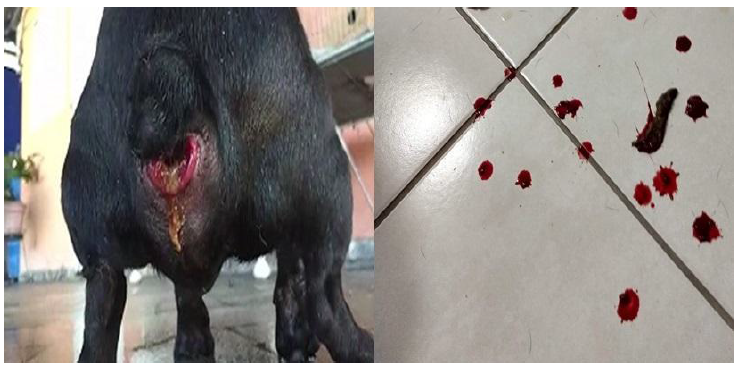
An 18-month-old, female, privately owned French bulldog, was attended in the Veterinary Hospital of Federal University of Viçosa presenting, during the last five months, chronic mucous-containing diarrhoea, haematochezia, increased frequency of defecation and tenesmus with a small rectal prolapse (Fig. 1). The most frequent sign (and most emphasized by the owners) was intense vocalization during defecation.
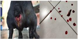
(A) A small rectal prolapse was present in the owner’s document. (B) Mucous-containing diarrhoea with serious haematochezia at the first clinical examination.
The patient was initially treated by a veterinary colleague with sulfasalazine, metronidazole, prednisone, and trimethoprim-sulfamethoxazole, without any improvement. The patient was alert, with a good body score (6/9), preserved appetite and normal physical parameters on clinical examination. Haematological, biochemical and coproparasitological analyses were performed, and normal results were obtained.
On ultrasound examination, increased echogenicity of the intestinal wall of the descending colon with a thickened wall measuring 0.51 cm, with preserved ultrasound appearance of the ascending and transverse colon, was observed. A colonoscopy examination was performed to collect biopsy samples and information on the appearance and integrity of the entire colon.
Prior to all colonoscopy exams, a complete blood count (CBC) and chemistry panel were performed, and the patient was prepared with eight hours of water and food fasting for the anaesthetic procedure. Under general inhalation anaesthesia, first a warm water enema was performed. A Veterinary Video Endoscope VET-8015 was used to acquire images and a biopsy forceps to obtain samples.
Enrofloxacin 5 mg/kg, prednisolone 0.5mg/kg, both every 12 hours, and commercial gastrointestinal diet therapy were started while awaiting the biopsy results. After the biopsy samples were obtained, all material was formalin-fixed and embedded in paraffin wax. Sections were cut to 4 μm and stained with the haematoxylin-eosin and periodic acid-Schiff (PAS) methods. Sections were analysed, and a standard form for the assessment of colonic mucosa was completed (Washabau et al., 2010). The biopsy samples were washed thoroughly in saline solution (NaCl 0.9%) and aseptically transferred to a disposable tissue grinder to release intracellular bacteria. The samples were streaked on MacConkey agar (HiMedia®) and Nutrient agar (HiMedia®). About five colonies were selected from each plate for biochemical identification using EPM, MILi and Simmons Citrate (Enterokit - PROBAC®), and Escherichia coli isolates were stored in Brain Heart Infusion (BHI - HiMedia®) with glycerol 20% (Merck®) at -80ºC for further assays. Histiocytic ulcerative colitis was confirmed by histopathological examination.
Fifteen days after the initial treatment, the patient had not improved, prednisolone was suspended and amoxicillin-clavulanate 15mg/kg and metronidazole 15mg/kg prescribed, every 12 hours, in combination with the enrofloxacin already prescribed for eight weeks.
After eight weeks of treatment, the patient showed no clinical improvement. However, there was weight loss and worsening of haematochezia. Another colonoscopy was performed to collect biopsy samples. The biopsy samples were washed thoroughly in saline solution (NaCl 0.9%) and sent for culture and antibiogram. In addition, haematological and biochemical tests were repeated, and mild anaemia (5.06×10¹²RBCs/L) with a low haematocrit (Hct) value (33.6%) and discrete hyperglobulinemia (2.61g/dL) were observed. After the procedure, empirical therapy with subcutaneous administration of amikacin 20mg/kg and faecal microbiota transplantation (FMT) from a dog from the same owner were prescribed, both once daily, and no improvement was achieved.
The antimicrobial susceptibility test was performed as recommended by the Clinical and Laboratory Standards Institute protocol (Performance standards for antimicrobial susceptibility testing, 2018a). The antimicrobials (Oxoid Ltd., Basingstoke, Hants, UK) and concentrations used for the disk diffusion test were aztreonam (ATM) - 30µg, cephazolin (CFZ) - 30µg, gentamicin (GEN) - 10µg, cefoxitin (CFO) - 30µg, trimethoprim-sulfamethoxazole (SXT) - 25µg, enrofloxacin (ENO) - 5µg, chloramphenicol (CHL) - 30µg, tetracycline (TET) - 30µg, imipenem (IPM) - 10µg, ampicillin (AMP) - 10µg, ciprofloxacin (CIP) - 5µg and azithromycin (AZI) - 15µg. The interpretation of the enrofloxacin results was based on CLSI (Performance standards for antimicrobial disk and dilution susceptibility tests for bacteria isolated from animals, 2018b). Detection of extended-spectrum beta-lactamase (ESBL) was performed by double-disk approximation test, using the following antimicrobials with the respective disk concentration: amoxicillin-clavulanate (AMC) - 30µg, ceftazidime (CAZ) - 30µg, cefepime (FEP) - 30µg and ceftriaxone (CRO) - 30µg. E. coli ATCC 25922 strain was used for quality control.
The protocol for the adhesion assay was adapted from Cravioto et al. (1979), with a six-hour incubation. HeLa cells were grown in 24-well plates to 80% confluence for the test. Bacteria 1.5×10⁸ were added to wells containing Dulbecco's Modified Eagle Medium (DMEM) supplemented with Methyl α-D-mannopyranoside (Sigma-Aldrich Ltd., Brazil) at a final concentration of 1%. The positive controls used were E. coli E2348/69 (EPEC) and E. coli 042 (EAEC), and E. coli DH5-α was used as a negative control.
The invasion assay was performed according to Chue-Gonçalves et al. (2018), by fluorescence detection. HeLa cells were cultured in a 96-well plate to 100% confluence for the test. E. coli O152 (EIEC) was used as a positive control and E. coli HB101 as a negative control.
Phylogenetic group determination was performed according to Clermont et al. (2013) by detection of the chuA, arpA, and yjaA genes and the TspE4.C2 DNA fragment. The diarrheagenic E. coli (DEC) virulence factors surveyed were eae, bfpA, aggR, elt, est, ipaH, stx1, stx2 and hlyA (Paton and Paton, 1998; López-Saucedo et al., 2003; Aranda et al., 2007).
Serotyping was performed by agglutination reactions with rabbit sera obtained for 188 O (somatic) and 53 H (flagellar) antigens.
The J774A.1 macrophage survival and replication assay was performed according to Glasser et al. (2001) with modifications. The positive and negative controls used were, respectively, Salmonella enterica serovar Typhimurium UK-1 and E. coli DH5α.
J774A.1 cells were cultured in 24-well plates with 1 mL of RPMI supplemented with 10% foetal bovine serum (FBS), bacterial culture was added at a multiplicity of infection (MOI) of 10 and the plate was incubated for 1 hour at 37ºC with 5% CO2. The wells were washed with PBS (pH 7.4), 1 ml of RPMI supplemented with 10% FBS and amikacin (200 µg/mL) was added, and the plate was incubated for 2 hours at 37ºC with 5% CO2. After incubation, the wells were washed with PBS, 1 mL of RPMI supplemented with 10% FBS and amikacin (20µg/mL) was added, and the plate was incubated at 37ºC with 5% CO2 until the stipulated times.
The lysis process, in which wells were washed with PBS, and 1mL of 0.1% Triton X-100 was added, was performed at times 2 and 24h. The contents were resuspended, diluted and plated on MacConkey agar for CFU (colony forming units)/mL counting.
The mucosal culture showed abundant multiplication of multidrug-resistant Escherichia coli, resistant to all fluoroquinolones tested. The bacteria had in vitro sensitivity to azithromycin; therefore, long-term therapy with commercial azithromycin (Azi® - Sigma Pharma - 200mg/5ml) 10mg/kg once daily was prescribed.
An improvement in clinical signs was observed in the first seven days of therapy with azithromycin. The patient showed reduction of the frequency of diarrhoea, tenesmus, haematochezia and vocalization during defecation. After eight weeks of treatment, the animal achieved considerable weight gain and showed no clinical signs of the disease. Furthermore, the patient did not present any side effects due to long-term azithromycin. At the third colonoscopy examination, a healthy and normal mucosa was observed, and the absence of macroscopic signs was achieved. Moreover, the biopsy showed a restructuring of the colonic mucosa. Therapy was discontinued on the basis of those colonoscopy and histopathology findings, as well as the absence of clinical signs.
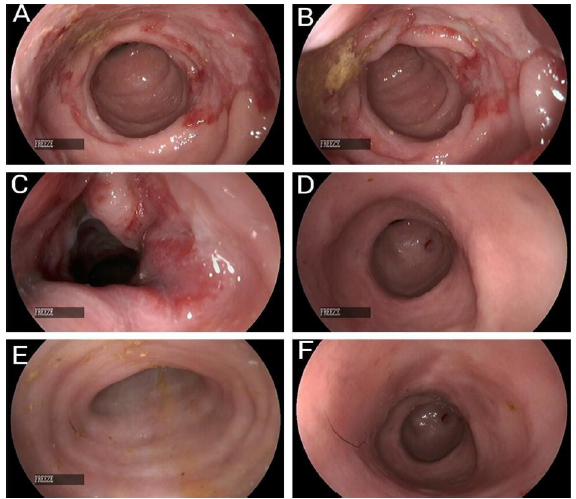
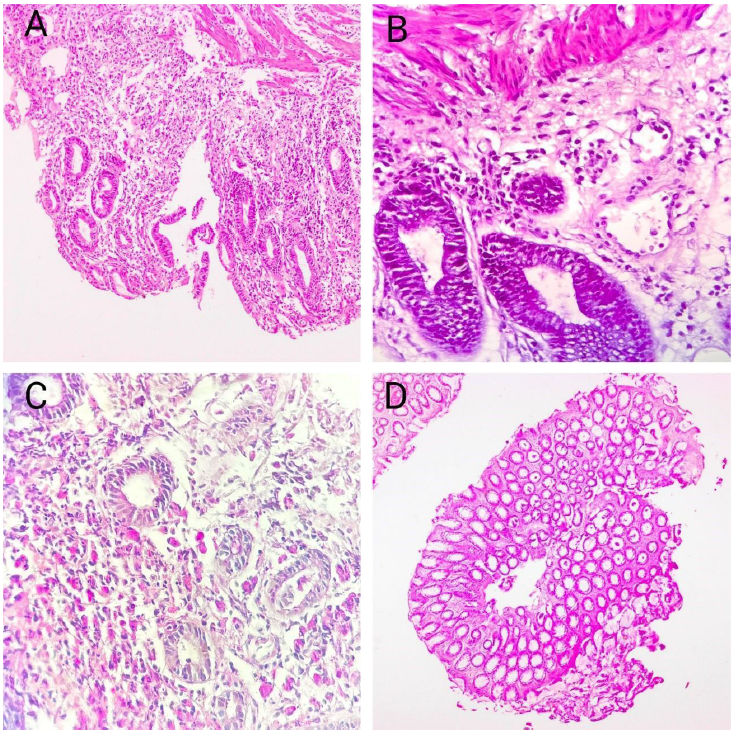
Initial endoscopic examination of the lower gastrointestinal tract revealed severe mucosal abnormalities, including marked hyperaemia, discoloration, friability, and irregular ulcerations (Fig. 2-A, 2-B, 2-C). These lesions extended approximately 5 cm along the distal descending colon and rectum, as measured by the endoscopic scale. Ulceration was associated with oedema and mucosal haemorrhage. Histopathological analysis of colonic biopsies obtained during the first colonoscopy showed irregular distribution of lesions. There was moderate surface epithelial damage, goblet cell depletion, mild crypt hyperplasia and dilation, and lamina propria infiltration with lymphocytes, plasma cells, neutrophils, and macrophages. Periodic acid-Schiff (PAS) staining revealed macrophages with foamy cytoplasm, consistent with the presence of histiocytic inflammation (Fig. 3).
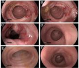
(A), (B), (C) show a severe hyperaemia with extensive, regular and friable ulceration on the mucosa of the descending colon during the first diagnostic colonoscopy. Figure (D), (E), (F) show a normal colonic mucosa with a good and healthy appearance after clinical macrolide treatment. Image Sources: Own authors.
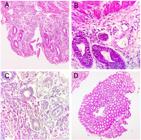
Photomicrographs of histologic sections from colonic endoscopic biopsies. All images were obtained by optical microscopy. (A) moderate epithelial injury and expansion of lamina propria and submucosa by inflammatory infiltrate - H&E stain, ×100. (B) Mild crypt dilatation, distortion and a paucity of goblet cells - H&E stain, ×400. (C) Foamy macrophages spread in lamina propria with staining of cytoplasm periodic acid-Schiff-positive - PAS+ stain, ×200. (D) Aspect of colonic mucosal after the macrolide treatment, indicating normal cellular architecture - H&E stain, ×40.
Following initial antimicrobial therapy (amoxicillin-clavulanate, enrofloxacin, and metronidazole), a second colonoscopy revealed disease progression, with lesion extension increasing to approximately 15 cm. After long-term treatment with azithromycin and subsequent clinical improvement, a third colonoscopy showed macroscopically normal mucosa, with a healthy and intact appearance (Fig. 2-D, 2-E, 2-F).
A total of 46 Escherichia coli isolates were recovered and identified via biochemical assays. All isolates (100%) were resistant to five antibiotics: ampicillin, enrofloxacin, chloramphenicol, ciprofloxacin, and trimethoprim-sulfamethoxazole. Additional resistance was observed against gentamicin (71%), tetracycline (67%), cefazolin (36%), amoxicillin-clavulanate (36%), and azithromycin (23%). Only isolate 1.4 produced extended-spectrum β-lactamase (ESBL) in phenotypic testing. All isolates met the criteria for multidrug resistance (MDR), as defined by Magiorakos et al. (2012), due to resistance to three or more antimicrobial classes.
Seven representative isolates (1.4, 3.4, 4.1, 5.2, 6.2, 6.5, and 7.1) were selected for further characterization based on distinct resistance profiles. Three isolates (1.4, 5.2, and 6.2) exhibited mannose-resistant adhesion to HeLa cells. Invasion assays revealed that six of the seven isolates demonstrated the ability to invade HeLa cells, with isolate 3.4 exhibiting the highest degree of invasiveness. Isolate 6.2 did not exhibit any invasive capacity. No known E. coli virulence genes were detected by PCR in any of the tested isolates. Serotyping identified isolates 5.2 and 6.2 as O132:HNM, and isolate 1.4 as O20:H4. The phylogenetic classification revealed distribution among groups A, B1, and B2.
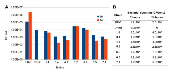
To assess survival and replication within macrophages, the seven selected isolates were tested in a murine macrophage cell line. Colony-forming unit (CFU) counts for isolates 1.4, 5.2, and 6.2 remained stable after 24 hours, with isolate 5.2 maintaining identical bacterial counts at both time points (2.8 × 10⁴ CFU/mL). The remaining isolates exhibited an approximate 1-log reduction in CFU counts over the incubation period. As expected, the negative control strain did not survive intracellularly (Fig. 4).
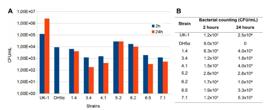
Survival and replication within J774A.1 macrophages. A: Graph of CFU/mL counting of the tested strains at times 2 and 24 hours. B: Table of bacterial counts of each tested strain at times 2 and 24 hours.
DISCUSSION
The emergence of microbial pathogens with invasive potential has been associated with inflammatory bowel diseases in both humans and animals, including adherent-invasive Escherichia coli (AIEC) (Shawki and McCole, 2017). Darfeuille-Michaud et al. (2004) defined AIEC strains by three main characteristics: absence of DEC virulence genes, adherence to and invasion of epithelial cells, and the ability to survive and replicate within macrophages. In this study, all isolates lacked DEC virulence genes. Three strains exhibited mannose-resistant adherence, and six out of seven demonstrated invasive potential in HeLa cells. One isolate showed particularly high invasiveness.
The increase in AIEC populations in the intestine, coupled with reduced microbial diversity, may contribute to epithelial barrier dysfunction and inflammation (Shawki and McCole, 2017). This mechanism is linked to the ability of AIEC to bind to CEACAM6 receptors on enterocytes via type 1 pili (Lee et al., 2019). According to previous studies, the capacity of E. coli to adhere to, invade, and replicate within intestinal cells is a potential virulence factor in the pathogenesis of HUC (Manchester et al., 2013; Argenta et al., 2018). The adherence of three isolates (1.4, 5.2 and 6.2) in the presence of mannose suggests the use of alternative adhesins, supporting the presence of multiple adherence mechanisms.
Regarding phylogeny, the most prevalent group was B1 (57%), followed by groups A (29%) and B2 (14%). Although phylogroup A has been described as predominant in AIEC strains (Conte et al., 2014), many characterized AIEC strains fall within the B2 group (Martinez-Medina et al., 2011; Lee et al., 2019). However, Lee et al. (2019) found no significant association between B2 and enhanced invasion or survival. This suggests that AIEC strains may belong to various phylogroups, particularly in non-human hosts. The Clermont phylogenetic classification, originally developed for human E. coli, may not fully capture the diversity of animal-derived isolates.
Multidrug resistance (MDR) poses a major concern in both human and veterinary medicine. The presence of MDR bacteria and the influence of those profiles on disease treatment should be emphasized. The United Nations Interagency Coordination Group (No time to wait: securing the future from drug-resistant infections, 2019) reported the occurrence of at least 700,000 worldwide deaths each year due to infections caused by MDR microorganisms, and MDR bacteria are well recognized as one of the most serious current public health problems.
In this study, all E. coli isolates were classified as MDR and exhibited resistance to five commonly used antimicrobials, including trimethoprim-sulfamethoxazole, fluoroquinolones, chloramphenicol, and ampicillin. One isolate (1.4) exhibited phenotypic ESBL production. MDR strains can act as reservoirs of resistance genes and pose a zoonotic risk, especially in close human-animal contact scenarios (Barrios-Villa et al., 2018).
Horizontal gene transfer and selective antibiotic pressure contribute to resistance dissemination. Resistance genes may be transferred between commensal and pathogenic bacteria (Guardabassi, 2004), especially under conditions of inappropriate antibiotic use or premature discontinuation of treatment (No time to wait: securing the future from drug-resistant infections, 2019; Van Hoek et al., 2011). Fluoroquinolone resistance, observed in all isolates, has been linked to widespread use in veterinary medicine (McEwen and Fedorka‐Cray, 2002), and its presence in HUC cases may lead to treatment failure and euthanasia (Craven et al., 2010).
One strain demonstrated ESBL production by phenotypic tests. It is important to emphasize that the ESBL-producing Enterobacteriaceae are a type of MDR bacteria that has been associated with a high zoonotic potential, which can be easily transferred from animals to humans and the environment by direct or indirect contact (Ewers et al., 2010; Huijbers et al., 2014; Blaak et al., 2015; Von Salviati et al., 2015; Dupouy et al., 2019). The Centers for Disease Control and Prevention (CDC) estimated that ESBL-producing E. coli caused over 9,000 deaths and $9.2 billion in healthcare costs in 2017 alone (Antibiotic resistance threats in the United States. Atlanta, 2019).
In this case, the initial clinical failure may be attributed to empirical treatment with enrofloxacin, despite its previous reported success in HUC cases (Hostutler et al., 2004; Mansfield et al., 2009; Manchester et al., 2013; Matsumoto et al., 2019). Given the increasing prevalence of MDR bacteria, biopsy, bacterial culture, and antimicrobial susceptibility testing should be performed early in suspected HUC cases.
Although beta-lactam resistance was not predominant in this study, this class of antibiotic usually results in none or weak intramacrophage activity (Chifiriuc et al., 2016). Effective therapy for HUC requires antimicrobials with both susceptibility in vitro and intracellular activity. Azithromycin, ciprofloxacin, clarithromycin, rifampicin, tetracycline, and trimethoprim are among the antibiotics that can penetrate macrophages (Subramanian et al., 2008). This supports the successful clinical outcome observed with azithromycin in this case.
To the authors’ knowledge, this is the first report of complete remission of HUC with azithromycin monotherapy, with no recurrence at the three-year follow-up.
The serotypes identified in this study (O132:HNM and O20:H4) are not typically associated with known E. coli pathotypes (Croxen et al., 2013). Similar serotypes have not been reported in previous studies of AIEC strains isolated from HUC (Simpson et al., 2006; Dogan et al., 2020; Manchester et al., 2021). However, the broad serotype diversity observed among AIEC strains may explain these findings (Martinez-Medina and Garcia-Gil, 2014). The O132:HNM serotype was described in Shiga toxin-producing E. coli (STEC) strains in humans and bovines/beef cattle (Gyles et al., 1998; Hussein and Sakuma, 2005). On the other hand, a recent study by Ori et al. (2019) reported the prevalence of the O132:H21 serotype in enteroinvasive E. coli (EIEC) isolated from human enteric infections in Brazil. In the same study, another EIEC isolate was identified as O132:HNM, suggesting a possible zoonotic link. Similarly, the O20:H4 serotype has also been associated with zoonotic potential in previous reports (Buvens and Piérard, 2012; Amézquita-López et al., 2016).
New serotypes of E. coli can be found in different animal species, such as dogs and cats. The absence of positive results in agglutination tests for somatic and flagellar antigens in four isolates may be an epidemiological characteristic of diarrheagenic E. coli (DEC). The bacterial invasion and phenotypic features (negative motility and lysine decarboxylase) are characteristic of the enteroinvasive E. coli (EIEC) pathotype. However, the absence of the ipa gene-commonly used to identify this pathotype-and the biochemical profile of the isolates may exclude this possibility. Instead, they support the hypothesis of an AIEC infection, consistent with previous reports describing histiocytic ulcerative colitis (HUC) associated with AIEC. Furthermore, EIEC infection in dogs is uncommon, having been reported primarily in humans and non-human primates (Croxen et al., 2013).
The tested strains showed a lower survival capacity in macrophages than those reported in previous studies. Glasser et al. (2001) evaluated the survival and replication capacity of 15 AIEC strains within J774A.1 macrophages and expressed the results as a survival percentage calculated at each time point, considering the bacterial count at 2 hours as 100%. All strains were able to replicate for up to 48 hours, with survival rates exceeding 100% within 24 hours. However, as the present study assessed bacterial survival at a single time point, a direct comparison with the results obtained by Glasser et al. (2001) was not possible.
Martinez-Medina et al. (2009) used the 24 hours survival percentage to classify strains as able to survive (% survival ≥100%) or to replicate (% survival ≥200%) within macrophages. Using this classification, only strain 5.2 was capable of surviving; however, the use of percentages in high values (exponential or log) would not be the best way to analyse bacterial survival, since all strains showed close CFU/mL values between 2 and 24 h, demonstrating a considerable ability to survive despite the low percentage. Importantly, the absence of 48-hour survival assays and whole-genome sequencing limits the ability to definitively confirm AIEC identity
Nonetheless, several isolates demonstrated adherence and invasiveness in HeLa cells, consistent with the AIEC phenotype. AIEC classification within DEC remains poorly defined.
Among the seven isolates, strains 1.4, 5.2, and 6.2 were notable due to similar log values in 2 hours and 24 hours. While 6.2 was non-invasive and 5.2 was susceptible to azithromycin, isolate 1.4 showed positive adherence and invasion, MDR profile, B2 phylogroup affiliation, and stable intracellular survival, supporting its classification as a potential AIEC strain.
CONCLUSION
The present study reports a case of histiocytic ulcerative colitis (HUC) in a dog associated with Escherichia coli isolates that exhibited phenotypic features suggestive of the adherent-invasive E. coli (AIEC) pathotype, including adherence and invasion of HeLa cells and survival in macrophages. Among the seven isolates tested, strain 1.4 showed the most consistent profile with previous AIEC descriptions, presenting adherence, invasion, survival at similar log CFU between 2 and 24 hours, and multidrug resistance, including an ESBL phenotype. However, the absence of DEC virulence genes, the lack of genomic sequencing, and the single time-point macrophage assay limited the definitive classification of these isolates as AIEC. These findings illustrate the diagnostic challenges in confirming AIEC infection and emphasize the importance of culture and antimicrobial susceptibility testing for guiding therapy in HUC cases.
REFERENCES
- AMÉZQUITA-LÓPEZ, B.A.; QUIÑONES, B.; SOTO-BELTRÁN, M. et al. Antimicrobial resistance profiles of Shiga toxin-producing Escherichia coli O157 and non-O157 recovered from domestic farm animals in rural communities in Northwestern Mexico. Antimicrob. Resist. Infect. Control., v.5, p.1, 2016.
- ANTIBIOTIC resistance threats in the United States. Atlanta: U.S. Department of Health and Human Services, Centers for Disease Control and Prevention, 2019. 113p.
- ARANDA, K.R.S.; FABBRICOTTI, S.H.; FAGUNDES-NETO, U.; SCALETSKY, I.C.A. Single multiplex assay to identify simultaneously enteropathogenic, enteroaggregative, enterotoxigenic, enteroinvasive and Shiga toxin-producing Escherichia coli strains in Brazilian children. FEMS Microbiol. Lett., v.267, p.145-150, 2007.
- ARGENTA, F.F.; SOUZA, S.O.; MEIRELLES, L.S. et al. Histiocytic ulcerative colitis in an American Staffordshire Terrier. J. Comp. Pathol., v.165, p.40-44, 2018.
- BARRIOS-VILLA, E.; CORTÉS-CORTÉS, G.; LOZANO-ZARAÍN, P. et al. Adherent/invasive Escherichia coli (AIEC) isolates from asymptomatic people: new E. coli ST131 O25:H4/H30-Rx virotypes. Ann. Clin. Microbiol. Antimicrob., v.17, p.42, 2018.
- BLAAK, H.; VAN HOEK, A.H.A.M.; HAMIDJAJA, R.A. et al. Distribution, numbers, and diversity of ESBL-producing E. coli in the poultry farm environment. PLoS One, v.10, p.e0135402, 2015.
- BUVENS, G.; PIÉRARD, D. Low prevalence of STEC autotransporter contributing to biofilm formation (Sab) in verocytotoxin-producing Escherichia coli isolates of humans and raw meats. Eur. J. Clin. Microbiol. Infect. Dis., v.31, p.1463-1465, 2012.
- CERQUETELLA, M.; SPATERNA, A.; LAUS, F. et al. Inflammatory bowel disease in the dog: differences and similarities with humans. World J. Gastroenterol., v.16, p.1050, 2010.
- CHIFIRIUC, M.C.; HOLBAN, A.M.; CURUTIU, C. et al. Antibiotic drug delivery systems for the intracellular targeting of bacterial pathogens. In: GRUMEZESCU, A.M. (Ed.). Smart drug delivery system. Rijeka: InTech, 2016.
- CHUE-GONÇALVES, M.; CUSTÓDIO, C.C.; PELAYO, J.S. et al. New approach for detection of Escherichia coli invasion to HeLa cells. J. Microbiol. Methods, v.152, p.31-35, 2018.
- CHURCHER, R.; WATSON, A. Canine histiocytic ulcerative colitis. Aust. Vet. J., v.75, p.710-713, 1997.
- CLERMONT, O.; CHRISTENSON, J.K.; DENAMUR, E.; GORDON, D.M. The Clermont Escherichia coli phylo-typing method revisited: improvement of specificity and detection of new phylo-groups. Environ.Microbiol. Rep., v.5, p.58-65, 2013.
- CONTE, M.P.; LONGHI, C.; MARAZZATO, M. et al. Adherent-invasive Escherichia coli (AIEC) in pediatric Crohn's disease patients: phenotypic and genetic pathogenic features. BMC Res Notes. v.7, p.748, 2014.
- CRAVEN, M.; DOGAN, B.; SCHUKKEN, A. et al. Antimicrobial resistance impacts clinical outcome of granulomatous colitis in Boxer dogs. J. Vet. Intern. Med., v.24, p.819-824, 2010.
- CRAVEN, M.; MANSFIELD, C.S.; SIMPSON, K.W. Granulomatous colitis of Boxer dogs. Vet. Clin. North Amer. Small Anim. Pract., v.41, p.433-445, 2011.
- CRAVIOTO, A.; GROSS, R.J.; SCOTLAND, S.M.; ROWE, B. An adhesive factor found in strains of Escherichia coli belonging to the traditional infantile enteropathogenic serotypes. Curr. Microbiol., v.3, p.95-99, 1979.
- CROXEN, M.A.; LAW, R.J.; SCHOLZ, R. et al. Recent advances in understanding enteric pathogenic Escherichia coli. Clin. Microbiol. Rev., v.26, p.822-880, 2013.
- DARFEUILLE-MICHAUD, A.; BOUDEAU, J.; BULOIS, P. et al. High prevalence of adherent-invasive Escherichia coli associated with ileal mucosa in Crohn’s disease. Gastroenterology, v.127, p.412-421, 2004.
- DARFEUILLE-MICHAUD, A.; NEUT, C.; BARNICH, N. et al. Presence of adherent Escherichia coli strains in ileal mucosa of patients with Crohn’s disease. Gastroenterology, v.115, p.1405-1413, 1998.
- DAVIES, D.; O’HARA, A.; IRWINN, P.; GUILFORD, W. Successful management of histiocytic ulcerative colitis with enrofloxacin in two Boxer dogs. Aust. Vet. J., v.82, p.58-61, 2004.
- DOGAN, B.; ZHANG, S.; KALLA, S.E. et al. Molecular and phenotypic characterization of Escherichia coli associated with granulomatous colitis of Boxer dogs. Antibiotics, v.9, p.540, 2020.
- DUPOUY, V.; ABDELLI, M.; MOYANO, G. et al. Prevalence of beta-lactam and quinolone/fluoroquinolone resistance in Enterobacteriaceae from dogs in France and Spain-characterization of ESBL/pAmpC isolates, genes, and conjugative plasmids. Front. Vet. Sci., v.6, p.279, 2019.
- EWERS, C.; GROBBEL, M.; STAMM, I. et al. Emergence of human pandemic O25:H4-ST131 CTX-M-15 extended-spectrum-β-lactamase-producing Escherichia coli among companion animals. J. Antimicrob. Chemother., v.65, p.651-660, 2010.
- GLASSER, A.L.; BOUDEAU, J.; BARNICH, N. et al. Adherent invasive Escherichia coli strains from patients with Crohn’s disease survive and replicate within macrophages without inducing host cell death. Infect. Immun., v.69, p.5529-5537, 2001.
- GUARDABASSI, L. Pet animals as reservoirs of antimicrobial-resistant bacteria: review. J. Antimicrob. Chemother., v.54, p.321-332, 2004.
- GYLES, C.; JOHNSON, R.; GAO, A. et al. Association of enterohemorrhagic Escherichia coli hemolysin with serotypes of Shiga-like-toxin-producing Escherichia coli of human and bovine origins. Appl. Environ. Microbiol., v.64, p.4134-4141, 1998.
- HOSTUTLER, R.A.; LURIA, B.J.; JOHNSON, S.E. et al. Antibiotic-responsive histiocytic ulcerative colitis in 9 dogs. J. Vet. Intern. Med., v.18, p.499, 2004.
- HUIJBERS, P.M.C.; GRAAT, E.A.M.; HAENEN, A.P.J. et al. Extended-spectrum and AmpC β-lactamase-producing Escherichia coli in broilers and people living and/or working on broiler farms: prevalence, risk factors and molecular characteristics. J. Antimicrob. Chemother, v.69, p.2669-2675, 2014.
- HUSSEIN, H.S.; SAKUMA, T. Invited review: prevalence of Shiga toxin-producing Escherichia coli in dairy cattle and their products. J. Dairy Sci., v.88, p.450-465, 2005.
- LEE, J.G.; HAN, D.S.; JO, S.V. et al. Characteristics and pathogenic role of adherent-invasive Escherichia coli in inflammatory bowel disease: potential impact on clinical outcomes. PLoS One, v.14, p.e0216165, 2019.
- LÓPEZ-SAUCEDO, C.; CERNA, J.F.; VILLEGAS-SEPULVEDA, N. et al. Single multiplex polymerase chain reaction to detect diverse loci associated with diarrheagenic Escherichia coli. Emerg.Infect. Dis., v.9, p.127, 2003.
- MAGIORAKOS, A.P.; SRINIVASAN, A.; CAREY, R.B. et al. Multidrug-resistant, extensively drug-resistant and pandrug-resistant bacteria: an international expert proposal for interim standard definitions for acquired resistance. Clin. Microbiol. Infect., v.18, p.268-281, 2012.
- MANCHESTER, A.C.; DOGAN, B.; GUO, Y.; SIMPSON, K.W. Escherichia coli-associated granulomatous colitis in dogs treated according to antimicrobial susceptibility profiling. J. Vet. Intern. Med., v.35, p.150-161, 2021.
- MANCHESTER, A.C.; HILL, S.; SABATINO, B. et al. Association between granulomatous colitis in French Bulldogs and invasive Escherichia coli and response to fluoroquinolone antimicrobials. J. Vet. Intern. Med., v.27, p.56-61, 2013.
- MANSFIELD, C.S.; JAMES, F.E.; CRAVEN, M. et al. Remission of histiocytic ulcerative colitis in Boxer dogs correlates with eradication of invasive intramucosal Escherichia coli. J. Vet. Intern. Med., v.23, p.964-969, 2009.
- MARTINEZ-MEDINA, M.; ALDEGUER, X.; LOPEZ-SILES, M. et al. Molecular diversity of Escherichia coli in the human gut: new ecological evidence supporting the role of adherent-invasive E. coli (AIEC) in Crohnʼs disease. Inflam. Bowel Dis., v.15, p.872-882, 2009.
- MARTINEZ-MEDINA, M.; GARCIA-GIL, J.; BARNICH, N. et al. Adherent-invasive Escherichia coli phenotype displayed by intestinal pathogenic E. coli strains from cats, dogs, and swine. Appl. Environ. Microbiol., v.77, p.5813-5817, 2011.
- MARTINEZ-MEDINA, M.; GARCIA-GIL, L.J. Escherichia coli in chronic inflammatory bowel diseases: an update on adherent invasive Escherichia coli pathogenicity. J. Gastrointest. World Jathophysiol., v.5, p.213, 2014.
- MATSUMOTO, I.; NAKASHIMA, K.; MORITA, H. et al. Escherichia coli-induced granulomatous colitis in a cat. J. Feline Med. Surg. Open Rep., v.5, p.205511691983653, 2019.
- MCEWEN, S.A.; FEDORKA‐CRAY, P.J. Antimicrobial use and resistance in animals. Clin. Infect. Dis., v.34, p.S93-S106, 2002.
- NO TIME to wait: securing the future from drug-resistant infections - report to the Secretary-General of the United Nations. Geneva: World Health Organization, 2019. (United Nations Interagency Coordination Group on Antimicrobial Resistance).
- NOLTE, A.; JUNGINGER, J.; BAUM, B.; HEWICKER-TRAUTWEIN, M. Heterogeneity of macrophages in canine histiocytic ulcerative colitis. Innate Immun., v.23, p.228-239, 2017.
- ORI, E.L.; TAKAGI, E.H.; ANDRADE, T.S. et al. Diarrhoeagenic Escherichia coli and Escherichia albertii in Brazil: pathotypes and serotypes over a 6-year period of surveillance. Epidemiol. Infect., v.147, p.e10, 2019.
- PATON, A.W.; PATON, J.C. Detection and characterization of Shiga toxigenic Escherichia coli by using multiplex PCR Assays for stx 1, stx 2, eaeA, enterohemorrhagic E. coli hlyA, rfb O111, and rfb O157. J. Clin. Microbiol., v.36, p.598-602, 1998.
- PERFORMANCE standards for antimicrobial disk and dilution susceptibility tests for bacteria isolated from animals. CLSI supplement VET08. 4.ed. Wayne: Clinical and Laboratory Standards Institute, 2018b.
- PERFORMANCE standards for antimicrobial susceptibility testing. CLSI supplement M100. Wayne: Clinical and Laboratory Standards Institute, 2018a.
- SHAWKI, A.; MCCOLE, D.F. Mechanisms of intestinal epithelial barrier dysfunction by adherent-invasive Escherichia coli. Cell. Mol. Gastroenterol. Hepatol., v.3, p.41-50, 2017.
- SIMPSON, K.W.; DOGAN, B.; RISHNIW, M. et al. Adherent and invasive Escherichia coli is associated with granulomatous colitis in Boxer dogs. Infect. Immun., v.74, p.4778-4792, 2006.
- STOKES, J.; KRUGER, J.; MULLANEY, T.; HOLAN, K.; SCHALL, W. Histiocytic ulcerative colitis in three non-boxer dogs. J. Am. Anim. Hosp. Assoc., v.37, p.461-465, 2001.
- SUBRAMANIAN, S.; ROBERTS, C.L.; HART, C.A. et al. Replication of colonic Crohn’s disease mucosal Escherichia coli isolates within macrophages and their susceptibility to antibiotics. Antimicrob. Agents Chemother., v.52, p.427-434, 2008.
- TANAKA, H.; NAKAYAMA, M.; TAKASE, K. Histiocytic ulcerative colitis in a French bulldog. J. Vet. Med. Sci., v.65, p.431-433, 2003.
- VAN HOEK, A.H.A.M.; MEVIUS, D.; GUERRA, B. et al. Acquired antibiotic resistance genes: an overview. Front. Microbiol., v.2, p.203, 2011.
- VAN KRUININGEN, H.J.; CIVCO, I.C.; CARTUN, R.W. The comparative importance of E. coli antigen in granulomatous colitis of Boxer dogs. APMIS, v.113, p.420-425, 2005.
- VAN KRUININGEN, H.J.; DOBBINS, W.O. Feline histiocytic colitis. Vet. Pathol., v.16, p.215-222, 1979.
- VAN KRUININGEN, H.J.; MONTALI, R.J.; STRANDBERG, J.D.; KIRK, R.W. A granulomatous colitis of dogs with histologic resemblance to Whipple’s disease. Pathol. Vet., v.2, p.521-544, 1965.
- VON SALVIATI, C.; LAUBE, H.; GUERRA, B. et al. Emission of ESBL/AmpC-producing Escherichia coli from pig fattening farms to surrounding areas. Vet. Microbiol., v.175, p.77-84, 2015.
- WASHABAU, R.J.; DAY, M.J.; WILLARD, M.D. et al. Endoscopic, biopsy, and histopathologic guidelines for the evaluation of gastrointestinal inflammation in companion animals. J. Vet. Intern. Med., v.24, p.10-26, 2010.
Data-in-article.