Microscopic and ultrastructural examination highlights the antioxidant and pancreatic protective effects of chitosan nanoparticles in diabetic rats
ABSTRACT
This research aimed to examine the impact of synthesized chitosan nanoparticles (ChN) on streptozotocin (STZ)-induced diabetic rats through metabolic, antioxidant, histopathological, histochemical, ultrastructural and immunohistochemical analyses. ChN were analyzed through Scanning Electron Microscopy (SEM), X-ray diffraction (XRD), and Fourier Transform Infrared (FTIR). Rats were allocated into the following groups: control, ChN (0.5mg/kg body weight), diabetics induced by streptozotocin (STZ, 65mg/kg body weight), and STZ-ChN (0.5 mg/kg body weight). SEM results indicated that ChN exhibited a uniform spherical morphology. Treatment with ChN led to a significant increase in insulin levels and a decrease in glucose and HbA1c, in contrast to the STZ group. ChN treatment significantly reduced the lipid profile. Significant increase in superoxide dismutase and nitric oxide levels, alongside a reduction in malondialdehyde levels was seen after ChN treatment. ChN therapy improved the histopathological changes of pancreatic islets, increasing β-cell density and insulin immunostaining expression. Pancreatic collagen deposition and TGF-β1 immunostaining were also decreased. Ultrastructural studies confirmed the protective effects of ChN on pancreatic cells (α, β, δ). These findings indicated that ChN may function as a potential therapeutic agent for diabetes mellitus management, emphasizing its role in modulating metabolic, antioxidant, and hypoglycemic disturbances while protecting pancreatic tissue.
Keyword:
chitosan nanoparticles; hypoglycemic; pancreas; streptozotocin; TEM
RESUMO
Esta pesquisa teve como objetivo examinar o impacto das nanopartículas de quitosana sintetizadas (ChN) em ratos diabéticos induzidos por estreptozotocina (STZ) por meio de análises metabólicas, antioxidantes, histopatológicas, histoquímicas, ultraestruturais e imuno-histoquímicas. As ChN foram analisadas por meio de microscopia eletrônica de varredura (MEV), difração de raios X (DRX) e transformada de Fourier no infravermelho (FTIR). Os ratos foram distribuídos nos seguintes grupos: controle, ChN (0,5 mg/kg de peso corporal), diabéticos induzidos por estreptozotocina (STZ, 65 mg/kg de peso corporal) e STZ-ChN (0,5 mg/kg de peso corporal). Os resultados da SEM indicaram que as ChN apresentavam uma morfologia esférica uniforme. O tratamento com ChN levou a um aumento significativo nos níveis de insulina e a uma diminuição na glicose e HbA1c, em contraste com o grupo STZ. O tratamento com ChN reduziu significativamente o perfil lipídico. Um aumento significativo nos níveis de superóxido dismutase e óxido nítrico, juntamente com uma redução nos níveis de malondialdeído, foi observado após o tratamento com ChN. A terapia com ChN melhorou as alterações histopatológicas das ilhotas pancreáticas, aumentando a densidade das células β e a expressão da imunocoloração da insulina. A deposição de colágeno pancreático e a imunocoloração de TGF-β1 também diminuíram. Estudos ultraestruturais confirmaram os efeitos protetores do ChN nas células pancreáticas (α, β, δ). Esses achados indicaram que o ChN pode funcionar como um agente terapêutico potencial para o tratamento do diabetes mellitus, enfatizando seu papel na modulação de distúrbios metabólicos, antioxidantes e hipoglicêmicos, ao mesmo tempo em que protege o tecido pancreático.
Palavras-chave:
nanopartículas de quitosana; hipoglicêmico; pâncreas; estreptozotocina; TEM
INTRODUCTION
Diabetes mellitus (DM) is a chronic condition marked by hyperglycemia, resulting from insufficient insulin production by the body or due to cellular resistance to the insulin that is generated or a combination of both (El-Baz et al., 2024). This disease accounts for 2.9 million fatalities globally each year, rendering it the third foremost cause of death (Diabetes…, 2008). The damage and failure of multiple organs are the long-term effects of this disease (Definition…, 1999). Elevated blood glucose levels result in the characteristic symptoms of polyuria (frequent urine), polydipsia (excessive thirst), and polyphagia (increased appetite) (Wild et al., 2004). Streptozotocin (STZ) is a broad-spectrum antibiotic that specifically triggers necrosis of β-cells in pancreatic islets (Lenzen, 2007). It is generally assumed that STZ is absorbed by the GLUT2 glucose transporter in the cell membrane, resulting in DNA and protein alkylation, which ultimately causes β-cell death. Additionally, it functions as a nitric oxide donor, potentially contributing to its cytotoxic effects (Szkudelski, 2001).
Pharmacological agents and modifications in food and lifestyle can reduce blood glucose levels and lipid profiles. Nonetheless, certain significant adverse effects are linked to the pharmacological therapy (Nyunaï et al., 2009). Recently, the synthesis of natural medicine nanoparticles has significantly contributed to the field of nanotechnology. The natural medicine nanoparticles have garnered attention for illness prevention and therapy in both animals and humans. In comparison to pure natural medication, the nanoparticle-based natural treatment enhanced drug stability, transport system, efficacy, and penetration capability (Wardani et al., 2022). Numerous studies indicated that antioxidants can both avert and remedy cellular damage caused by elevated free radicals in the body. Exogenous antioxidants, such as natural products, have been utilized to prevent free radical generation in streptozotocin-induced heart cell injury in diabetic rats (Yu et al., 2017; Wang et al., 2020). One of the antioxidants found in natural products is chitosan. Chitosan is extensively utilized in pharmaceutical, industrial, and medicinal applications. due to its diverse biological activities linked to its antioxidant properties. In addition, it possesses pharmacological activities including hepatoprotective, nephroprotective, antiulcer, anticancer, immunostimulant, and antioxidant effects (Ogawa et al., 2000). It modulates the function of antioxidant enzymes and diminishes lipid peroxidation (Zubaidah et al., 2017).
Therefore, this research intended to examine the potential antidiabetic, antioxidant and anti-fibrotic effects of ChN in induced diabetic rat models with streptozotocin.
ETHICAL ASPECT
The research was submitted to the Ethics Committee on Animal Use of the Kafrelsheikh University, and approved under the number KFS-IACUC/248/2025.
MATERIAL AND METHODS
ChN was synthesized as previously described by Borai et al. (2017). A 0.1% chitosan solution was produced in acetic acid. A 400 µl volume of chitosan solution was continuously agitated at ambient temperature with 40 ml of sodium bis (2-ethylhexyl) sulfosuccinate (0.04 M) serving as a surfactant. The solvent mixture was evaporated using a rotary vacuum evaporator. A volume of 4 ml of calcium chloride was introduced to precipitate the surfactant. The resultant mixture was centrifuged at 4°C and 6000 rpm for 15 minutes. The separated aqueous layer was eliminated and subjected to centrifugation. Thereafter, the entire aqueous dispersion of ChN was subjected to dialysis and freeze-drying to get a powdered form.
The synthesized ChN was examined using scanning electron microscopy (SEM) to assess its shape and dimensions. The crystal structure of ChN was also evaluated by the XRD technique utilizing Shimadzu - XRD 6000, X-ray diffractometer. The functional groups of ChN were identified using Fourier Transform Infrared (FTIR) analysis on JASCO, FTIR- 6800 Spectrometer.
Male Sprague Dawley albino rats (225±25 grams) had about 2.5-3 months old were involved in the study. The rats were held in plastic cages in well-ventilated room that had a temperature-controlled set at 25±2°C for 7 days to be acclimatized. The periods of darkness and light were alternated every 12 hours. Rats were freely accessed into a commercial standard food pellets and water ad libitum.
Streptozotocin (STZ) was used for induction of diabetes. STZ was dissolved in citrate buffer (1.0 M; pH 4.5). Rats were allocated into two groups; control and diabetic model (50 rats/ group). All rats were allowed to be fasted for 16 h. Control rats were injected intraperitoneally (IP) with 0.5 ml citrate buffer (Sigma Aldrich, USA)/rat. Injection of diabetic induced rats' models was achieved intraperitoneally with a dosage of 65 mg/ kg body weight (Ali and Agha, 2009). To prevent initial hypoglycemic death, STZ- inoculated rats were administered 5% glucose (10 ml/rat) for 24 hours. One week later, blood samples were obtained from the rats' tail vein to measure the serum glucose level with a glucometer (IME-DC GmbH Co., Hof, Germany). Rats exhibiting fasting serum glucose levels exceeding 250mg/dL were classified as diabetic and involved in this study (Sathaye et al., 2020).
The study design involved 40 rats. They were randomly distributed into 4 equal groups, each had 10 rats. Group І (Control) was orally inoculated with 0.9 % normal saline. Group Π (ChN) was non diabetic-administered orally with 0.5 mg/kg bodyweight of chitosan nanoparticles (Borai et al., 2017). Group Ш (STZ) was IP injected with STZ. Group IV (STZ-ChN) was IP injected with STZ and treated orally with ChN at 0.5 mg/kg bodyweight. Seven days post diabetic induction, ChN was inoculated orally once a day for 21 days.
Rats were scarified by cervical decapitation and blood was collected. A small quantity of blood was taken on EDTA for measuring Hemoglobin A1c (HbA1c) using DCA 2000 analyzer (Bayer, Elkhart, IN). Another quantity was collected in non-anticoagulated tubes and centrifuged at 2000-3000 rpm for 10 min to obtain serum. The serum samples were used for measurement levels of glucose and insulin. Also, lipid profile (total cholesterol (TC), triglycerides (TG), high-density lipoproteins-cholesterol (HDL-C), and low density lipoproteins-cholesterol (LDL-C)) was measured using diagnostic Biomed kits, Egypt. All these serum biochemicals were performed according to the manufacturer instructions using Rayto Rt-9700 semi-auto chemistry analyzer, China.
Concentrations of serum antioxidant enzymes, including Superoxide dismutase (SOD) (Nishikimi et al., 1972), nitric oxide (NO) (Tarpey et al., 2004), and malondialdehyde (MDA) (Preuss et al., 1998), were measured in accordance with the manufacturer's instructions. The enzyme levels were quantified spectrophotometrically utilizing Bio-diagnostic kits, Egypt.
Pancreatic tissue samples were collected and processed normally as per Suvarna et al. (2018), then fixed in 10% neutral buffered formalin for 48 hours. Tissues underwent dehydration in alcohol, were cleaned using xylene, and were imbedded in molten paraffin wax. Thin sections, approximately 4-5 μm in thickness, were stained with hematoxylin and eosin for histological examination or with Masson's trichrome to monitor changes in the content and distribution of collagen fibers using a light microscope (Olympus BX 41, Japan).
Samples of pancreas were collected and quickly cut into 1 mm3 pieces for TEM examination. The tissues were fixed in 2.5% glutaraldehyde, buffered in 0.1 M sodium cacodylate at 4°C, post-fixed in osmium tetroxide for one day, dried in ethyl alcohol, embedded in EPON, and sectioned by ultramicrotome. Ultrathin sections (60-70 nm) were stained with uranyl acetate and lead citrate on copper grids (Reynolds, 1963). The JOEL JEM-1010 TEM was used to examine and photograph sections at the Electron Microscope Unit, Faculty of Science, Alexandria University, Egypt.
Formaldehyde-fixed, paraffin-embedded sections of pancreas and kidney tissues were detected by immunohistochemistry for detection of insulin and TGF-β1 expression in pancreatic tissue. Tissue slices (5 µm) were embedded in paraffin then deparaffinized and rehydrated. The tissue slices were treated with 0.3% H2O2 for 10 min for inhibition of Endogenous peroxidase, then were incubated with insulin (monoclonal insulin antibody, Cell Signaling@, USA) and anti-TGF-β1 (polyclonal rabbit anti- TGF-β1 antibody, BPS Bioscience, Inc.) overnight at 4°C. The slides were then washed three times with phosphate buffer and incubated with biotinylated secondary antibody, which was followed by the Avidin Biotin Complex (ABC) Method for visualization. The slides were washed and stained with 3, 3 diaminobenzidine tetrahydrochloride (DAB) as chromogen using kits (R&D Systems, Inc., USA) followed by washing, dehydration, mounting, and covering with coverslips for detection under the light microscopy (Olympus BX 41, Japan).
The data are presented as means ± SD using SPSS 26.0 Software, CA. The data were assessed using a one-way analysis of variance. Duncan's multiple comparison tests were used to show statistical differences. The values were regarded as statistically significant when the P-value was less than 0.05.
RESULTS
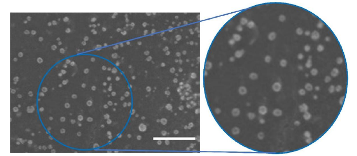
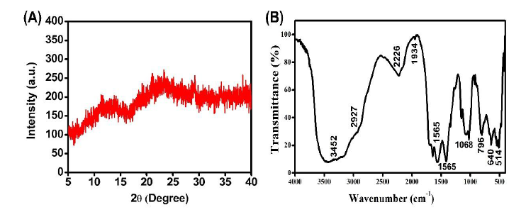
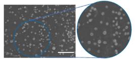
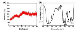
SEM micrographs indicated that the synthesized ChN particles exhibited a uniformly spherical, dense structure and displayed a fairly uniform particle size distribution, with an average size of 18.55±3.29 nm (Fig. 1). Fig. 2 demonstrates the XRD pattern of ChN with two broad peaks at 2Ɵ of 11° and 22°. The FTIR spectrogram of ChN showed a major absorption bands at a wavelength of 3452, 2927, 2226, 1934, 1565, 1390, 1068, 796, 640 and 514 cm−1 (Fig 2, Tab. 1). Alcohol was confirmed by a strong, broad band at 3452 cm−1 that suggested O-H stretching. The existence of amine salt was indicated by the strong, broad band at 2927 cm−1, which indicated N-H stretching. The existence of nitrile was indicated by the band at 2226 cm−1, which suggested C≡N stretching. C=C=C stretching at 1934 cm−1 verified that allene was present. C=C stretching at the 1565 cm−1 medium band permitted cyclic alkene to be present. The bands that verified the presence of phenol, amine, 1,4-disubstituted, alkene and halo compound were 1390 cm−1 (O-H bending), 1068 cm−1 (C-N stretching), 796 cm−1 (C-H bending), 640 cm−1 (C = C bending) and 514 cm−1 (C-I stretching) (Table 1).
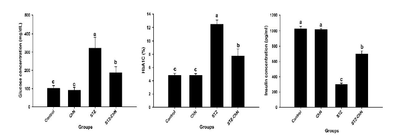
Comparing the STZ group to the control rats, the average serum glucose level and HbA1c (%) rose while insulin levels dramatically decreased (P < 0.05). The STZ-ChN group had a substantial increase in insulin concentration (P < 0.05) and a decrease in HbA1c and insulin levels compared to the STZ group (Fig. 3).
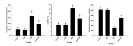
Modulating effect of ChN on glucose, HbA1C, and insulin of diabetic rats. Results are shown as means± standard deviation. Different indicated lowercase letters demonstrated significant alterations between groups at P values < 0.05
Significant increases (P < 0.05) in TC, TG, and LDL-C were observed in the STZ group, but HDL-C was not significantly (P ˃ 0.05) affected, when compared to the control group. However, the STZ-ChN revealed significant (P < 0.05) decreases in TC, TG, and LDL-C levels and nonsignificant reductions in HDL-C against the STZ group (Table 2).
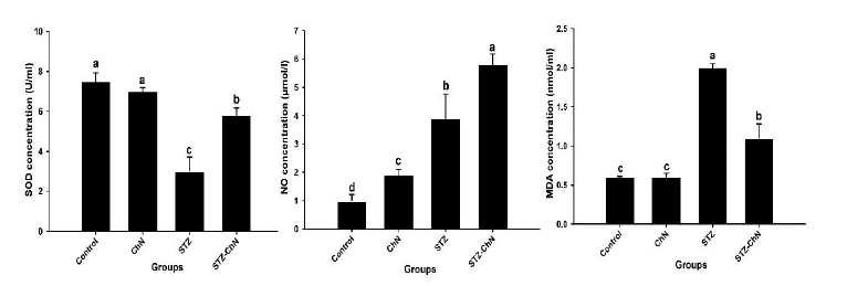
The STZ group showed a remarkable significant increase in NO and MDA, but a significant (P < 0.05) decrease in serum SOD concentration (Fig. 4). Comparing the STZ-ChN group to the STZ group, however, after therapeutic treatment with ChN, SOD and NO increased, while MDA reduced with a significant value (P < 0.05) (Fig. 4).
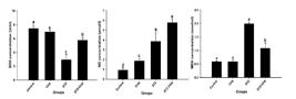
Modulating effect of ChN on antioxidant enzymes of diabetic rats. Results are shown as means± standard deviation. SOD; superoxide dismutase, NO; nitric oxide, MDA; malondialdehyde. Different indicated lowercase letters demonstrated significant alterations between groups at P values < 0.05.
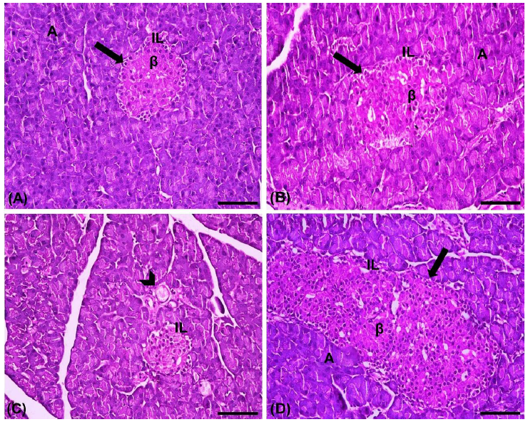
The histopathological analysis of the pancreas in the control group demonstrated normal histology of the pancreatic acini and islets of Langerhans. The islets featured a concentrated core of β-cells encircled by dark nuclei on the periphery (α-cells) (Fig. 5A). The ChN group appeared with very similar morphology to the control group with normal islets and acinar structure (Fig. 5B). On the other hand, the pancreas from the STZ group exhibited cytoplasmic degenerative alterations in most islets with its endocrine cells, particularly in the core of the islet with an irregular outlining of the islet (Fig. 5C). The use of ChN in STZ-ChN group resulted in enlargement in the islets of Langerhans with normal distribution of centrally placed β-cells and peripheral α-cells, which seen embedded within the acinar cells (Fig. 5D).
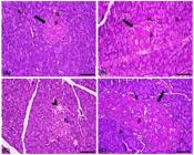
Photomicrographs of H& E stained pancreatic tissue from diabetic treated rats with ChN. (A, B) Control and ChN groups showing normal islets of Langerhans (IL) centrally placed beta-cells (β) encircled by peripheral α-cells (arrow) embedded within acini cells (A). (C) Diabetic group revealing degenerative alterations in the islets (IL) with its inside endocrine cells and blood vessel (arrowhead) with an irregular outlining of the islet, (D), Diabetic-ChN showing enlargement in the islets (IL) with normal distribution of central β-cells and peripheral α-cells (arrow), surrounded by acinar cells (A). Scale Bar= 50µm.
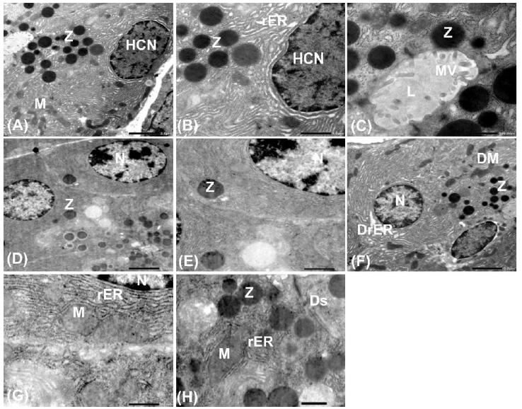
Electron micrographs of the pancreas from the control group showed normal exocrine pancreatic acini with an obvious heterochromatic nucleus that surrounded by rough endoplasmic reticulum, zymogen granules, mitochondria and lumen. The lumen appeared inside microvilli (Fig. 6A- C). The pancreas of rats administrated with ChN appeared with normal pancreas exocrine part with obvious nucleus surrounded by zymogen granules (Fig. 6D, E). Whilst the STZ group revealed damaged rough endoplasmic reticulum and abnormal condensing mitochondria revealed damaged rough endoplasmic reticulum and abnormal condensed mitochondria (Fig. 6F). Additionally, the exocrine portion of the acini in the STZ-ChN showed normal nucleus with an apparent normal zymogen granule, rough endoplasmic reticulum, mitochondria with remarkable desmosomes (Fig. 6G, H).
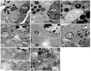
Ultrastructural micrographs of pancreatic exocrine acinar cells from diabetic treated rats with ChN. (A, B, C) Control group revealing normal exocrine acini with an obvious heterochromatic nucleus (HCN) that is surrounded by rough endoplasmic reticulum (rER), zymogen granules (Z), mitochondria (M) and lumen (L) with inside microvilli (MV) (Bar= 2, 1µm, 500nm). (D, E) ChN group showing normal nucleus (N) surrounded by zymogens (Z) (Bar= 2, 1µm). (F) STZ group indicating nucleus (N), zymogens (Z) with damaged rough endoplasmic reticulum (DrER) and damaged mitochondria (DM) (Bar= 2µm). (G, H) STZ-ChN showing nearly normal nucleus (N) with apparent normal zymogen granules (Z), rough endoplasmic reticulum (rER), mitochondria (M) with remarkable desmosomes (Ds) (Bar= 2, 1µm).
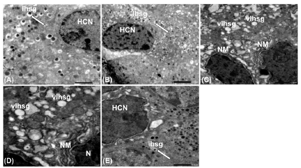
The pancreatic beta-cells (β-cells) from diabetic rats treated with ChN are depicted in Fig. 6. Both the control and ChN control groups exhibited normal β-cells characterized by typical heterochromatic nuclei and intact insulin secretory granules (Ihsg) with a distinct clear zone (Fig. 7A, B). In contrast, the STZ group had significantly vacuolated Ihsg and an unevenly delineated nuclear membrane (Fig. 7C, D). The STZ-ChN group displayed heterochromatic nuclei with somewhat enhancement in nuclear membrane irregularity. Furthermore, the Ihsg, which again exhibited a distinct zone, appeared more characteristic (Fig. 7E).
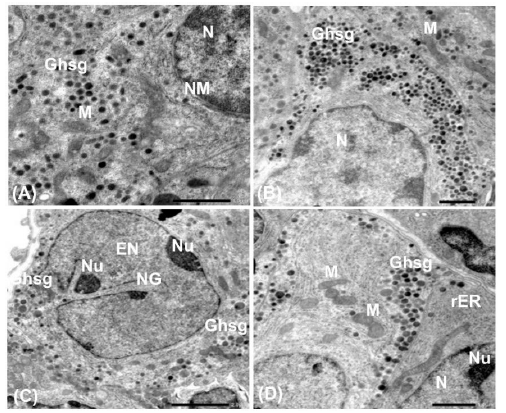
Fig. 8. illustrates the pancreatic alpha-cells (α-cells) from diabetic rats treated with ChN. The control group cells exhibited a normal nucleus, enclosed by a double-layered nuclear membrane, and mitochondria surrounded by healthy glucagon hormone secretory granules (Ghsg) (Fig. 8A). A portion of a standard nucleus is noted in the ChN treatment group, displaying a double nuclear membrane and characteristic Ghsg (Fig. 8B). The STZ group exhibited an euchromatic nucleus, distinguished by nuclear grooves and two nucleoli, surrounded by Ghsg (Fig. 8C). The STZ-ChN group demonstrated an improvement in the ultrastructure of α-cells, evidenced by the presence of a normal nucleus with a nucleolus, accompanied by rough endoplasmic reticulum and normal mitochondria, as well as normal Ghsg (Fig. 8D).
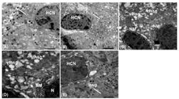
Ultrastructural micrographs of endocrine pancreatic beta-cells (β-cells) from diabetic treated rats with ChN. (A) Control group demonstrates part of the heterochromatic nucleus (HCN), insulin hormone secretory granules (Ihsg) characterized by electron dense core surrounded by a clear zone (arrow) (Bar= 1 µm). (B) ChN group showing normal heterochromatic nuclei (HCN) and normal insulin secretory granules (Ihsg) with a clear zone (arrow) (Bar= 2 µm). (C, D) STZ group showing heterochromatic nuclei (N) with irregular separated nuclear membrane (NM) and highly vacuolated insulin hormone secretory granules (vIhsg) (Bar= 1 µm). (E) STZ-ChN group showing heterochromatic nuclei (HCN) that has slight improvements in its irregular nuclear membrane and insulin hormone secretory granules (Ihsg) with a clear zone (arrow) (Bar= 2, 1µm).
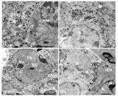
Ultrastructural micrographs of endocrine pancreatic alpha-cells (α-cells) from diabetic treated rats with ChN. (A) Control group revealing normal nucleus (N) surrounded by normal glucagon hormone secretory granules (Ghsg) (Bar= 1µm). (B) ChN group showing part of normal nucleus (N) and double nuclear membrane (NM) and normal glucagon hormone secretory granules (Ghsg) (Bar= 1µm). (C) STZ group showing euchromatic nucleus (EN) with nuclear grooves (NG) and two nucleoli (Nu) surrounded by glucagon hormone secretory granules (Ghsg) (Bar= 2µm). (D) STZ-ChN group exhibiting part of normal nucleus (N) with nucleolus (Nu), surrounded by rough endoplasmic reticulum (rER) enclosing normal mitochondria (M) and normal glucagon hormone secretory granules (Ghsg) (Bar= 1µm).
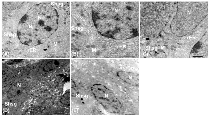
The pancreatic delta-cells (δ-cells) from diabetic rats treated with ChN are shown in Fig. 9. The control group cells exhibit a characteristic nucleus with an associated nucleolus, encircled by rough endoplasmic reticulum, mitochondria, and somatostatin hormone secretory granules (Shsg) (Fig. 9A, B). In the ChN therapy group, the cells have a prominent nucleus surrounded by rough endoplasmic reticulum, mitochondria, and Shsg (Fig. 9C). The STZ group exhibited a small nucleus with a limited amount of Shsg (Fig. 9D). The STZ-ChN group exhibited normal nuclei and a limited presence of Shsg, which suppresses the production of insulin and glucagon hormones (Fig. 9E).
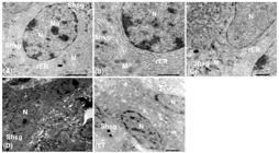
Ultrastructural micrographs of endocrine pancreatic delta-cells (δ-cells) from diabetic treated rats with ChN. (A, B) Control group showing normal nucleus (N) with nucleolus (Nu) surrounded by rough endoplasmic reticulum (rER), mitochondria (M) and somatostatin hormone secretory granules (Shsg) (Bar= 1 µm). (C) ChN group indicating large nucleus (N) enclosed by rough endoplasmic reticulum (rER), mitochondria (M) and somatostatin hormone secretory granules (Shsg) (Bar= 1 µm). (D) STZ group exhibiting small nucleus (N) with very few somatostatin hormone secretory granules (Shsg) (Bar= 2 µm). (E) STZ-ChN group showing normal nuclei (N) with few somatostatin hormone secretory granules (Shsg) (Bar= 1 µm).
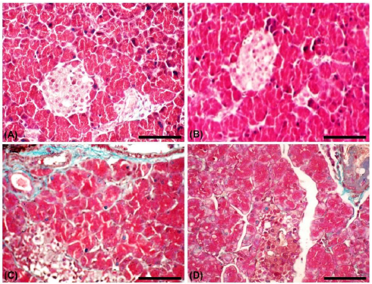
This study shows Paraffin slices of the control and ChN rat pancreas exhibited negative staining of islet Langerhans cells, with pale blue thin fibers encircling blood vessels and the absence of basement membranes in acinar glands (Fig. 10A, B). Whilst the pancreas of STZ rats exhibited negative staining of necrotic and pyknotic islet Langerhans cells, accompanied by moderate blue fibers encircling blood vessels, interlobular ducts, and most basement membranes of acinar glands (Fig. 10C). Moreover, the pancreas of STZ-ChN rats exhibited islet Langerhans cells characterized by dark red nuclei and pink cytoplasm, minor blue staining in most acinar gland basement membranes, and moderate blue fibers surrounding interlobular ducts and areas of lymphocytic infiltration (Fig. 10D).
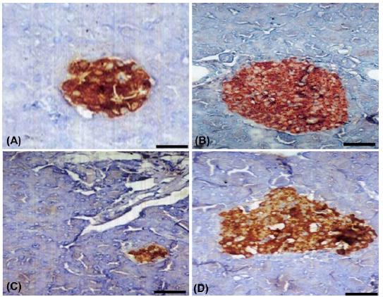
In the control group, the insulin-secreting cells, or beta cells, constituted the predominant cell population of the islets, primarily located in the central zone. Insulin expression was positively observed as dark brown granules within the cytoplasm of β-cells (Fig. 11A). The immuno-stained β-cells from ChN group resembled those of the control group (Fig. 11B). In contrast, STZ group exhibited marked decrease in area of β-cells with a significant decline in insulin expression (Fig. 11C). Following treatment with ChN, the insulin expression was obviously increased in the pancreatic islets core with normal density, differentiated by blood capillaries and blue color area of absent insulin in area of α and δ-cells (Fig. 11D).
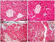
Paraffin photomicrographs of Masson's trichrome stained sections from diabetic-treated rats with ChN. (A) Control group and (B) ChN group shows negative staining of islet Langerhans cells, with pale blue thin fibers encircling blood vessels. (C) STZ group reveals negative staining of necrotic and pyknotic islet Langerhans cells, accompanied by moderate blue fibers encircling blood vessels, interlobular ducts, and most basement membranes of acinar glands. (D) STZ-ChN group shows minor blue staining in most acinar gland basement membranes, and moderate blue fibers surrounding interlobular ducts and areas of lymphocytic infiltration. Scale Bar= 50µm.
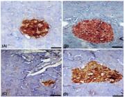
Immunohistochemical staining of pancreatic islets of Langerhans showing insulin expression in diabetic treated rats with ChN. (A) Control group showing strong immunoreactivity of insulin in beta-cells, which occupy most of the islet. (B) ChN group showing substantial positive insulin expression in the cytoplasm of beta-cells. (C) STZ group showing marked reduction in the immunohistochemical expression of insulin in β-cells. (D) STZ-ChN showing an evident elevation in the insulin expressing β-cells in the islet core with normal density differentiated by blood capillaries and blue color area of absent insulin in area of α and δ-cells. Scale bar= 50 µm.
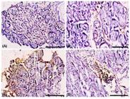
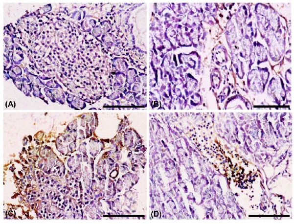
Paraffin photomicrographs of TGF-β1 protein immuno-expression in diabetic treated rats with ChN. (A) Control group, (B) ChN group showing mild positive expression (+1) on β-cell membranes, with a negative blue coloration for the nuclei. (C) STZ group demonstrated strong immuno-expression (+3) in β-cell membranes and multiple blood capillaries, alongside pronounced diffuse brown expressions in many proliferating β-cells. (D) STZ-ChN group exhibited mild expression (+1) in β-cell membranes and considerable diffuse brown in endothelial cells, accompanied by negative blue nuclei and light blue in necrotic islets. Scale Bar= 50µm.
DISCUSSION
DM comprises a collection of metabolic illnesses marked by persistent hyperglycemia, resulting from inadequate insulin synthesis, impaired insulin action, or a combination of both (Unuofin and Lebelo, 2020). Polysaccharides are a bioactive class of macromolecules obtained from either plant sources or crustacean shells, demonstrating various biological characteristics such as immunomodulatory, anticancer, antioxidant, and hypoglycemic effects (Ji et al., 2021). Therefore, this research was conducted to examine the impact of chitosan nanoparticles (ChN) on streptozotocin (STZ)-induced diabetic rats. In the present study, The TEM analysis revealed that the synthesized ChN particles exhibited a uniformly spherical and dense structure with an average size of 18.55 ± 3.29 nm. This uniformity in particle size is crucial for their application in drug delivery, as it can enhance bioavailability and efficacy. The stability and consistent morphology of the particles suggest that they are suitable for therapeutic applications (Danaei et al., 2018). The current results also showed that ChN with two broad peaks at 2Ө of 11° and 22°. The appearance of these peaks indicates the semi-crystalline nature of ChN (Fletes-Vargas et al., 2023). Similar FTIR obtained results in this study were similar to those described by Varma and Vasudevan (2020).
In the herein study, significant rise in serum glucose and HbA1c levels was observed in the STZ group, compared to the control group and this highlights the hyperglycemic state induced by STZ. The decrease in insulin levels further corroborates the destructive impact of STZ on pancreatic β-cells. These results are consistent with those reported by Abdel-Rahman et al. (2019). The observed changes in glucose and insulin levels in STZ-treated rats are associated with the death of pancreatic β-cells, resulting in hyperglycemia, hyperlipidemia, and weight loss (Bayramoglu et al., 2014). Furthermore, insulin insufficiency has been linked to hypercholesterolemia and hypertriglyceridemia due to the inactivation of lipases responsible for hydrolyzing these fats (Oyedemi et al., 2011). Conversely, the STZ-ChN group demonstrated a marked increase in insulin concentration and a reduction in glucose and HbA1c levels, indicating that ChN treatment effectively restored β-cell function and improved glycemic control. This suggests that ChN may exert a protective effect on the pancreatic islets, potentially enhancing insulin secretion and reducing glucose levels. Similarly, Farid et al. (2024) demonstrated that chitosan nanoparticles loaded with bee venom reduced blood glucose levels and elevated insulin levels. Tzeng et al. (2022) showed that both high and low molecular weight chitosan administration diminished insulin resistance by suppressing lipid buildup in the liver and adipose tissue.
The lipid profile analysis of the current study revealed significant elevations in total TC, TG, and LDL-C in the STZ group in relation to the control group. Similarly, Oyedemi et al. (2011) showed that streptozotocin-induced diabetic mice suffering from hypercholesterolemia that characterized by elevated levels of triglycerides, total cholesterol, and LDL-cholesterol, attributed to enhanced cholesterol production and intestinal absorption. It is widely recognized that diabetes is frequently associated with various possible risk factors for cardiovascular events, including increased TC (Rhee et al., 2017) and TG (Kompoti et al., 2006). Bopanna et al. (1997) associated the elevation of serum lipids with enhanced mobilization of free fatty acids from peripheral adipose stores, where the esterification of free fatty acids is equilibrated within the lipolysis cycle. Notably, ChN treatment in this study led to significant reductions in these lipid parameters, indicating its potential to ameliorate lipid metabolism disturbances associated with diabetes. On the other hand, the lack of significant change in HDL-C suggests that while ChN may improve overall lipid profiles, it may not influence HDL-C levels directly. Nanoparticles have hypocholesterolemic capabilities by enhancing lipid profiles, resulting in a reduction of antioxidant enzymes, and safeguarding against harm to the liver and kidneys (Gutiérrez et al., 2022).
The oxidative stress markers in this study highlighted a notable increase in NO and MDA levels in the STZ group, alongside a significant decrease in SOD concentrations. These findings underscore the oxidative stress present in diabetic conditions. Comparable findings indicated markedly elevated NO end products in the serum of diabetes patients (Alghazeer et al., 2022). Oxidative stress and tissue damage may exacerbate the symptoms of diabetes, as STZ-induced diabetic mice exhibited elevated liver tissue MDA levels and reduced NO compared to controls (Elmetwalli et al., 2022). This NO elevation result from the excessive generation of NO linked to elevated oxidative stress levels in diabetes patients (Assmann et al., 2016). The gradual glycation of enzymatic proteins results in a reduction in antioxidant enzymatic system activity in diabetes. In individuals with diabetes, approximately 50% of SOD is glycated, resulting in diminished enzyme activity (Bikkad et al., 2014). On the other hand, in the current study, the restoration of SOD levels and the reduction in MDA and NO in the STZ-ChN group suggest that ChN not only improves glycemic control but also enhances antioxidant defenses, thereby potentially protecting pancreatic β-cells from oxidative damage. These findings corroborate those of Farid et al. (2024), who indicated that chitosan nanoparticles loaded with bee venom diminished oxidative stress by lowering MDA levels and elevating antioxidant enzyme SOD levels.
Histopathological analysis results of this study revealed normal pancreatic architecture in the control and ChN groups, while the STZ group exhibited significant degenerative changes in the islets. These results were confirmed using the ultrastructural studies that provided additional insights into the cellular changes induced by STZ. The control and ChN groups exhibited typical pancreatic morphology, while the STZ group showed damaged pancreatic islets with vacuolation in β-cells. Histopathological data obtained from the current study were also consistent with Hadi et al. (2016) and Elkotby et al. (2018) who reported that the size and number of pancreatic islets were decreased in diabetic rats in comparison to normal rats. Remarkably, the present results indicated that STZ-ChN group displayed enlarged islets with a preserved architecture, indicating that ChN treatment may reverse STZ-induced morphological alterations. This restoration of islet structure is critical, as it reflects improved β-cell health and function. Moreover, the ultrastructural study revealed that after ChN treatment, there was notable improvement in the ultrastructure of both β-cells, α-cells and δ-cells, suggesting that ChN not only protects but also promotes the restoration of cellular integrity in the pancreatic tissue. Comparable findings demonstrated that Langerhans islets exhibited substantial enhancement, while pancreatic acini displayed normal cytoarchitecture with hyperpigmented nuclei when treated with zinc oxide nanoparticles (Sewelam and Shehata, 2021).
The current histochemical data indicated that STZ-treated rats exhibited a considerable increase in collagen deposition in pancreatic tissues, as demonstrated by Masson’s Trichrome staining. Sanai et al. (2000) indicated that diabetes causes the accumulation of type IV collagen fibers leading to renal fibrosis. However, in this study, the use of ChN against STZ group resulted in minor presence of collagen in pancreatic tissue.
Immunohistochemical findings further supported these observations, showing a marked decrease in insulin expression in the STZ group. Similar results were reported by Abdel-Rahman et al. (2020) and Ayuob et al. (2021). On the other hand, the enhanced presence of insulin-secreting β-cells post-treatment with ChN is indicative of a functional recovery of the pancreatic islets, thereby reinforcing the therapeutic potential of ChN in diabetes management. TGF-β1 is a pivotal profibrotic cytokine that governs the production of extracellular matrix constituents, enhances cell-matrix interactions, and diminishes matrix breakdown (Zhang et al., 2014). In this study, the STZ induced diabetic group showed strong TGF-β1 immuno-expression in β-cell membranes and multiple blood capillaries, alongside pronounced diffuse brown expression in many proliferating β-cells. There is substantial data indicating that TGF-β significantly contributes to the enhancement of fibrosis in the diabetic kidney (Ghavimishamekh et al., 2018). On the other hand, the using of ChN in this study, had resulted in mild TGF-β1 immunostaining protein expression (+1) in β-cell membranes and considerable diffuse brown in endothelial cells, accompanied by negative blue nuclei and light blue in necrotic islets. Sutthasupha and Lungkaphin (2020) reported that chitosan could improve kidney structure and function in diabetic rats by reducing the production of fibrotic proteins, particularly TGF-β1. Moreover, Chitosan can regulate the secretion of the cytokine TGF-β1, augment the activity of antioxidative enzymes, and exhibits anti-inflammatory characteristics (Qiao et al., 2018).
CONCLUSION
In summary, the findings from this study demonstrated that ChN possesses potent antidiabetic properties, evidenced by improved glycemic control, ameliorated lipid profiles, enhanced antioxidant defenses, and preservation of pancreatic architecture and function. These results warrant further investigation into the mechanisms underlying ChN's protective effects and its potential as a therapeutic agent for diabetes management. Future studies could explore the long-term effects of ChN treatment and its applicability in clinical settings.
ACKNOWLEDGMENTS
This study was supported by Princess Nourah bint Abdulrahman University Researchers Supporting Project number (PNURSP2026R96), Princess Nourah Bint Abdulrahman University, Riyadh, Saudi Arabia and the Ongoing Research Funding program (ORF-2026-655), King Saud University, Riyadh, Saudi Arabia.
REFERENCES
- ABDEL-RAHMAN, M.A.; MOHAMMED, A.K.; AHMED, S.H. et al. Antidiabetic effect of the scorpion Scorpio maurus palmatus body extract using alloxan-induced diabetic mice model. J. Taibah Univ. Sci., v.13, p.504-513, 2019.
- ABDEL-RAHMAN, R.F.; EZZAT, S.M.; OGALY, H.A. et al. Ficus deltoideaextract down-regulates protein tyrosine phosphatase 1B expression in a rat model of type 2 diabetes mellitus: a new insight into its antidiabetic mechanism. J. Nutrit. Sci., v.9, p.1-18, 2020.
- ALGHAZEER, R.; ALGHAZIR, N.; AWAYN, N. et al. Biomarkers of oxidative stress and antioxidant defense in patients with type 1 diabetes mellitus. Ibnosina J. M. Biol. Sci., v.10, p.198-204, 2022.
- ALI, M.M.; AGHA, F.G. Amelioration of streptozotocin‐induced diabetes mellitus, oxidative stress and dyslipidemia in rats by tomato extract lycopene. Scand. J. Clin. Lab. Invest., v.69, p.371-379, 2009.
- ASSMANN, T.S.; BRONDANI, L.A.; BOUÇAS, A.P. et al. Nitric oxide levels in patients with diabetes mellitus: A systematic review and meta-analysis. Nitric Oxide, v.61, p.1-9, 2016.
- AYUOB, N.; AL-SHATHLY, M.R.; BAKHSHWIN, A. et al. p53 Rather Than β-Catenin mediated the combined hypoglycemic effect of cinnamomum cassia (L.) and zingiber officinale roscoe in the streptozotocin-induced diabetic model. Front. Pharmacol., v.12, p.664248, 2021.
- BAYRAMOGLU, G.; SENTURK, H.; BAYRAMOGLU, A. et al. Carvacrol partially reverses symptoms of diabetes in STZ-induced diabetic rats. Cytotechnology, v.66, p.251-257, 2014.
- BIKKAD, M.D.; SOMWANSHI, S.D.; GHUGE, E.H. et al. Oxidative stress in type 2 diabetes mellitus. Biomed. Res. Tokyo., v.25, p.84-87, 2014.
- BOPANNA, K.N.; KANNAN, J.; SUSHMA, G. et al. Antidiabetic and antihyperlipidaemic effects of neem seed kernel power on alloxan diabetic rabbits. Indian J. Pharmacol., v.29, p.162-167, 1997.
- BORAI, I.H.; HANAFI, N.; ELGAWISH, M.A. et al. Role of chitosan nanoparticles in reducing pancreatitis in rats treated with L-arginine. Int. J. Adv. Sci. Tech. Res., v.7, p.44-58, 2017.
- DANAEI, M.; DEHGHANKHOLD, M.; ATAEI, S. et al. Impact of particle size and polydispersity index on the clinical applications of lipidic nanocarrier systems. Pharmaceutics, v.10, p.57, 2018.
- DEFINITION, diagnosis and classification of diabetes mellitus and its complications: report of a WHO consultation. Part 1, Diagnosis and classification of diabetes mellitus. Rome: WHO, 1999.
- DIABETES Fact Sheet. n. 312, Rome: WHO, 2008.
- EL-BAZ, Y.G.; MOUSTAFA, A.; ALI, M.A. et al. Green synthesized silver nanoparticles for the treatment of diabetes and the related complications of hyperlipidemia and oxidative stress in diabetic rats. Exp. Biol. Med., v.248, p.2237-2248, 2024.
- ELKOTBY, D.; HASSAN, A.K.; EMAD, R. et al. Histological changes in islets of langerhans of pancreas in alloxan-induced diabetic rats following egyptian honey bee venom treatments. Int. J. Pure Appl. Zool., v.6, p.1-6, 2018.
- ELMETWALLI, A.; HASSAN, J.; ALAA, H. et al. Nanoparticle zinc oxide obviates oxidative stress of liver cells in induced-diabetes mellitus model. Med. J. Viral Hepat., v.7, p.8-12, 2022.
- FARID, A.; MOHAMED, A.; AHMED, A. et al. Preparation of bee venom-loaded chitosan nanoparticles for treatment of streptozotocin-induced diabetes in male Sprague Dawley rats. Beni Suef Univ. J. Basic Appl. Sci., v.13, p.97, 2024.
- FLETES-VARGAS, G.; ESPINOSA-ANDREWS, H.; CERVANTES-UC, J.M. et al. Porous chitosan hydrogels produced by physical crosslinking: physicochemical, structural, and cytotoxic properties. Polymers, v.15, p.2203, 2023.
- GHAVIMISHAMEKH, A.; ZIAMAJIDI, N.; DEHGHAN, A. et al. Study of insulin-loaded chitosan nanoparticle effects on TGF-Β1 and fibronectin expression in kidney tissue of type 1 diabetic rats. Indian J. Clin. Biochem., v.34, p.418-426, 2018.
- GUTIÉRREZ, R.; GÓMEZ, J.; URBY, R. et al. Evaluation of diabetes effects of selenium nanoparticles synthesized from a mixture of luteolin and diosmin on streptozotocin-induced type 2 diabetes in mice. Molecules, v.27, p.5642, 2022.
- HADI, M.; ZAIDAN, H.K.; AL-SAADI, A.H. Histopathological changes of pancreatic tissues in hyperglycemic male rats treated with mixture of plants extracts. Int. J. Chem. Tech. Res., v.9, p.501-513, 2016.
- JI, X.; ZHOU, Y.; LI, Q. et al. Protein-Mimicking nanoparticles for a cellular regulation of homeostasis. ACS Appl. Mater. Interfaces, v.13, p.31331-31336, 2021.
- KOMPOTI, M.; MARIOLIS, A.; ALEVIZOS, A. et al. Elevated serum triglycerides is the strongest single indicator for the presence of metabolic syndrome in patients with type 2 diabetes. Cardiovasc. Diabetol., v.5, n.1, 2006.
- LENZEN, S. The mechanisms of alloxan- and streptozotocin-induced diabetes. Diabetologia, v.51, p.216-226, 2007.
- NISHIKIMI, M.; RAO, N.A.; YAGI, K. The occurrence of superoxide anion in the reaction of reduced phenazine methosulfate and molecular oxygen. Biochem. Biophys. Res. Commun., v.46, p.849-854, 1972.
- NYUNAÏ, N.; NJIKAM, N.; ABDENNEB, E. et al. Hypoglycaemic and antihyperglycaemic activity of Ageratum conyzoides L. in rats. Afr. J. Tradit. Compl. Altern. Med., v.6, p.123-130, 2009.
- OGAWA, N.; SATSU, H.; WATANABE, H. et al. Acetic acid suppresses the increase in disaccharidase activity that occurs during culture of Caco-2 Cells. J. Nutr., v.130, p.507-513, 2000.
- OYEDEMI, S.O.; YAKUBU, M.T.; AFOLAYAN, A.J. Anti-diabetic activities of aqueous leaves extract of Leonotis leonurus in streptozotocin induced diabetic rats. J. Med. Plants Res., v.5, p.119-125, 2011.
- PREUSS, H.G.; JARRELL, S.T.; SCHECKENBACH, R. et al. Comparative effects of chromium, vanadium and gymnema sylvestre on sugar-induced blood pressure elevations in SHR. J. Am. Coll. Nutr., v.17, p.116-123, 1998.
- QIAO, J.; LIU, Y.; JIANG, Z. et al. Preparation and renoprotective effects of carboxymethyl chitosan oligosaccharide on adriamycin nephropathy. Carbohydr. Polym., v.201, p.347-356, 2018.
- REYNOLDS, E.S. The use of lead citrate at high ph as an electron-opaque stain in electron microscopy. J. Cell Biol., v.17, p.208-212, 1963.
- RHEE, E.; HAN, K.; KO, S. et al. Increased risk for diabetes development in subjects with large variation in total cholesterol levels in 2,827,950 Koreans: a nationwide population-based study. PLoS ONE, v.12, p.e0176615, 2017.
- SANAI, T.; SOBKA, T.; JOHNSON, T. et al. Expression of cytoskeletal proteins during the course of experimental diabetic nephropathy. Diabetologia, v.43, p.91-100, 2000.
- SATHAYE, S.; KAIKINI, A.; DHODI, D. et al. Standardization of type 1 and type 2 diabetic nephropathy models in rats: Assessment and characterization of metabolic features and renal injury. J. Pharm. Bioallied Sci., v.12, p.295, 2020.
- SEWELAM, A.; SHEHATA, M. Nano-zinc oxide induced pancreatic toxicity and the ameliorating role of Naringenin: histological and immunohistochemical study Egypt J. Histol., v.44, p.1081-1097, 2021.
- SUTTHASUPHA, P.; LUNGKAPHIN, A. The potential roles of chitosan oligosaccharide in prevention of kidney injury in obese and diabetic conditions. Food Funct., v.11, p.7371-7388, 2020.
- SUVARNA, K.S.; LAYTON, C.; BANCROFT, J.D. Bancroft's theory and practice of histological techniques. [Amsterdã]: Elsevier Health Science, 2018.
- SZKUDELSKI, T. The mechanism of alloxan and streptozotocin action in B cells of the rat pancreas. Physiol. Res., v.50, p.537-546, 2001.
- TARPEY, M.M.; WINK, D.A.; GRISHAM, M.B. Methods for detection of reactive metabolites of oxygen and nitrogen: in vitro and in vivo considerations. Am. J. Physiol. Regul. Integr. Comp. Physiol., v.286, p.R431-R444, 2004.
- TZENG, H.; LIU, S.; CHIANG, M. Antidiabetic properties of chitosan and its derivatives. Mar. Drugs, v.20, p.784, 2022.
- UNUOFIN, J.O.; LEBELO, S.L. Antioxidant effects and mechanisms of medicinal plants and their bioactive compounds for the prevention and treatment of Type 2 diabetes: an updated review. Oxid. Med. Cell Longev., v.2020, p.1-36, 2020.
- VARMA, R.; VASUDEVAN, S. Extraction, characterization, and antimicrobial activity of chitosan from horse mussel modiolus modiolus. ACS Omega, v.5, p.20224-20230, 2020.
- WANG, Y.; SUN, H.; ZHANG, J. et al. Streptozotocin-induced diabetic cardiomyopathy in rats: ameliorative effect of PIPERINE via Bcl2, Bax/Bcl2, and caspase-3 pathways. Biosci. Biotechnol. Biochem., v.84, p.2533-2544, 2020.
- WARDANI, G.; NUGRAHA, J.; MUSTAFA, M.R. et al. Antioxidative stress and antiapoptosis effect of chitosan nanoparticles to protect cardiac cell damage on Streptozotocin-Induced diabetic rat. Oxid .Med. Cell. Longev., v.2022, p.1-9, 2022.
- WILD, S.H.; ROGLIC, G.; GREEN, A. et al. Global prevalence of diabetes: estimates for the year 2000 and projections for 2030. Diabetes Care, v.27, p.2569, 2004.
- YU, L.; SHI, W.; GUO, X. Cardio-protective role of gingerol along with prominent anti-diabetic cardiomyopathy action in a streptozotocin-induced diabetes mellitus rat model. Cell J., v.19, p.469-475, 2017.
- ZHANG, H.; ZHAO, T.; GONG, Y. et al. Attenuation of diabetic nephropathy by Chaihuang-Yishen granule through anti-inflammatory mechanism in streptozotocin-induced rat model of diabetics. J. Ethnopharmacol., v.151, p.556-564, 2014.
- ZUBAIDAH, E.; PUTRI, W.D.R.; PUSPITASARI, T. et al. The effectiveness of various salacca vinegars as therapeutic agent for management of hyperglycemia and dyslipidemia on diabetic rats. Int. J. Food Sci., v.2017, p.1-7, 2017.
The research data are available within the article itself.