Familial cutaneous asthenia in a population of mixed-breed domestic cats - a case report
ABSTRACT
Cutaneous asthenia, or Ehlers-Danlos syndrome, is a rare disease of domestic animals, especially cats, that culminates in hyperextensibility and fragility of the skin in addition to possible ligament laxity. It is a hereditary disease of the connective tissue that occurs with inadequate synthesis of collagen along with alterations in the formation of collagen fibers, associated with mutations in the genes that encode collagen or the enzymes responsible for its organization. The objective of this article is to report a case of hereditary cutaneous asthenia in a domestic cat, of undefined breed, and in the respective litter of four kittens. The mother cat suffered from an extensive and sudden skin lesion located in the dorsolumbar region. The animal received a definitive diagnosis of cutaneous asthenia through histopathological evaluation and use of Masson's trichrome staining, followed by application of tilapia fish skin for the management and treatment of the wound.
Keywords:
feline; dermatopathy; skin fragility; hyperextensibility and tilapia skin
RESUMO
A astenia cutânea, ou síndrome Ehlers-Danlos, é uma doença rara na medicina felina, que culmina em hiperextensibilidade da pele e fragilidade cutânea, além de possível frouxidão articular. A astenia cutânea consiste em uma doença hereditária do tecido conjuntivo, que cursa com síntese inadequada de colágeno, bem como alterações na formação de fibras colágenas. Ademais, é uma doença de origem genética associada a mutações nos genes que codificam o colágeno ou nas enzimas responsáveis pela sua organização. Objetiva-se, com este trabalho, relatar um caso de astenia cutânea hereditária em um gato doméstico, sem raça definida, e em sua respectiva ninhada de quatro filhotes. A felina apresentou lesão cutânea extensa e súbita, localizada em região dorsolombar. Diante disso, o animal recebeu diagnóstico definitivo de astenia cutânea, mediante avaliação histopatológica e utilização de coloração de tricrômico de Masson, seguido do emprego da pele de peixe de tilápia no manejo e tratamento da ferida em questão.
Palavras-chave:
felinos; dermatopatia; fragilidade cutânea; hiperextensibilidade; pele de tilápia
INTRODUCTION
Cutaneous asthenia (CA) is a rare inherited disease of the connective tissue that occurs due to inadequate collagen synthesis (Hansen et al., 2015), as well as abnormal processing of collagen fibers (Szczepanik et al., 2006). This pathology is observed in domestic animals, such as cats, dogs, horses, cattle and sheep, as well as in humans (McElroy et al., 2023). CA is also defined as a collagenopathy analogous to Ehlers-Danlos syndrome (EDS), referring to a heterogeneous group of inherited connective tissue diseases (Hansen et al., 2015).
Collagen dysplasia impairs the resistance and functional integrity of connective tissue, and can therefore affect the integumentary tissue, locomotor system (joints; bones), blood vessels, visceral organs and other body structures (Hansen et al., 2015). In domestic cats, it culminates in changes involving the integumentary system, with hyperextensibility of the skin and cutaneous fragility being the main findings. However, possible joint laxity, joint hypermobility and vascular changes are also occasionally observed (Hansen et al., 2015; Szczepanik et al., 2006).
Cutaneous asthenia is a term generally restricted to animals, and is similar to EDS dermatosparaxis in humans, in which the main clinical and pathological manifestations are restricted to the skin (Hansen et al., 2015; McElroy et al., 2023). In felines, mutations in the genes that encode collagen or in the enzymes responsible for its organization have been documented as being autosomal dominant or autosomal recessive (Caramalac et al., 2022). The disease mainly afflicts Burmese and Himalayan felines but has also been reported in mixed-breed cats (MBCs), although less frequently (Hansen et al., 2015).
This article reports a case of CA in a domestic cat with no defined breed and in its respective litter of four kittens. Our aim is to highlight CA as a differential diagnosis of cats with dermatopathies, with the condition possibly occurring in the offspring as well, given its hereditary and familial nature.
ETHICAL ASPECTS
This work was not submitted to the Ethics Committee on the Use of Animals, as it is a case report.
CASUISTRY
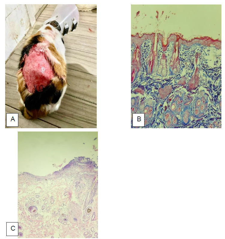
A female domestic mixed-breed cat, approximately two years old and weighing 3.60kg, was brought for treatment at the Small Animal University Hospital of Federal Rural University of Rio de Janeiro (UFRRJ), located in the city of Seropédica, Rio de Janeiro State, with the main symptom being extensive skin lesion/laceration in the dorsolumbar region, with no previous underlying cause (Fig. 1).
The cat lived in a shelter with its respective litter of four kittens. However, it was kept in a separate cattery together with a conflicting feline. On physical examination, an extensive skin lesion was observed along the dorsolumbar spine, measuring approximately 16cm x 11 cm, with a non-exudative and non-scaly appearance, in addition to hyperextensibility, skin fragility and a sad/crestfallen facial appearance. No other pathological changes were observed in the clinical evaluation. The glycemic level was 130mg/dL and the cat tested negative for feline immunodeficiency virus (FIV) and feline leukemia virus (FeLV).
Neither the laboratory tests nor the ultrasound imaging showed any noteworthy changes. The animal underwent surgical-anesthetic intervention to perform wound debridement and to collect a skin fragment for biopsy, for the purpose of histopathological evaluation, fungal culture and bacterial culture/antibiogram.
In the tissue fragment culture, no fungal structures were observed, but coagulase-negative Staphylococcus, Corynebacterium spp., gram-negative fermenting bacillus and Bacillus spp. were isolated in the bacterial culture. In the histopathological evaluation, atrophy of the epidermis and skin appendages was observed through hematoxylin-eosin staining, as well as disorganization and atrophy of the collagen fibers. Through Masson's trichrome staining (with labeling for collagen fibers), some collagen fibers with intense blue color were observed, in addition to ruptured, segmented and disorganized fibers with moderate red marking (imperfect fibers) (Fig. 1).
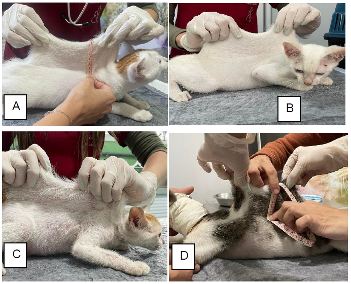
After the diagnostic conclusion of CA in the mother through histopathological evaluation, we examined her four kittens due to the familial nature of the disease. They also presented signs of hyperextensibility, skin fragility and sad facial appearance, in addition to chronic pruritus, so we submitted them to evaluation of the skin extensibility index (Fig. 2), which consists of the relationship between the length of the skin fold and the body length from the occipital crest to the base of the tail. The following indices were obtained: kitten 1 (F1) 28% (7/25); kitten 2 (F2) 25% (7/28); kitten 3 (F3) 22% (7/31); and kitten 4 (F4) 21% (5.5/26).
Dorsolumbar lesion in a cat with CA, photographed immediately after the occurrence of the skin laceration, accompanied by its histopathology. A: Extensive skin laceration. B: Histopathological findings, with hematoxylin-eosin staining, suggestive of atrophy of the epidermis, disorganization and atrophy of the collagen fibers. C: Histopathological findings suggestive of collagen dysplasia evidenced by Masson's trichrome staining (marking for collagen fibers). Source: Personal collection and collection of Gabriela Cid.
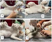
Cutaneous extensibility index (CSI) performed on the offspring of the mother with CA. A and C: Note the CSI level, by measuring the skin fold value, thus, F1 with CSI of 28% (A) and F3 with CSI of 22% (C). B and D: Note the skin hyperextensibility, thus, F2 with CSI of 25% (B) and F4 with CSI of 21% (D). Source: Personal collection.
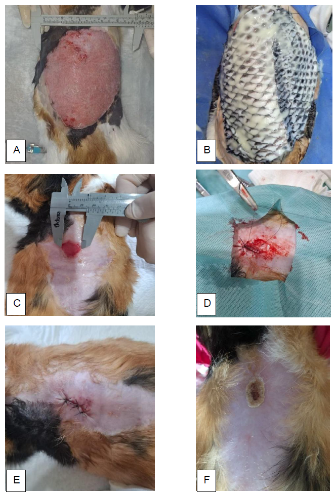
The wound was treated using commercially obtained tilapia fish skin (TFS) (Fig. 3), which was subjected to a sterilization procedure and kept in a sealed container and frozen. The TFS was applied to the wound in a surgical environment, using aseptic techniques, with each kitten under anesthesia. The TFS was then prepared by washing with sterile saline solution for five minutes, three times in a row, to remove any trace of impurities and promote the removal of excess glycerol. The wound was washed with sterile saline solution (250 mL). Hydrogel (Dersani Hydrogel®, without alginate) was applied to the wound bed, followed by fixation of the TFS with a simple suture followed by application of a non-adherent absorbent dressing (Kruuse®). An average interval of four days was used between dressings, making a total of eight dressings performed with TFS. After 30 days of wound management with TFS, adequate granulation tissue and significant lesion reduction were noted. After this period, due to obstacles in the preparation and management of TFS, the use of this dressing was discontinued. Thus, only polyhexamethylene biguanide (PHMB) solution (Prontosan®), hydrogel (Dersani Hydrogel®, without alginate), and non-adherent absorbent dressing (Kruuse®) were employed, maintaining the interval of four days. Four months after treatment, the lesion in the dorsolumbar region measured approximately 1.5 cm x 1.5 cm. However, stagnation of healing was observed, so surgical debridement of the wound and healing by primary intention were instituted, using nylon thread and non-continuous suture. Three months after suturing, healing of the lesion was observed, so the dermatological interventions were stopped (Fig. 3).
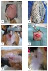
Wound in the dorsolumbar region of a cat suffering from CA. A: Extensive skin laceration in the dorsolumbar region, measuring approximately 16 cm x 11 cm. B: Use of tilapia skin on the wound bed, with an overlap of 1 cm more than the edge of the lesion. C, D and E: Wound in the dorsolumbar region, measuring approximately 1.5 cm x 1.5 cm, undergoing healing by first intention. F: Wound three months after the procedure to approximate the edges of the wound. Source: Personal collection.
In the long term, we recommend the nails of the cats in this report be trimmed regularly to avoid possible trauma, as well as other management alternatives. These included the use of surgical clothing by handlers, assiduously controlling weight, and coexistence only between non-conflicting cats, in addition to sterilization, periodic use of antiparasitic drugs to mitigate the occurrence of dermatopathies, and constant attention to episodes of pruritus, which should be promptly investigated by a veterinarian. Furthermore, given the chronic pruritus of the entire group, a hypoallergenic diet was instituted together with clinical management, with no clinical improvement during its use or after discontinuation. Therefore, we suspected that the offspring had CA concomitant with the mother’s feline atopic skin syndrome (FAS). Coating and fixing of polyvinyl chloride (PVC) on the nails of all offspring was also recommended to minimize the aggravating effects of possible self-injury.
DISCUSSION
CA is particularly common in Burmese and Himalayan cats (Hansen et al., 2015), but is also sometimes found in mixed-breed cats (Caramalac et al., 2022); as was the case of the five felines described in this case report. However, the etiology of CA in the mentioned breeds is correlated with previous vascular alterations in the dermal region, which triggers the possible presence of purpura, alopecia, necrosis and skin laceration, without the strict need for a minimal underlying trauma. Thus, the stretching of blood vessels due to skin hyperextensibility and the reduction of collagen around the vessels have been proposed as the basis for the occurrence of lesions in Burmese cats (Hansen et al., 2015). In contrast, this report describes a skin laceration without evidence of vasculopathy in its etiology, because the domestic cat was of mixed breed.
In felines with CA, skin lacerations can generally be observed after minimal trauma, mainly on the back and head regions (Hansen et al., 2015). This is consistent with the clinical symptoms of the animals reported here. We believe the mother cat’s wound was caused by trauma resulting from a conflict in the shelter. The clinical abnormalities of the five animals mentioned were restricted to the integumentary system.
Feline acquired skin fragility syndrome (FSFS) is a rare disease with multifactorial etiology that, like CA, causes skin to tear easily, although FSFS is associated with hypercortisolism (spontaneous or iatrogenic), hepatic lipidosis, cholangiohepatitis, multicentric lymphoma, feline infectious peritonitis and other diseases. However, hyperextensibility is typically not observed (Furiani et al., 2017). Therefore, we ruled out FSFS, given that the entire family group was affected and hyperextensibility was evident. In the mother, we disregarded the mentioned comorbidities based on history, anamnesis, clinical evaluation and complementary laboratory and imaging tests, which did not reveal any noteworthy changes.
According to Szczepanik et al. (2006), the definitive diagnosis can only be established when histopathological examination with Masson's trichrome staining reveals irregular structures of collagen fibers. Histopathological characteristics include changes in the arrangement and appearance of collagen fibers, which tend to be reduced in number, fragmented, shortened and disorganized (abnormal orientation), with irregular sizes (Hansen et al., 2015). This was corroborated by the histopathological findings in the mother cat described here. Furthermore, the histopathological evaluation of animals with CA using Giemsa staining has shown marked presence of cutaneous mast cells (McElroy et al., 2023). This finding predisposes chronic pruritus and consequently lesions due to self-injury.
The chronic pruritus observed in all the offspring of the mother has often been reported in cats with CA. According to McElroy et al. (2023), chronic mast cell activation can occur with chronic pruritus, which is a condition evident in humans with EDS, through mast cell activation syndrome. According to Kiener et al. (2022), a domestic MBC with CA and concomitant FAS exhibited evident chronic pruritus, which was partially responsive to histamine therapy and completely responsive to steroids. Those authors also described the exclusion of flea bite allergy dermatitis and food allergy dermatitis, in addition to the involvement of dermatological infectious and parasitic agents. Therefore, we suspect the offspring had chronic pruritus as a manifestation of FAS, due to the assiduous control of ectoparasites and a specific diet and might also have mast cell activation syndrome as a comorbidity.
Genetic analysis (genome sequencing) is also used for diagnosis and to further elucidate possible variations in the genes involved in CA. The feline COL5A1 variant has been suggested as a possible causative agent of CA in domestic cats (McElroy et al., 2023). In addition, other independent causal variants of the COL5A1 gene have also been characterized in cases of feline CA (Kiener et al., 2022).
The skin extensibility index (SEI) is obtained by the ratio between the length of the dorsal lumbar skin fold (a) and the body length from the occipital crest to the base of the tail (b), where (a)/(b) x 100% should be greater than 19% in cats with cutaneous asthenia (Hansen et al., 2015). All the offspring of the mother in the current study presented extensibility indices greater than 19%, so they probably suffered from CA. Based on the history and SEI results of the offspring, there was a basis for diagnosis of probable CA in the offspring, so we did not perform dermatological biopsy for histopathological examination, given the invasive nature of the procedure, especially due to the potential for generating a new wound and associated risks of the underlying disease. It is important to emphasize that the sad facial appearance of the affected animals is directly related to the excessive skin tissue, in turn due to the cutaneous hyperextensibility and the underlying collagenopathy.
The application of tilapia fish skin (TFS) is a current strategy for wound treatment. It promotes healing by ensuring moisture and resistance to bacterial colonization, as well as by supplying type 1 collagen to the wound bed and reducing pain sensitivity (Faraji et al., 2022). Thus, the application of PPT made it possible to offset the lack of collagen in the wound bed and enabled faster regeneration and reduced treatment time, all of which are important aspects given the extensive laceration in the dorsolumbar region of the animal in question.
CA involves atrophic healing in humans (Malfait et al., 2017). Thus, inadequate healing has been described in human patients and various animal species suffering from CA. However, it is not a current finding in the literature on feline CA, which corroborates the favorable clinical response to the treatments used to heal the extensive wound of the mother cat.
For animals with CA, we recommend environmental management, including measures to minimize possible skin trauma, including self-injury (by the use of polyvinyl chloride coating on the nails of felines with CA). Another measure is careful weight control, since excess subcutaneous adipose tissue promotes skin stretching and predisposes laceration, (Hansen et al., 2015). Finally, given the genetic nature of the disease, population control through sterilization/castration is important.
CONCLUSION
Reports of CA in felines are rare, making this report important. As observed, CA is a condition that should be considered in feline medicine, especially as a differential diagnosis in cats with dermatopathies, especially to treat extensive lesions without obvious causes, aggravated by the possible occurrence in offspring. The current study innovates by reporting familial CA in domestic MBCs, with the mother being diagnosed by histopathological examination and her offspring presumptively. Furthermore, our findings open doors for new genetic investigations to improve the understanding of CA in cats. Finally, the originality of wound management with TFS is a promising alternative for the treatment of extensive lesions in animals with CA.
REFERENCES
- CARAMALAC, S.M.; CARAMALAC, S.M.; BARBOSA, F.C. et al. Ehlers-Danlos syndrome in a crossbreed cat. Ciênc. Rural, v.52, p.e20210160, 2022.
- FARAJI, N.; GOLI, R.; GHALANDARI, M. et al. Treatment of severe extravasation injury in a newborn by using tilapia fish skin: a case report. Int. J. Surg. Case Rep., v.91, p.106759, 2022.
- FURIANI, N.; PORCELLATO, I.; BRACHELENTE, C. Reversible and cachexia‐associated feline skin fragility syndrome in three cats. Vet. Dermatol., v.28, p.508-e121, 2017.
- HANSEN, N.; FOSTER, S.F.; BURROWS, A.K. et al. Cutaneous asthenia (Ehlers-Danlos-like syndrome) of Burmese cats. J. Feline Med. Surg., v.17, p.954-963, 2015.
- KIENER, S.; APOSTOLOPOULOS, N.; JENNIFER SCHISSLER, J. et al. Independent COL5A1 variants in cats with Ehlers-Danlos syndrome. Genes, v.13, p.797, 2022.
- MALFAIT, F.; FRANCOMANO, C.; BYERS, P. et al. The 2017 international classification of the Ehlers-Danlos syndromes. Am. J. Med. Genet. C Semin. Med. Genet., v.175, p.8-26, 2017.
- MCELROY, A.; EDWARDS, H.G.; COGHILL, L.M.; LYONS, L.A. Precision medicine using whole genome sequencing in a cat identifies a novel COL5A1 variant for classical Ehlers‐Danlos syndrome. J. Vet. Intern. Med., v.37, p.1716-1724, 2023.
- SZCZEPANIK, M.; WILKOLEK, P.; POPIEL, J. et al. Ehlers-Danlos syndrome (cutaneous asthenia)-A report of three cases in cats. Bull. Vet. Inst. Pulawy, v.5v, p.609, 2006.
Data-available.