Assessing the in vivo therapeutic properties of pear (Pyrus communis L.) as a natural substitute for lowering lipids in animals
ABSTRACT
This study aimed to assess the in vivo therapeutic potential of pear powder in the treatment of hyperlipidemia. Hyperlipidemic male albino rats were divided into three groups (n=10). The first group (G0) was considered as the control, while the second and third groups (G1 and G2) received 300 and 600mg/kg pear powder along with the basal diet, respectively, for 2 months. The experimental results showed that there was a significant reduction in the weight (235.61-210.52g) of the rats (from G0 to G2) after two months of study. Water intake and nutrient digestibility were higher in G2 than in G0. The reduction of the total cholesterol from (69.02 to 59.43mg/dL), triglycerides from (160.24 to 63.45mg/dL), and LDL from (50.83 to 17.08mg/dL) was noted, when G0 were compared with G2. Improvements in HDL levels were also recorded. Reductions in aspartate aminotransferase and alanine aminotransferase levels were also recorded. Histopathological screening of control and pear powder-fed rats revealed a significant reduction in kidney and liver injury in the G2 group. Therefore, it can be concluded that pear powder, which is rich in bioactive substances, can be used as a medicinal food to prevent hyperlipidemia.
Keywords:
Pyrus communis; hyperlipidemia; antioxidants; hepatoprotective; fiber
RESUMO
Este estudo teve como objetivo avaliar o potencial terapêutico in vivo do pó de pera no tratamento da hiperlipidemia. Ratos albinos machos hiperlipidêmicos foram divididos em três grupos (n=10). O primeiro grupo (G0) foi considerado o controle, enquanto o segundo e o terceiro grupos (G1 e G2) receberam 300 e 600mg/kg de pó de pera, respectivamente, juntamente com a dieta basal, durante dois meses. Os resultados experimentais mostraram uma redução significativa no peso (de 235,61 para 210,52g) dos ratos (do G0 para o G2) após dois meses de estudo. A ingestão de água e a digestibilidade dos nutrientes foram maiores no G2 do que no G0. Observou-se redução do colesterol total (de 69,02 para 59,43mg/dL), dos triglicerídeos (de 160,24 para 63,45mg/dL) e do LDL (de 50,83 para 17,08mg/dL) quando o G0 foi comparado ao G2. Também foram observadas melhorias nos níveis de HDL. Além disso, houve redução nos níveis de aspartato aminotransferase e alanina aminotransferase. A análise histopatológica de ratos do grupo controle e dos ratos alimentados com pó de pera revelou uma redução significativa nas lesões renais e hepáticas no grupo G2. Portanto, pode-se concluir que o pó de pera, rico em substâncias bioativas, pode ser utilizado como alimento medicinal para a prevenção da hiperlipidemia.
Palavras-chave:
Pyrus communis; hiperlipidemia; antioxidantes; hepatoprotetor; fibra
INTRODUCTION
Native Americans have adapted their use of plants to suit their dietary practices, customs, and folk songs. Tribals rely heavily on traditional medicine for medicinal and spiritual purposes. According to estimates from the World Health Organization, 80% of traditional primary health care treatments in underdeveloped nations involve the use of herbal medicinal items because of the presence of secondary metabolites. Secondary metabolites include phenols, terpenes, and other nutraceuticals present in herbs, spices, vegetables, and fruits (Hong et al., 2021).
One of the most prevalent medical disorders in humans, hyperlipidemia, is caused by a disturbance of lipid metabolism in the body, which elevates serum lipid concentrations to normal levels. Hyperlipidemia can be classified into two categories: primary hyperlipidemia, caused by a high intake of fatty foods, and genetic flaws. Secondary hyperlipidemia: Metabolic illness is the primary cause. In Pakistan, smoking and sedentary lifestyle are the main causes of hyperlipidemia (Stewart et al., 2020). The pear fruit (Pyrus communis L.) belongs to the family Rosaceae, subfamily Maloideae or Spiraeoideae, and genus Pyrus. It is typically cultivated in temperate zones worldwide. Pears contain a lot of fiber and less salt, which aids in obesity as well as low caloric content, and contain pectin-bound fatty acids that help in weight loss and lower cholesterol. The ability of haptoglobin to bind with the hemoglobin-forming complex (haptoglobin-hemoglobin) and protect against oxidative damage can be supported by free hemoglobin synthesis by the liver. The methanol present in the seed extract of pears improves the synthesis of haptoglobin and other proteins. Dietary fibers adsorb around lipid droplets and form a protective coating that prevents these intestinal enzymes from coming into contact with the lipid substrate inside the droplets, thereby reducing their capacity to absorb lipids, including cholesterol (Nie and Luo, 2021).
Cardiovascular disease, a major cause of death globally, claims the lives of almost 17 million individuals. Hyperlipidemia is the main cause, while sedentary lifestyles and illogical eating habits are other causes. Eating rich pear fibers can lower the risk of stroke and coronary heart disease. By binding cholesterol molecules in its matrix and preventing cholesterol from entering the bloodstream, pectin-rich pears, a water-soluble fiber, can reduce LDL and cholesterol levels. While insoluble fiber lowers cholesterol levels by slowing down intestinal absorption or preventing the production of clotting factors, soluble fiber lowers blood LDL cholesterol levels and improves insulin resistance (Naser et al., 2021).
Consuming pears improves the lipid profile by elevating the levels of high-density lipoprotein cholesterol and decreasing the levels of triglycerides, total cholesterol, and LDL cholesterol. Together, the bioactive substances found in pear, including flavonoids, steroids, alkaloids, sugars, tannins, and phenolic compounds, help control blood sugar and lipids. By reducing intestinal α-glucosidase activity, four compounds, rutin, vanillic acid, ferulic acid, and chlorogenic acid, have hypoglycemic and hypolipidemic effects by delaying the absorption and digestion of carbohydrates (Hong et al., 2021; He et al., 2024).
Conventional drugs are associated with significant side effects and can lead to other health ailments. Therefore, these drugs could be replaced with natural food ingredients that possess great potential to not only mimic the risks but also the capacity to treat the disease. The investigation of an increasing number of natural ingredients has been a great priority that would be used in our common households and should explore their benefits. Therefore, the purpose of this study was to explore the role of pear powder in the presence of nutritional and bioactive contents, and to discover its medicinal role in animal models, by feeding rats a high-fat diet, in order to check the antihyperlipidemic potential of this fruit. For this purpose, serum lipid profiles and hepatic anatomy were screened in different groups of rats fed different quantities of pear powder and compared with the untreated rat groups.
ETHICAL ASPECTS
We used rats as a study model; therefore, ethical approval was granted by the Ethics Committee, and all experiments were performed with the approval of the ethical review board of “The University of Faisalabad”, Punjab, Pakistan (Ref. No. TUF/IRB/212/23).
MATERIALS AND METHODS
This research work was carried out during March 2023 to August 2023 in Government College University Faisalabad, Punjab, Pakistan. When conducting the animal study in this work, the ARRIVE guidelines (https://arriveguidelines.org/arrive-guidelines) were followed for the animal study.
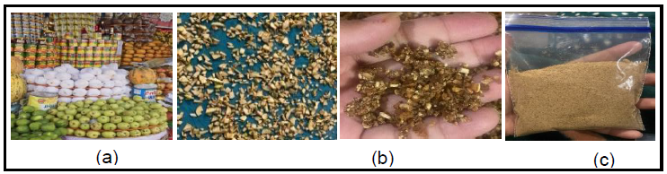
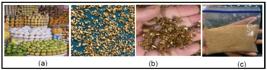
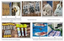
Pear fruit was collected from a local market shop in Faisalabad, Pakistan, during March, 2023, and washed to remove dirt and dust. The fruits were selected based on uniform color and shape with the same maturity level. After botanical identification, fruits were peeled and diced into small pieces for drying. After that, the fruit slices were allowed to air dry in a microwave oven (Model R-3556M, 2450 MHz, Sharp Electronics Ltd., UK) at 800 W power and ground into a fine powder using a mortar and pestle and ceramic knives in a manual grinder, sieved through an 80 mesh size plastic sieves, and was finally packed into a zip lock bag for further use. Fig. 1 presents the powder collection, drying, and powder form of pear.
Pear powder was evaluated for moisture, ash, crude fat, fiber, crude protein content, and (nitrogen-free extract) NFE according to the relevant standard procedures given by AOAC (Methods…, 2005) standards. Ash content of pear powder was examined according to AOAC (Methods…, 2005) method No. 923.03, moisture content was determined following the method No. 925.10. For protein content Kjeldahl’s method was used, for fat contents Soxhlet method was used, and fiber content was determined by method No 962.09. The NFE was calculated using the difference method as given below; [100 - (fat% + protein% + ash% + moisture%)].
According to Saquet et al. (2019), inductively coupled plasma optical emission spectrometry (ICP-OES; IRIS Intrepid II, XSP Radial, Thermo Fisher Scientific Inc., Waltham, MA, USA) was used to measure P, K, Ca, and Mg in pear powders after the samples were dissolved in acid. Calibration solutions were prepared using aliquots of ICP multielement standard solution (10 to 50mg/L Merck) that contained elements including Na, K, Mg, and P. The stock standard solutions were diluted to the required concentration in 1% HNO3 to create working standard solutions. The five-point calibration curve ranges were chosen to correspond to the anticipated concentrations of each mineral in the pear powder sample that was examined using ICP-OES. For every case, the correlation coefficient (r2) was 0.9999. On a dry mass basis, mineral concentrations were represented as mg/kg.
The DPPH assay was performed to assess the radical scavenging activity of ethanolic extracts of dried pear powders (Wang et al., 2021). In a 96-well plate, 40 µL of sample extract and 260µL of 0.1 M DPPH radical methanol solution were combined, and the mixture was incubated for 30 min at 25 °C. Absorbance was measured at 517 nm using a spectrophotometer (Thermo Fisher Scientific, Waltham, MA, USA). Ascorbic acid was used to create a standard curve, and the outcome was reported as the percentage of inhibition.
With a few modifications, the reduction capacity of the pear powder extract samples was ascertained using Wang et al. (2021) methodology. A 10:1:1 (v/v/v) mixture of sodium acetate solution (300mM), TPTZ solution (10 mM), and ferric chloride (20mM) was added to the freshly prepared FRAP reagent. Subsequently, a 96-well plate was filled with 20µL of sample extract and 280µL of FRAP dye solution. The plates were incubated for 10 min at 37°C. Absorbance was measured at 593 nm using the same spectrophotometer. Ascorbic acid was used to create a standard curve, and the results were reported as a percentage of the reducing power.
With slight modifications, the spectrophotometric technique of Hussain et al. (2021) was used to evaluate the TPC of ethanolic extracts of pear powder. A 96-well plate (Costar, Corning, NY, USA) was added to the sample extracts (25µL), Folin-Ciocalteu reagent solution (25µL, diluted with water 1:3), and Milli-Q water (200µL). After incubation (25°C, 5 min), 25µL of 10% (w/w) sodium carbonate was added and the mixture was incubated for 60 min in the dark. Absorbance was measured at 764 nm using a spectrophotometer (Thermo Fisher Scientific, Waltham, MA, USA). Each sample was quantified using a standard curve created by combining gallic acid with ethanol. The outcome is expressed as (mg GAE/mL).
As described by Hussain et al. (2021), the aluminum chloride method was modified to quantify the TFC of pear extracts. The 96-well plate was filled with the sample extract (80µL), 2% aluminum chloride (80 µL, w/v, diluted with ethanol), and sodium acetate solution (120µL, 50g/L). The plate was incubated for 60 min at room temperature in the dark. The absorbance was measured at 440 nm. The TFC of each sample was calculated using a catechin standard curve, and the results were expressed as µg CE/mL.
The total soluble solids (TSS) were determined by the method proposed by Siddique et al. (2024). Briefly, a pear powder sample (4g) was obtained, and 20mL of distilled water was used to dilute it five times. The prism of the refractometer was filled with a few drops of sample. The TSS of the sample was then viewed on the Brix scale of the refractometer, with the dark band multiplied by a dilution factor corrected to 20°C.
The pH was determined using a digital pH meter, according to the guidelines provided by the AOAC (Methods…, 2005) method, as detailed by Siddique et al. (2024). Briefly, after removing 50 mL of distilled water in a glass beaker, 5g of the pear powder sample was added, and the pH of the mixture was determined after each sample was homogenized for 30 s.
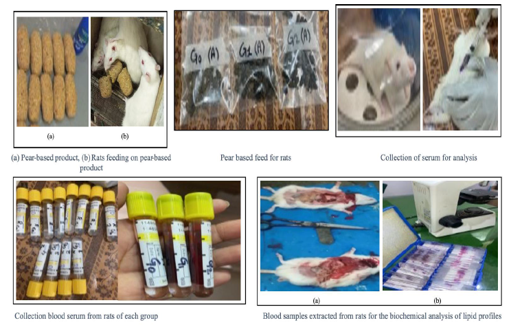
The biological activity analysis was carried out to explore the effectiveness of pear powder on hyperlipidemia following the standard procedure adopted by Velmurugan and Bhargava (2013). Male albino rats with uniform average weights and sizes were obtained from the University of Agriculture, Faisalabad, during April, 2023. The animal study was carried out during April, May, and June 2023, dividing rats in three groups: the control group (G0), treatment group 1 (G1), and treatment group 2 (G2). Each group contained ten albino rats used in the experimental trials. The rats were administered a high-saturated diet (including vegetable ghee and coconut oil 50g and 100g, respectively) for one month to induce hyperlipidemia. Throughout the trial, environmental conditions were carefully controlled such as temperature (23±2°C), relative humidity (55±5%) along with 12 hours light-dark period were maintained. At the initiation of the study, some rats were subjected to anesthesia and were euthanized to obtain the baseline values, following the veterinary guidelines provided by American Veterinary Medical Association. Pentobarbital sodium at a dose rate of 50mg/kg was administrated for anesthesia. Biochemical tests were performed before the initiation of the treatment plan and on the day after the termination of the treatment plan to determine the potential of pear powder in hyperlipidemic rats. Table 1 presents the treatment plan for the animal model used in the trials, and pear powder was added to the basal normal diet of rats at different substitution levels as given in Table 1. Fig. 2 presents a graphical overview of the steps involved in this study. All experiments were performed with the approval of the ethical review board of “The University of Faisalabad”, Punjab, Pakistan (Ref. No. TUF/IRB/212/23).
Water consumption and weight gain of the rats were measured before starting and ending the trial to check the change and gain in total body weight. Water was delivered via graded drinking bottles and daily intake was tracked (You et al., 2017).
At the end of the trial, feces of the rats were collected to evaluate total nutrient digestibility using the method described by You et al. (2017). Briefly, rats from all three groups had their droppings collected before and after treatment. The samples were weighed, collected, and stored at -20°C. The fecal samples were then subjected to chemical (proximate) examination using AOAC techniques, as mentioned earlier. The difference between specific nutrients in the rat meal and feces was used to compute the apparent nutrient digestibility of dry matter, fat, protein, fiber, and NFE using the following formula:
Blood was collected from the different rat groups before and after the trial to analyze ALT, AST, total protein, urea, and creatinine levels, and blood samples were centrifuged at 3000 rpm for 5 min to collect serum for further analysis, as described by Palipoch and Punsawad (2013). ALT and AST activities were determined using a colorimetric Randox diagnostic kit, whereas total protein levels were determined using the Biuret reaction method (Ajilore et al., 2016).
At the end of the trial, the rats were analyzed using biochemical tests of the lipid profile, as explained by Ajilore et al. (2016). For this purpose, blood samples were collected from overnight-fasted rats, and commercial kits were used to determine the LD, HDL, total cholesterol, and triglyceride levels.
At the end of the trials, when the rats were sacrificed and their organs were removed, after being preserved in 10% neutral buffered formalin solution for 24 h, the liver and kidneys were cleaned with 70% ethanol. The tissues were then placed in tiny metal jars, agitated using a magnetic stirrer, and dehydrated using a series of alcohols ranging from 70% to 100% alcohol before being embedded in paraffin with the aid of an embedding machine. Histopathological evaluation of the liver and kidneys of rats was conducted using a Leica DM750 microscope interfaced with a Leica ICC 50 camera, following the protocols described by Palipoch and Punsawad (2013).
All analyses in this study were performed three times and are presented as mean ± standard deviation. Subjected treatments were studied for both one-way analysis of variance (ANOVA), using STATISTIX (Version 8.1) software, where Duncan’s multiple range test, significance and non-significant behavior were checked at a level of P ≤ 0.05.
RESULTS AND DISCUSSION
Proximate analysis is used to estimate food and food ingredient quantities such as moisture, crude protein, total fat, NFE, and fiber. The moisture content of the pear powder is (1.17% Table 2) in pear powder. Furthermore, the results revealed that crude protein (10.63%), crude fiber (9.77%), NFE (73.96%), crude ash (2.33%), and fat (1.81%) were present in the pear powder. The analysis of the research showed that the crude protein and crude fiber content was high in the pear powder, while the crude fat and ash contents were very low. These findings are also in close agreement with the values provided by Hussain et al. (2013), who reported crude fiber content of 5-10% and ash content 1.56 - 1.86% in different varieties of pear fruits grown in Pakistan. In another study, the moisture content of pear powder was found to be low, with a moisture content of 1.16%. The study found high crude protein and fiber contents, while crude fat and ash contents were very low.
The pear fruit powder was analyzed for important macromineral and results are given in Table 3, indicating low levels of phosphorus (22.30±3.19mg/kg) and potassium (22.29±3.23mg/kg), which were observed when compared with recommended dietary allowance. According to Li et al. (2016), the mineral composition of pear fruit powder is important for different physiological functions of the fruit-consuming body. The low amount of phosphorus in the sample may have contributed to bone formation. Also, low potassium may still reduce the risk of stroke, while high sodium (43.18±2.89mg/kg) content may add value to osmotic regulation of the body fluids and transmission of nerve impulses. Magnesium is relatively high (52.18±3.35mg/kg) as compared to sodium, potassium, and phosphorus, and can help muscles and nerves to work properly, and maintain the sugar level and blood pressure at the right level. In one study, the analysis of pear fruit powder revealed low levels of phosphorus and potassium, which may contribute to bone formation and stroke risk, while high sodium content may improve fluid regulation and nerve impulse transmission. Magnesium, on the other hand, helps maintain proper muscle and nerve function (Wang et al., 2021). The present findings regarding the presence of these macrominerals were in line with the results provided by Saquet et al. (2019), who determined mineral contents in different portions of pear fruit.
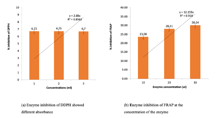
The DPPH (2, 2-diphenyl-1-picrylhydrazyl) assay is a valid, accurate, simple, and cost-effective approach to assess the radical scavenging ability of antioxidants. The ferric reducing antioxidant potential (FRAP) test offers an efficient, affordable, and flexible method to evaluate the overall antioxidant activity in a meal, as well as in plasma or urine following food (Ajilore et al., 2016). The DPPH and FRAP assay showed different absorbance levels at different concentrations of pear powder in this research work after the analysis (Fig. 3). The ethanolic extracts derived from pear fruit powders in this study were found to possess moderate antioxidant activities, as can be seen from the inhibition of DPPH and FRAP. The current results regarding the antioxidant activity of pear fruit powders are in line with the findings of Wang et al. (2021), who also used both DPPH and FRAP assays to estimate the antioxidant capacities of different pear fruit varieties. Antioxidant activity was greater in dried fruit powder samples of pears than in fresh fruit. The antioxidant content of the dried pear samples ranged from 31.74% to 66.55%, and the amounts of vitamin C, TPC, and TFC increased dramatically. These results closely matched the most recent research on the antioxidant capacity of pear powder. Furthermore, Kolniak-Ostek (2016) also confirmed similar antioxidant results using both DPPH and FRAP assays when they compared the antioxidant activities of different fractions of pear plants.
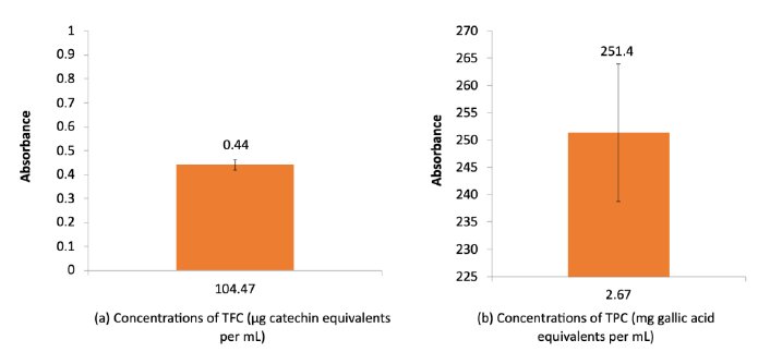
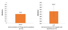
The results of TPC and TFC analysis showed that the absorbance (251.4) of TPC was recorded at 2.67 mg GAE/mL concentration of TPC and absorbance (0.44) of TFC was recorded at 104.47 concentration µg catechin/mL of TFC (Fig. 4). The present results are strongly in line with the findings of Wang et al. (2021), who also found similar amounts of TPC and TFC in different pear fruit varieties. The TPC and TFC contents of pears are sufficiently high to be regarded as bioactive substances needed for medicinal purposes. These contents can be significantly varied using different extraction solvents, such as n-hexane, ethyl acetate, ethanol, and methanol, and the drying conditions of the fruit also affect the bioactive substances involved in antioxidant activities. Research on natural antioxidant sources has increased because people adopt healthier lifestyles and consume more natural products. This research focused on fruits as a whole or on their constituent parts, particularly those that are medically significant. Thus, TPC and TFC have evolved as important bioactive substances in pear fruits, which have been linked to different health claims. Current research results regarding TPC and TFC were also confirmed by Kolniak-Ostek (2016), who explored leaves, peels, fruit, and seeds of pear, and reported fruit powder to be a good source of TPC and TFC, although they have lower amounts than those present in seeds and leaves.
The results of TPC and TFC analysis showed that the absorbance (251.4) of TPC was recorded at 2.67 mg GAE/mL concentration of TPC and absorbance (0.44) of TFC was recorded at 104.47 concentration µg catechin/mL of TFC (Fig. 4). The present results are strongly in line with the findings of Wang et al. (2021), who also found similar amounts of TPC and TFC in different pear fruit varieties. The TPC and TFC contents of pears are sufficiently high to be regarded as bioactive substances needed for medicinal purposes. These contents can be significantly varied using different extraction solvents, such as n-hexane, ethyl acetate, ethanol, and methanol, and the drying conditions of the fruit also affect the bioactive substances involved in antioxidant activities. Research on natural antioxidant sources has increased because people adopt healthier lifestyles and consume more natural products. This research focused on fruits as a whole or on their constituent parts, particularly those that are medically significant. Thus, TPC and TFC have evolved as important bioactive substances in pear fruits, which have been linked to different health claims. Current research results regarding TPC and TFC were also confirmed by Kolniak-Ostek (2016), who explored leaves, peels, fruit, and seeds of pear, and reported fruit powder to be a good source of TPC and TFC, although they have lower amounts than those present in seeds and leaves.
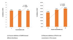
(a) Inhibition of DDPH showed different absorbance, (b) enzyme inhibition of FRAP at the concentration of the enzyme.
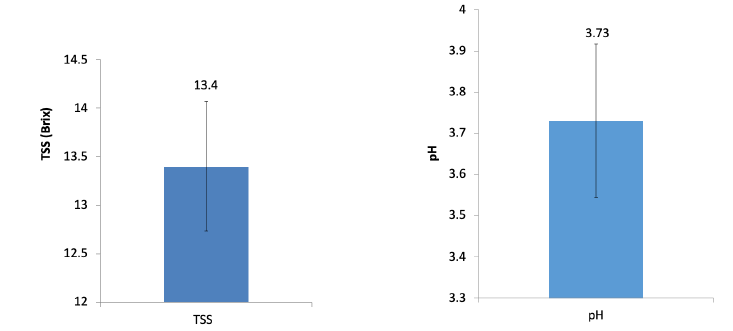
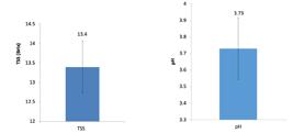
The results given in Fig. 5 showed that the mean value of TSS was 13.4 brix, and the mean value of pH was 3.73 for the pear powder. TSS is used to determine the amount of sugar present in any solution. The components were measured using a refractor. The refractive index can be used to calculate the total soluble solid content of the solution. The pH scale, which ranges from 1 to 14, quantifies the degree of acidity or alkalinity of a food or solution. One is the most acidic, 7 is neutral, and anything over 7 is basic or alkaline. Foods that have been acidified have a pH of less than or equal to 4.6. These results were in line with the findings of Hussain et al. (2013) as they reported pH of different varieties of pear fruits ranging from 4.12 - 5.24 and TSS ranging from 11.03 - 14.42 brix.
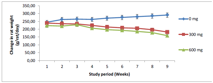
The weight of the rats was measured on a weekly basis during the two-month trial, from the first week to the 8th week, and the overall results showed significant changes, as presented in Table 4. From these data, the overall mean weight of rats in all groups at the start of the study was 235.61g, which was reduced to 210.52g at the end of the study, showing a significant decrease in weight in the studied rats. As the lipid profile of the rats decreased, weight reduction was also observed, which was a positive aspect of this study. The current trial results showed that there was an increase in the weight of rats during the first four weeks, which revealed that pear powder treatment was not effective in reducing the weight of the rats. However, the later a four-week study duration revealed a significant reduction in weight from the start of the trial (270.43±3.14g) to the end of the trial (198.96±3.23g), in the rat weight over the study period compared to G1 (300mg dose rate) and G0 (0 mg dose rate), while the results revealed that G2 (600mg) resulted in a remarkable weight reduction (Fig. 6). A similar clinical study demonstrated the anti-obesity effects of fresh pear consumption. After 12 weeks of fresh pear fruit consumption, users can lower leptin concentrations and waist circumference (Hong et al., 2021). The present findings regarding the decrease in weight of pear-fed rats were also confirmed from the experimental results provided by Velmurugan and Bhargava (2013), who noted a decrease in body weight of the rats that were fed with pear extracts rich in dietary fibers and polyphenols. A similar decrease in the body weight of rats was also reported by You et al. (2017) when water extracts from pear pomace were administered at 200 and 400 mg/kg, which were compared to the control rats with hyperlipidemia.
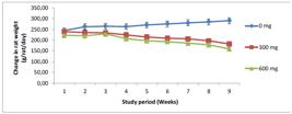
Variation in rat weight changes over the study period, fed by pear powder fortified lipid diet.
The change in water intake in bio-assayed rats fed a pear powder-incorporated diet throughout the study period varied significantly over the two-month period, as shown in Table 5. The current results revealed that G2 group (600mg pear powder) resulted in a remarkable increase in water intake (10.04mL/rat/day), after two months of study period, compared to G1 (300 mg pear powder) and G0 (0 mg pear powder), where values of water intake were 9.50 and 5.45mL/rat/day, respectively. The results showed that there was an increase in water intake i.e., 6.02 - 10.04mL/rat/day of the bio-assayed rats from 0 to 2 months in the G2 group. Water intake per rat per day in rats was found to be influenced by the increased feeding amount of pear powder, as a clear trend shown in Figure 7. A similar increase in water intake in pear extract-treated rats was also reported by Velmurugan and Bhargava (2013). In another relevant work, You et al. (2017) also observed increased water intake by rats fed with aqueous extracts of pear pomace at 200 and 400mg/kg.
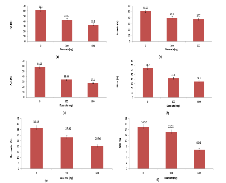
Digestibility refers to the amount of nutrients absorbed by the rat and is generally calculated as the quantity of nutrients consumed minus the quantity of nutrients retained in feces. It has been reported in different studies that it takes 1 - 2 h for the transit of contents to reach the cecum and 4 - 6 h to transit from the stomach to the colon in 16-month-old rats. After a trial period of two months, the nutrient digestibility study was performed after collecting feces from different groups of rats. The current result outcomes (Fig. 8) showed that the lowest dry matter (20.36%) was recorded in case of 600 mg while the highest in dry matter (36.43%) in the control group, lowest fat (33.30%) was recorded in case of 600mg while highest fat (61.30%) in control group, lowest protein (37.70%) was recorded in case of 600 mg, while highest protein (50.66%) in control group, lowest ash (27.10%) was recorded in case of 600 mg, while highest ash content (58.03%) in control group, lowest fiber (34.5%) was recorded in case of 600 mg while highest fiber (64.20%) in control group, the lowest NFE (6.36%) was recorded in case of 600mg while highest NFE content (14.52%) in control group. These results showed that as the dose of pear powder increased from 300mg to 600 mg, the digestibility of the nutrients increased as significantly lower amounts of nutrients were found in the feces of the G2 group rats compared to the G1 and G0 group rats.
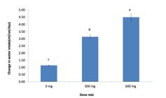
Effects of different treatments on mean water intake (mL/rat) in bio-assayed rats at different dose rates of pear powder.
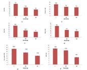
Effects of different treatments on mean nutrient intake (mg/rat) in bio-assayed rats at different dose rates of pear powder (a) fat, (b) protein, (c) ash, (d) fiber, (e) dry matter, (f) NFE.
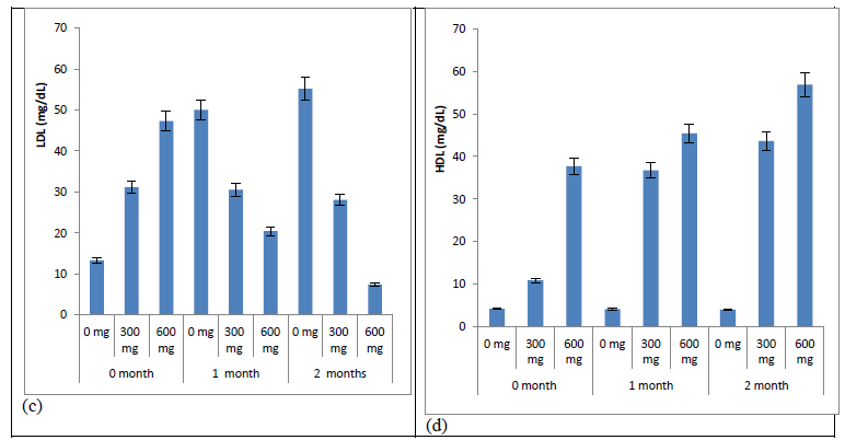
The outcome of the current study revealed that comparatively greater lipid contents in the control group, except HDL, were recorded compared with G1 and G2, which showed that the intake of pear powder in the diet reduced the lipid content in the bio-assayed rats. From the data presented in Fig. 9, at the start of the study, cholesterol, triglycerides, and LDL were significantly high in the rats’ blood, whereas during the first month of feeding pear powder, they were significantly reduced in a dose-dependent manner in the rats. However, a more significant decrease in these lipid profiles was observed during the second month of feeding pear powder, as a 600 mg dose of pear powder provided the most significant results. However, the results were reversed for HDL (beneficial lipids), as these were found to be increased because of feeding pear powder to the rats. High blood lipid levels are referred to as hyperlipidemia. It is a metabolic disorder rather than an illness, and can be caused by several illnesses, including cardiovascular conditions. However, fruits such as pears, which contain bioactive chemicals, have been shown to help lower the risk of hyperlipidemia. Moreover, another study conducted by You et al. (2017) showed that the consumption of pear displayed a reduction in triglycerides by 6.8%, LDL-C by 17.4%, and total concentration of cholesterol by 14.6% in bio-assayed rats; a similar decrease was recorded in the current study. According to some views, the lipid-lowering effects of pears seem to be related to the components such as catechin, which are more condensed in peels and pulp (He et al., 2024). Pear extracts rich in polyphenols were also found to be effective in reducing blood triglyceride, total cholesterol, and LDL levels when clinical trials were performed by Velmurugan and Bhargava (2013). According to a study, the method by which pear fruit extracts decrease cholesterol is due to the ability of cholesterol to quench the exogenous fluorescence of polyphenols through a static mechanism. The thermodynamic interaction results showed that hydrogen bonding and hydrophobic contacts play a major role in the spontaneous interaction between polyphenols and cholesterol (He et al., 2024).
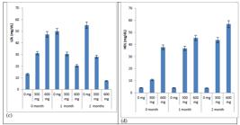
Effects of different treatments on mean biochemical profile (mg/rat) in bio-assayed rats at different dose rates of pear powder (a) cholesterol, (b) triglycerides, (c) LDL, (d) HDL
Pear powder decreased the levels of creatinine and urea in the rats, and this effect was more prominent after two months as compared to the first month and the start of the study, when the pear powder was fed to the rats (Table 6). The results of a two-month trial showed that the lowest urea (32.45 mg/dL) and creatinine (0.56mg/dL) levels were recorded after the exposure period of 2 months of feeding pear powder to the rats, whereas these levels of urea and creatinine were comparatively high in the control rats. Similarly, there was also a significant decrease in ALT and AST levels in rats fed with 600mg pear powder. The lowest ALT levels (9.09U/I) of the bio-assayed rats were observed after two months of exposure, while the ALT level (18.83U/I) was recorded after an exposure period of one month. The highest ALT content (48.43U/I) was recorded in the control group. Pear powder also decreased the AST content in the rats, and this effect was more prominent after two months of feeding, compared to one month 0 months. The lowest AST level (2.24U/I) of the bio-assayed rats was noted after a two-month exposure period, while a greater AST level (126.67U/I) was recorded after an exposure period of one month. The highest AST content (132.47U/I) was recorded at the start of the experiment (0 months). Whenever intoxication or hyperlipidemia is introduced in animal models, there are abnormalities found in different organ functions due to changes in the levels of different chemicals and enzymes in the body; in particular, Alt and AST are increased in animals (Palipoch and Punsawad, 2013; You et al., 2017). The oxidative damage of ROS on the membrane components of liver cells and the subsequent escape of intracellular contents into the circulation may be the cause of the significant decrease in creatinine and urea levels observed in the serum of the rats fed pear powder (600mg/kg) and the significant increase in the serum of the control rats. Singh et al. (2021) evaluated the ethanolic extract of pear fruit powder for its ability to protect rat livers from hepatotoxicity caused by carbon tetrachloride. Biochemical markers, such as total albumin, total protein, serum ALT, and AST, were measured to screen for hepatoprotection. Rats administered 400mg/kg of pear fruit extracts showed significantly decreased levels of ALT, AST, and total protein, which is consistent with the current findings. The present study results were also validated when Ajilore et al. (2016) observed the hepatoprotective potential of pear fruit seeds in cadmium-toxicated Wistar albino rats, as the levels of ALT, AST, total protein, and haptoglobin were significantly reduced in rats fed with 500mg/kg body weight pear seeds. The loss of hepatocyte membrane structure and function, as well as the release of the enzymes into the bloodstream, are indicated by the significant increases in serum activities of these liver marker enzymes in control rats and the significant decreases in AST and ALT activities in the rats fed 600mg/kg pear powder. In contrast, the antioxidant properties of pear powder may have minimized the loss of structure by shielding liver cells from damage.
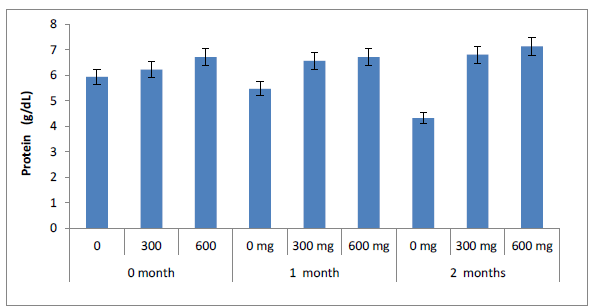
From the results presented in Fig. 10, the protein levels in the control rats were lower than those in the rats fed 300 and 600mg pear powder. A similar study has shown that the methanol extract from the pomace of pear can protect against liver injury. Cadmium was administered to the rats to induce injury, and the methanol extract from pomace of pear improved the synthesis of haptoglobin (synthesized in the liver) and other proteins that reverse liver injury due to their antioxidant properties, which ultimately reduce oxidative stress. Therefore, the methanol extract of pears restored haptoglobin expression and provided protection against severe heavy metal-induced liver injury (You et al., 2017). Present results were also in line with the findings of Ajilore et al. (2016), when cadmium toxicated rats were investigated for total protein level and it was found high (7.58g/dL) in the rats fed with 500mg/kg body weight pear seeds, as compared to cadmium toxicated rats (6.46mg/dL).
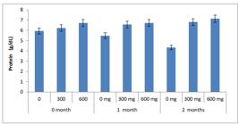
Change in protein values at various exposure periods and dose rates of pear powder intake by rats.
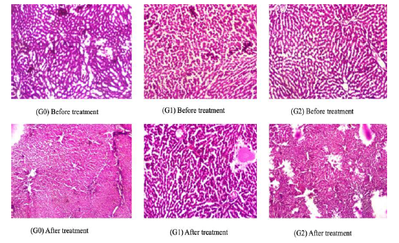
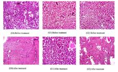
Pears have been shown to have anti-inflammatory, blood cholesterol-lowering, and renal and liver-protective properties in a variety of in vitro, in vivo, and human studies. These pharmacological properties have been linked to several active compounds, including flavonoids, triterpenoids, and phenolic acids (Hong et al., 2021). The histopathology results showed that the hepatic tissue of control rats had preserved architecture (Fig. 11). However, this hepatic tissue encloses an occasional portal triad, which reveals extensive fibrosis. The limiting plates were intact. There was no spillover of the inflammatory cells into the adjacent parenchyma. Centrilobular fibrosis was observed in the control rats before treatment (Fig. 11). Pear powder was administered to the rats over the course of two months, and after two months of trial, mild focal centrilobular degeneration was observed. No fibrosis was present and the limiting plates were intact (Figure 11). The results clearly indicated that pear powder reduced centrilobular necrosis of the hepatic lobule tissue and inflammation. A similar study was conducted by Singh et al. (2021) to evaluate the hepatoprotective and in vitro antiradical activities of an ethanolic extract of pear fruit powder against rat liver hepatotoxicity induced by carbon tetrachloride. Their findings were consistent with the present findings, showing that in rats intoxicated with CCl4, the ethanolic extract (200 mg/kg and 400 mg/kg body weight) had a significant hepatoprotective effect in a dose-dependent manner. Active plant substances called polyphenols are widely distributed in pears and are said to have the ability to reduce or even completely eradicate the harmful effects of free radicals, thus preserving the structural integrity of vital organs, such as the liver. This study demonstrated the hepatoprotective properties of this plant by examining the normal liver histoarchitecture of rats fed a high dose of pear powder. This might be a result of the phytochemicals in the pear fruit extract, which have been previously shown to have anti-inflammatory and antioxidant properties.
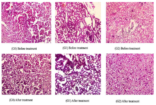
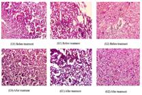
Similarly, extensive renal atrophy was observed before treatment in the control rats (Fig. 12). There was extensive infiltration of chronic inflammatory cells comprising lymphocytes and plasma cells. The histopathological results of the current study showed extensive infiltration of chronic inflammatory cells comprising lymphocytes and plasma cells. The intersititum reveals mild fibrosis, focal areas of hemorrhage, and amp, and is infiltrated by mild-to-moderate chronic inflammatory cell infiltrate. However, when pear powder was administered to the rats for two months, the results were significantly different, as well-preserved hepatic architecture and mild focal centrilobular degeneration were observed. No fibrosis was observed and the limiting plates were intact (Fig. 12). The histopathological findings of the current study indicated that the pear powder at 600mg/kg dose rate showed significant effects on renal function compared to the 300mg/kg dose rate. These beneficial effects on health were most likely caused by the bioactive antioxidant chemicals found in pears. Thomas et al. (2010) stated that substantial concentrations of anthocyanins, pigments that confer their therapeutic properties when eaten, are present in pear fruit. However, rats fed pear powder may have benefited from the protective effects of anthocyanins on the liver and kidneys.
CONCLUSION
Pears (Pyrus communis L.), which grow in temperate regions of Earth, are extensively distributed worldwide. Customers prefer this fruit because of its flavor and alleged health advantages. Many bioactive substances, such as phenolic and flavonoid compounds, have been found in pear fruits and are thought to be responsible for these health benefits. The therapeutic efficacy of feeding pear powder for hypolipidemic effects in rats was studied in this work, and findings have shown encouraging results in the treatment of hyperlipidemia, owing to the high number of dietary fibers, antioxidants, and bioactive substances. Pear powder analysis revealed high contents of protein and fiber, low contents of fat, ash, and macrominerals (sodium, potassium, magnesium, and phosphorus), and the presence of significant amounts of TPC and TFC, whereas DPPH and FRAP assays showed prominent antioxidant activities. When biochemical assays were performed, as compared to the control group (G0) and rats fed pear powder at 300 mg/kg body weight (G1), rats fed 600 mg/kg body weight (G2) showed significantly decreased amounts of urea, creatinine, ALT, and AST. Furthermore, in vivo screening of pear powder was found to increase the blood HDL and significantly decrease the total cholesterol, LDL, and triglyceride levels in the blood of fed rats, thereby improving overall lipid profiles and lowering the risk of cardiovascular disease. Finally, at the end of the study, the histopathological screening of the liver and kidney tissues of these rats showed proper and improved functionalities in the rats fed with 600 mg/kg body weight pear powder, as compared to the control group rats and rats fed with 300 mg/kg pear powder. Thus, pear powder containing bioactive substances can be consumed as a health-promoting food, especially with hypolipidemic potential.
REFERENCES
- AJILORE, B.; FALOLU, I.; OLANIYAN, O. Effect of Pyrus communis (Common Pear) seeds on selected parameters of liver function in rats treated with cadmium. Am. Sci. Res. J. Eng. Technol., v.23, p.41-53, 2016.
- HE, X.; CHEN, L.; PU, Y. et al. Evaluation of cholesterol and cholates binding capacity and mechanism exploration of'Yali'Pear polyphenol extracts: in vitro. Food Innov. Adv., v.3, p.268-278, 2024.
- HONG, S.Y.; LANSKY, E.; KANG, S.S.; YANG, M. A review of pears (Pyrus spp.), ancient functional food for modern times. BMC Compl. Med. Ther., v.21, p.1-14, 2021.
- HUSSAIN, A.; KAUSAR, T.; DIN, A. et al. Determination of total phenolic, flavonoid, carotenoid, and mineral contents in peel, flesh, and seeds of pumpkin (Cucurbita maxima). J. Food Proc. Preserv., v.45, p.e15542, 2021.
- HUSSAIN, S.; MASUD, T.; ALI, S.; BANO, R.; ALI, A. Some physico-chemical attributes of pear (Pyrus communis L.) cultivars grown in Pakistan. Int. J. Biosci, v.3, p.206-215, 2013.
- KOLNIAK-OSTEK, J. Chemical composition and antioxidant capacity of different anatomical parts of pear (Pyrus communis L.). Food Chem., v.203, p.491-497, 2016.
- LI, X.; LI, X.; WANG, T.; GAO, W. Nutritional composition of pear cultivars (Pyrus spp.). In: SIMMONDS, M.S.J.; PREEDY, V.R. Nutritional composition of fruit cultivars. London: Academic Press, 2016. p.573-608.
- METHODS of analysis. 18.ed. Arlington, VA, USA.: AOAC, 2005.
- NASER, I.H.; ALKAREEM, Z.A; MOSA, A.U. Hyperlipidemia: pathophysiology, causes, complications, and treatment. A review. Karbala J.n Pharm. Sci., v.1, n.19, 2021.
- NIE, Y.; LUO, F. Dietary fiber: An opportunity for a global control of hyperlipidemia. Oxid. Med. Cell. Long., v.2021, p.5542342, 2021.
- PALIPOCH, S.; PUNSAWAD, C. Biochemical and histological study of rat liver and kidney injury induced by cisplatin. J. Toxicol. Pathol., v.26, p.293-299, 2013.
- SAQUET, A.A.; STREIF, J.; ALMEIDA, D.P. Mineral composition and distribution within ‘Rocha’pear in relation to internal storage disorders. Postharvest Biol. Technol., v.158, p.111002, 2019.
- SIDDIQUE, F.; HUSSAIN, A.; MAHDI, A.A. et al. Comparison of chemically treated, pasteurized, and microwave‐treated (at different time durations) chia seeds added to mango‐whey beverage, during different storage periods, for physicochemical and sensory parameters. J. Food Quality, v.2024, p.6688945, 2024.
- SINGH, S.;; BAJPAI, M.; MISHRA, P. Pharmacognostic profile, In-vitro antioxidant and hepatoprotective potential of ethanolic fruit extract of pyrus communis Linn. Curr. Bioactive Comp., v.17, p.49-59, 2021.
- STEWART, J.; MCCALLIN, T.; MARTINEZ, J.; CHACKO, S.; YUSUF, S. Hyperlipidemia. Pediatr. Rev., v.41, p.393-402, 2020.
- THOMAS, L.A.; SEHATA, M.J.; DU PREEZ, M.G.; REES, J.G.; NDIMBA, B.K. Establishment of proteome spot profiles and comparative analysis of the red and green phenotypes of ‘Bon Rouge’pear (Pyrus communis L.) leaves. Afr. J. Biotechnol., v.9, p.4334-4341, 2010.
- VELMURUGAN, C.; BHARGAVA, A. Anti-diabetic and hypolipidemic activity of fruits of Pyrus communis L. in hyperglycemic rats. Asian J. Pharm. Clin. Res., v.6, p.108-111, 2013.
- WANG, Z.; BARROW, C.J.; DUNSHEA, F.R.; SULERIA, H.A. A comparative investigation on phenolic composition, characterization and antioxidant potentials of five different australian grown pear varieties. Antioxidants, v.10, p.151, 2021.
- YOU, M.K.; RHUY, J.; KIM, H.A. Pear pomace water extract suppresses hepatic lipid peroxidation and protects against liver damage in rats fed a high fat/cholesterol diet. Food Sci. Biotechnol., v.26, p.801-806, 2017.
All the research data are available within the article.