Transitional nasal carcinoma - case report
ABSTRACT
A six-year-old female Siberian Husky was presented to the Veterinary Hospital of the State University of Londrina (UEL) with a clinical history of persistent sneezing and coughing, accompanied by right-sided unilateral epistaxis. Initial therapeutic management included inhaled corticosteroids and a broad-spectrum antibiotic. Despite treatment, the patient showed progressive clinical deterioration. Advanced diagnostic procedures and associated incisional biopsy were undertaken. Histopathological analysis was inconclusive; however, tomography revealed a well-defined amorphous mass with irregular margins, extending from the middle to the caudal third of the right nasal cavity, associated with signs of local bone degeneration. The condition progressed to generalized seizures, which proved refractory to anticonvulsant therapy, and ultimately resulted in death. Post-mortem examination revealed an infiltrative malignant epithelial sinonasal neoplasm with invasion of the cerebral cortex, supporting neurogenic shock as the cause of death. This case highlights the diagnostic challenges posed by nasal tumors in advanced stages, particularly due to the nonspecific nature of clinical signs and limitations of initial diagnostic methods. It reinforces the importance of considering nasal neoplasia as a differential diagnosis in cases of chronic respiratory disease in dogs and underscores the need for comprehensive and timely diagnostic approaches to enable early detection and improve clinical outcomes.
Keywords:
computed tomography; necropsy; neoplasm; postmortem diagnosis; rhinoscopy
RESUMO
Um cão, Husky Siberiano, fêmea, seis anos de idade, foi atendido no Hospital Veterinário da Universidade Estadual de Londrina (UEL) com histórico de espirro e tosse persistentes, além de epistaxe unilateral direita. O tratamento inicial incluiu corticosteroide inalatório e antibiótico de amplo espectro. Após algumas semanas, o animal apresentou piora do quadro clínico. Foram realizados exames avançados de imagem e coleta de amostras por biópsia incisional. O exame histopatológico não demonstrou qualquer diagnóstico conclusivo; a tomografia revelou uma formação amorfa de limites definidos e margens irregulares, localizada do terço médio ao terço caudal da cavidade nasal direita, com sinais de degeneração óssea. O caso evoluiu para convulsões não responsivas aos tratamentos anticonvulsivantes e seguinte óbito. Ao exame post-mortem, foram evidenciados achados anatomopatológicos de neoplasia epitelial sinonasal maligna infiltrativa em córtex cerebral, os quais favorecem o choque neurogênico como causa mortis. Este relato destaca a dificuldade em identificar tumores nasais em estágios avançados devido à natureza inespecífica dos sinais clínicos e à falta de exames conclusivos realizados durante o acompanhamento. Enfatiza-se a importância de considerar neoplasias nasais no diagnóstico diferencial de doenças respiratórias crônicas em cães, reforçando a necessidade de uma investigação mais detalhada em busca de um diagnóstico precoce.
Palavras-chave:
diagnóstico pós-morte; necropsia; neoplasia; rinoscopia; tomografia computadorizada
INTRODUCTION
Transitional nasal carcinoma is a rare and aggressive neoplasm that represents approximately 4 to 5% of neoplasms in dogs and cats (Bracho-Villalobos et al., 2012). It affects the upper respiratory tract and is most diagnosed at advanced stages due to the non-specific nature of its clinical signs (Thrall, 2018). Symptoms such as persistent sneezing, nasal discharge, epistaxis, and dyspnea are frequently misattributed to inflammatory or infectious diseases, potentially delaying a definitive diagnosis. In more advanced cases, it may lead to facial deformity, severe respiratory distress and, in some instances, neurological alterations because of central nervous system invasion (Leal, 2017).
The tumor originates from the transitional epithelium lining the upper airways and is characterized by aggressive behavior, with potential for bone degeneration and invasion of adjacent structures such as the nasal bones, paranasal sinuses, and cranial cavities. These characteristics make treatment particularly challenging and in most cases the prognosis is reserved (Mondal et al., 2015; Aljanabi and Sheibani, 2019).
Computed tomography (CT) is the diagnostic method of choice for assessing the extent of nasal neoplasia and its invasion into adjacent structures, as it provides detailed images of nasal anatomy and assists in guiding additional diagnostic procedures such as rhinoscopy with biopsy sampling. However, definitive diagnosis relies on histopathological examination, and, in some cases, it is only achievable postmortem following necropsy, especially in advanced stages where neoplastic cells have reached clinically inaccessible regions (Mondal et al., 2015).
The aim of this case report is to present the clinical case of a dog with a clinical history suggestive of chronic respiratory disease, in which a diagnosis of transitional nasal carcinoma was confirmed only postmortem.
ETHICAL ASPECTS
Our institution do not require approval from the Ethics Committee on Animal Use for case reports.
CASUISTRY
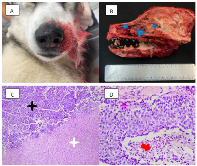
A six-year-old female Siberian Husky was presented to the Veterinary Hospital of the State University of Londrina (UEL) with persistent sneezing, unilateral nasal discharge, and intermittent episodes of epistaxis (Fig. 1A), predominantly affecting the right nostril. Initially, inhaled fluticasone propionate (Seretide®, GSK, Brazil) was administered to alleviate clinical symptoms. Blood was subsequently collected via venipuncture for hematological analysis, where no signs of thrombocytopenia or anemia were identified to justify the bleeding. Laboratory findings included elevated hepatic enzyme levels, hyperproteinemia and hyperglobulinemia. The peripheral blood PCR test did not detect the presence of Ehrlichia spp..
Due to the persistence of clinical signs and progressive clinical deterioration, a rhinoscopy examination was requested. It revealed mild to moderate rhinitis in the left nostril and moderate rhinitis in the right nostril, associated with a mass extending into the oropharynx (right choana) and dorsal nasal meatus/turbinates. A biopsy sample was collected during this procedure; however, histopathology yielded an inconclusive result, revealing fragments of respiratory epithelium-frequently flattened and ulcerated-with infiltration of intact and degenerated neutrophils and rare lymphocytes, findings consistent with mild to moderate suppurative and hemorrhagic rhinitis. So, a broad-spectrum antimicrobial (amoxicillin + clavulanic acid) was prescribed.
Persistent clinical signs progressed to neurological symptoms, including recurrent seizures that were refractory to anticonvulsant therapy (diazepam and phenobarbital), raising suspicion of a nasal cavity neoplasm with possible central nervous system commitment.
Thoracic radiography (which revealed no abnormalities) and abdominal ultrasonography were conducted. The latter indicated moderate splenomegaly with a heterogeneous nodule of unknown malignancy, moderate biliary sludge, and kidneys with increased cortical echogenicity, though with preserved architecture.
Subsequently, cranial computed tomography was performed. The scan report described an amorphous, heterogeneous soft-tissue-density mass (measuring 9.0cm in length × 3.3 cm in height × 2.2cm in width), with defined boundaries and irregular margins. The lesion extended from the middle to the caudal third of the right nasal cavity, causing destruction of nasal turbinates, leftward deviation of the nasal septum, and discrete foci of osteolysis in the lateral portion of the right maxillary bone. The mass extended into the right nasopharyngeal meatus and the ventral portion of the right ethmoidal sinus, leading to destruction of the associated ethmoturbinates. The formation induced osteolysis of the right ventral portion of the cribriform plate and invaded the right rostral cranial fossa, with no cleavage plane between the mass and the right olfactory bulb, causing leftward deviation of the cerebral falx.
The patient exhibited progressive neurological deterioration, with increased frequency and duration of convulsive episodes refractory to pharmacological intervention. The animal ultimately died following one of these seizure episodes. Necropsy revealed a malignant sinonasal epithelial neoplasm with characteristics consistent with transitional nasal carcinoma, which had extended beyond the nasal cavity, infiltrating the ethmoid bone and invading the cerebral cortex-findings that explained the convulsions. Histopathological analysis confirmed the diagnosis of transitional nasal carcinoma with central nervous system invasion (Fig. 1B), demonstrating the aggressive and locally invasive nature of the tumor.
A: Autopsy of a female adult Siberian Husky showing bilateral rhinorrhagia; B: Sagittal section of the skull reveals an obliterative neoplastic mass in the right nasal cavity extending to the frontal telencephalic cortex. The mass appears multilobulated rostrally (arrow) and infiltrates the ethmoturbinates caudally (arrowhead). Neoplastic infiltration is observed within the cranial cavity, compressing and replacing the olfactory bulb and a focally extensive area of the frontal telencephalic cortex (asterisk); C: Histological section of the sinonasal mass-neoplastic proliferation arranged in folded cords of non-keratinized stratified epithelium with an indistinct fibrovascular core (black asterisk), infiltrating the neuroparenchyma (white asterisk), 40x magnification, hematoxylin and eosin (H&E) staining; D: Histological section of the sinonasal mass-cuboidal cells with round to oval nuclei, aggregated chromatin, and moderate anisocytosis, anisokaryosis, and pleomorphism. Intraluminal suppurative exudate is observed (arrow), 400x magnification, H&E staining.
The necropsy suggested neurogenic shock as the cause of death, secondary to tumor invasion of the central nervous system, which resulted in failure of autonomic neurological control and consequent death.
DISCUSSION
Although rare, transitional nasal carcinoma in dogs is an aggressive condition with a reserved prognosis. This type of neoplasm, which originates in the transitional epithelium of the nasal cavity, often exhibits invasive behavior, including local bone degeneration and infiltration into adjacent structures such as the central nervous system, as observed in the present case (Mondal et al., 2015; Aljanabi and Sheibani, 2019). The main challenge in early diagnosis is the nonspecific nature of the clinical signs-such as sneezing, nasal discharge, and epistaxis-which are easily mistaken for common respiratory conditions such as infections or chronic rhinitis, delaying appropriate treatment (Park et al., 2015). Additional clinical signs may include seizures, mydriasis, vestibular dysfunction, blindness, and facial deformities, as malignant epithelial neoplasms tend to exhibit invasive behavior and lead to destruction of bony structures in the region (Gorza et al., 2021).
In this case, the initial clinical signs were treated as a common inflammatory disease, using symptomatic therapies. However, combined rhinoscopy and computed tomography (CT) revealed an expansive mass with irregular margins and bone degeneration-features typical of advanced-stage nasal neoplasms. CT is considered the imaging modality of choice for assessing nasal tumors due to its ability to provide precise anatomical detail, allowing for better delineation of tumor extent, particularly in cases with suspected invasion of adjacent structures such as the paranasal sinuses and the brain (López et al., 2013). Although not used in the present case, some studies suggest that immunohistochemistry with general cytokeratin and cytokeratin-19 antibodies can assist in diagnosing epithelial-origin neoplasms (Gorza et al., 2021).
In many cases of transitional nasal carcinoma, a definitive diagnosis is only achieved postmortem, as in this case, where necropsy and histopathological examination revealed the true nature of the lesion (Aljanabi and Sheibani, 2019; Mondal et al., 2015).
In the reported case, the progressive worsening of clinical signs was due to intracranial invasion of the tumor. While primary intracranial tumors are relatively common in dogs, secondary intracranial neoplasms may arise from distant metastases or from local extension and compression of the brain by adjacent tissues. Among nasal tumors leading to secondary intracranial invasion, carcinomas represent the most frequently diagnosed type (Gorza et al., 2021).
Currently, there is no evidence suggesting a specific predisposition to nasal carcinoma in the Siberian Husky breed. However, dolichocephalic breeds are generally more susceptible to developing nasal tumors due to increased exposure of the airways to irritants and pollutants. The etiology of these tumors remains poorly understood in veterinary medicine. Studies suggest that exposure to urban pollutants (e.g., cigarette smoke, topical insecticides) and advanced age, particularly between 10 and 15 years, may increase the risk of developing this type of cancer in dogs (Gorza et al., 2021; Reif et al., 1998).
Surgical treatment is often limited by the anatomical location of the tumor, which may prevent effective resection without causing significant damage, especially in cases where vital structures are involved (Leal, 2017). Palliative radiotherapy has been used as a treatment option to alleviate symptoms and improve quality of life, although the prognosis remains reserved (Rancilio, 2021). Studies suggest that the average survival time for dogs with nasal carcinoma ranges from 3 to 6 months, even with therapeutic intervention, depending on the stage of disease at diagnosis. In a study by Henry et al. (1998), dogs treated with radiotherapy showed significantly better survival times compared to those treated with chemotherapy and/or surgical resection. More recent studies by London et al. (2012) and Park et al. (2015) have reported that metronomic chemotherapy combined with toceranib phosphate has shown promise in treating nasal cavity neoplasms.
Recent reports of primary nasal neoplasms with subsequent brain invasion in dogs, as well as detailed descriptions of their clinical findings, remain scarce. This scarcity hinders the development of more precise therapeutic protocols for such cases (Gorza et al., 2021).
CONCLUSIONS
This case report highlights the importance of early diagnosis and underscores the need for advanced imaging examinations-such as computed tomography-and early biopsy sampling in patients presenting with chronic respiratory signs. The fatal progression of the disease illustrates the invasive nature of transitional nasal carcinoma and reinforces the importance of including nasal neoplasms in the differential diagnosis of dogs with persistent respiratory symptoms. Therefore, environmental factors and age should be considered when evaluating the risk of nasal tumors in the breed described in this study, as well as in other dolichocephalic breeds. Faster and more accurate diagnostics could improve treatment options and potentially increase survival and quality of life for affected patients.
REFERENCES
- ALJANABI, K.S.K.; AL SHEIBANI, S.M.H. Rare case of transitional cell carcinoma of the sino nasal tract: a diagnosis with multiple synonyms. Review of literatures. J. Otolaryngol. Rhinol., v.1, n.5, 2019.
- BRACHO-VILLALOBOS, G.A.; CASADO, B.A.; DANIEL, C. Contribution to the study of carcinoma of nasal cavity in dogs: review of cases from 2005 to 2011. Rev. Colegio Med. Vet. Estado Lara, v.2, n.1, 2012.
- GORZA, L.L.; OLIVEIRA, A.R.; PORTO, M.G. et al. Clinical, anatomopathological, and immunohistochemical findings of a transitional cell carcinoma from nasal cavity, frontal and ethmoidal sinus with meningoencephalic invasion in a dog. Open Vet. J., v.11, p.1-5, 2021.
- HENRY, C.J.; BREWER JR, W.G.; TYLER, J.W. et al. Survival in dogs with nasal adenocarcinoma: 64 cases (1981-1995). J. Vet. Intern. Med., v.12, p.436-439, 1998.
- LEAL, S.M.R. Neoplasias sinonasais caninas: revisão bibliográfica e estudo de casos. 2017. Dissertação (Mestrado Integrado em Medicina Veterinária) - Universidade de Lisboa, Lisboa, PRT.
- LONDON, C.; MATHIE, T.; STINGLE, N. et al. Preliminary evidence for biologic activity of toceranib phosphate (Palladia®) in solid tumours. Vet. Comp. Oncol., v.10, p.194-205, 2012.
- LÓPEZ, F.; LLORENTE, J.L.; COSTALES, M. et al. Molecular characterisation of sinonasal carcinomas and their clinical implications. Acta Otorrinolaringol., v.64, p.289-296, 2013.
- MONDAL, M.; DEY, A.; BANDYOPADHYAY, J.; BANERJEE, D. Transitional cell carcinoma of the sinonasal tract: a rare entity. Clin. Cancer Invest. J., v.4, p.77-80, 2015.
- PARK, H.; KIM, J.; JEONG, S. et al. Adjuvant metronomic chemotherapy in a dog with nasal transitional carcinoma. J. Vet. Clin., v.32, p.183-186, 2015.
- RANCILIO, N. Conventional versus stereotactic radiotherapy. Today’s Vet. Pract., n.Nov./Dec., 2021.
- REIF, J.S.; BRUNS, C.; LOWER, K.S. Cancer of the nasal cavity and paranasal sinuses and exposure to environmental tobacco smoke in pet dogs. Am. J. Epidemiol., v.147, p.488-492, 1998.
- THRALL, D.E. Textbook of veterinary diagnostic radiology. 7.ed. St. Louis: Elsevier, 2018.
The research data are available within the article itself.