Liposomal Encapsulation of Two Novel Lytic Bacteriophages targeting Klebsiella pneumoniae: Preliminary Ex Vivo Evaluation of a Protective Delivery Platform for Pulmonary Infections
Abstract
Multidrug resistance, exacerbated by inappropriate antibiotic use and limited development of new antimicrobials, represents a critical global health challenge. Klebsiella pneumoniae, characterized by its protective capsule and strong colonization capacity, is a major cause of hospital-acquired pneumonia, emphasizing the urgency of alternative therapeutic strategies. Phage therapy has emerged as a promising option, and a novel “Trojan Horse” approach using liposome-encapsulated phages (LEP) may enhance treatment by enabling phage delivery while evading immune detection. This study investigated the encapsulation of a lytic bacteriophage cocktail within liposomes to target multidrug-resistant K. pneumoniae. LEP presented a negative Zeta potential similar to that of K. pneumoniae cells, suggesting potential electrostatic repulsion between LEP and bacterial surfaces. Antimicrobial susceptibility testing confirmed that the evaluated strain was sensitive only to amikacin, gentamicin, and tetracycline. Physicochemical characterization showed that LEP exhibited a 21% smaller diffusion coefficient than empty liposomes, consistent with the increased size of the loaded structures. In vitro, LEP demonstrated sustained phage release and effectively prevented bacterial regrowth after 9 hours of treatment. Ex vivo assays using artificially contaminated canine lung tissue revealed that LEP achieved a maximal bacterial reduction of 1.04 log CFU/mL after 12 hours, markedly outperforming free phages, which allowed bacterial resurgence after 9 hours. The limited phage diffusion within the solid lung tissue matrix likely reduced phage-bacterium interactions, indicating that higher multiplicity of infection may be required to enhance therapeutic efficacy in structured biological environments.
Keywords:
Bacteriophage; Klebsiella pneumoniae; liposomes; lung infections.
HIGHLIGHTS
• Liposome-encapsulated phages (LEP) effectively target multidrug-resistant K. pneumoniae
• LEP prevent bacterial regrowth longer than free phages in ex vivo lung assays
• Liposomal delivery provides a “Trojan Horse” mechanism for pulmonary phage therapy
• Encapsulation enhances phage stability and enables sustained virion release
INTRODUCTION
The widespread misuse and overuse of antibiotics have accelerated the emergence of bacterial strains resistant to conventional treatments, creating major challenges for the management of infectious diseases and contributing to the rise of highly resistant “superbugs” [1]. Respiratory infections remain a leading cause of global morbidity and mortality, and the growing prevalence of antibiotic-resistant Gram-negative pathogens-including Pseudomonas aeruginosa, Klebsiella pneumoniae, and Acinetobacter baumannii-further complicates the treatment of both communityand hospital-acquired pneumonia, as well as infections in patients with chronic obstructive pulmonary disease (COPD) [2-4]. Poor clinical outcomes associated with multidrug-resistant (MDR) bacteria are often linked to the inappropriate use of empirical antibiotic therapies [3], a situation exacerbated by the limited pipeline of new antimicrobials [5,6].
The World Health Organization (WHO) has identified antimicrobial resistance as one of the most pressing threats to global health, highlighting pathogens grouped under the “ESKAPE” acronym, among which K. pneumoniae is of particular concern [7]. This organism possesses a robust polysaccharide capsule and efficiently colonizes the upper respiratory and gastrointestinal tracts, enabling dissemination to other body sites and contributing to severe infections. In the United States, it is one of the main causes of hospital-acquired pneumonia. The increasing resistance of K. pneumoniae highlights the urgent need for alternative therapeutic strategies [8].
Phage therapy is increasingly recognized as a promising and safe approach for treating bacterial infections, relying on bacteriophages that specifically infect and lyse bacterial cells [9-11]. Phage-host interactions involve complex and dynamic molecular processes, often resulting in rapid bacterial and phage adaptation [9,12]. A key advantage of phage therapy lies in its self-amplifying nature: phages replicate as long as susceptible bacteria are present and naturally decline once the infection is cleared [9,13]. Despite its potential, critical knowledge gaps remain, including phage pharmacokinetics, host immune interactions, and effective strategies for targeted delivery [14-16].
Advances in liposome technology have facilitated the development of inhaled antimicrobial therapies, many currently under clinical evaluation. Liposomes are safe, biodegradable carriers capable of encapsulating a variety of therapeutic agents, enhancing their stability, lung residence time, and patient tolerability [17,18]. Moreover, a “Trojan Horse” strategy using liposome-encapsulated bacteriophages may provide stealth properties that help phage particles evade immune detection [19,20].
In this context, the present study evaluates the encapsulation of a cocktail of two lytic bacteriophages within the aqueous core of liposomes as an innovative approach to combat MDR K. pneumoniae. We describe the physicochemical characteristics of the liposome-encapsulated phages and their performance in ex vivo inactivation assays using artificially contaminated canine lung tissue.
MATERIAL AND METHODS
Biological material
The Klebsiella pneumoniae strain NCTC-13439 used as the phage host was obtained from the National Collection of Type Cultures (UK Health Security, United Kingdom). Two lytic bacteriophages were employed: KpnS01BRG, isolated from sewage samples from the Veterinary Hospital of the University of Sorocaba (UNISO, Brazil), and KpnS02SCE, recovered from pooled wastewater samples from various environmental sources in Nsukka and surrounding areas (Enugu State, Nigeria). Both phages were previously characterized. For ex vivo inactivation assays, canine lungs from animals that died naturally were provided by the Anatomy Laboratory of the UNISO Veterinary Hospital and stored at -20 °C. Fresh cultures of K. pneumoniae NCTC-13439 were maintained on Tryptic Soy Agar (TSA, HiMedia, India) at 4 °C. Prior to assays, a single colony was inoculated into 25 mL of Tryptic Soy Broth (TSB) and incubated overnight at 37 °C. A 100 µL aliquot was then transferred to 10 mL of fresh TSB and incubated again overnight at 37 °C to obtain an OD₆₀₀ of 1.0 (≈109 CFU/mL).
Chemicals
Tryptic Soy Agar (TSA) and Tryptic Soy Broth (TSB) were obtained from Sigma-Aldrich Brazil. “Enterokit B” (EPM, MILi, Simmons Citrate and Kovacs reagent) was purchased from PROBAC do Brasil. For liposome preparation, phosphatidylcholine type X, cholesterol, PEG 8000, NaCl, MgSO₄ and chloroform were acquired from Sigma-Aldrich, and PEG 1500 from Labsynth. Sterile filtration was performed using Sartolab®-RF 250 units (0.22 µm PES membrane) from Sartorius. Ultrapure water (18.18 MΩ·cm; 0.05 µS·cm⁻1) was produced using a Master System All MS2000 (Gehaka, Brazil).
Antibiogram of Klebsiella pneumoniae NCTC-13439
Bacterial cultures were grown to OD₆₁₀ ≈ 0.5 and evenly spread on TSA plates using a sterile swab. Antibiotic-impregnated discs were placed on the agar surface with sterile tweezers, ensuring adequate spacing. Plates were incubated at 37 °C for 18 h. After incubation, antibiotic efficacy was determined by measuring the inhibition zone diameters around the discs.
Phage PEG-precipitation
Phage suspensions (1010 PFU/mL) were added with the sterile mixture of polyethylene glycol (PEG) 8000 (Sigma-Aldrich, St. Louis MO, USA) (10%, w/w) and NaCl (1 M) (Sigma-Aldrich, St. Louis MO, USA), in a volumetric proportion of 2:1, respectively. The resulting suspensions were incubated overnight at 4 °C and then centrifuged at 11000 rpm (4 °C, 45 min). The supernatant was then discarded, and the pellet was resuspended and homogenized in 5 mM MgSO4 (Sigma-Aldrich, St. Louis MO, USA).
Liposome synthesis with encapsulated phage virions
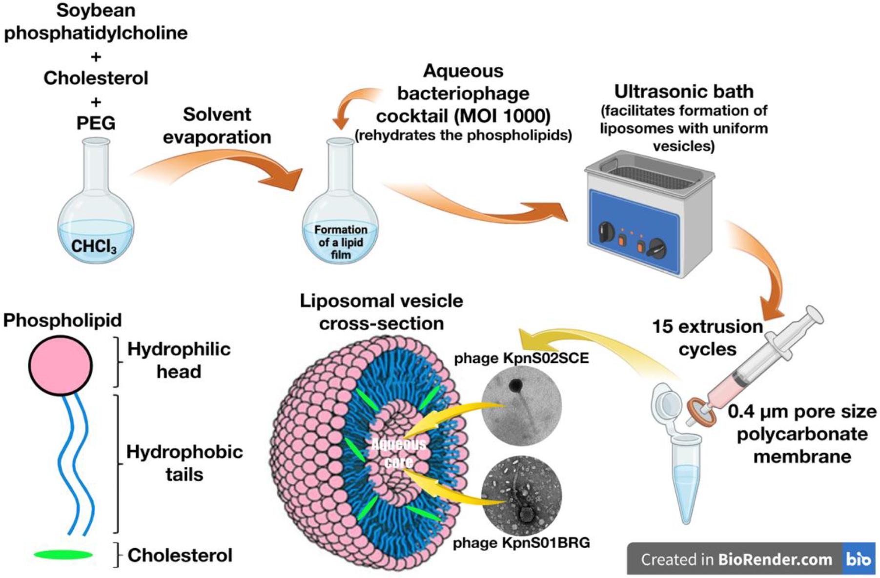
Bacteriophages KpnS01BRG and KpnS02SCE were encapsulated as a cocktail at MOI 1000. Soybean phosphatidylcholine (SPC), cholesterol (CH) and PEG 1500 were separately dissolved in chloroform. Appropriate volumes of each solution were combined in an amber vial, and the solvent was evaporated overnight to form a lipid film. The film was then hydrated with 5 mL of the phage cocktail, allowing spontaneous liposome formation. The suspension was subjected to an ultrasonic bath at 25 °C for 30 min and extruded 15 times through a 0.4 µm polycarbonate membrane to obtain uniform vesicles. Final liposome preparations were stored at 4 °C [21,22]. Figure 1 shows the encapsulation strategy, and Table 1 presents the lipid composition.
Composition of the liposomal formulation integrating a cocktail of phages KpnS01BRG and KpnS02SCE virions.
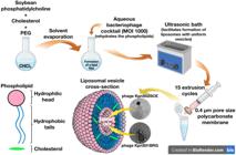
Schematic representation of phage encapsulation in liposomes and the putative lipid bilayer structure.
Liposome characterization via dynamic laser light scattering
Liposomal formulations, with and without encapsulated phages, were analyzed by DLS. Samples were diluted 1:30 (v/v) in ultrapure water, within the equipment’s recommended range. Fifty microlitres of each formulation were diluted in 20 mL of water, homogenized, thermostatted at 25 °C for 120 s, and measured in triplicate using a ZetaPALS NanoBrook 90PlusPALS system (661 nm, 90° angle). Mean hydrodynamic size, polydispersity index, and Zeta potential were obtained using PALS-based measurements, with data processed by Particle Solutions software (v3.5).
Ex vivo phage treatment in artificially contaminated lung tissue
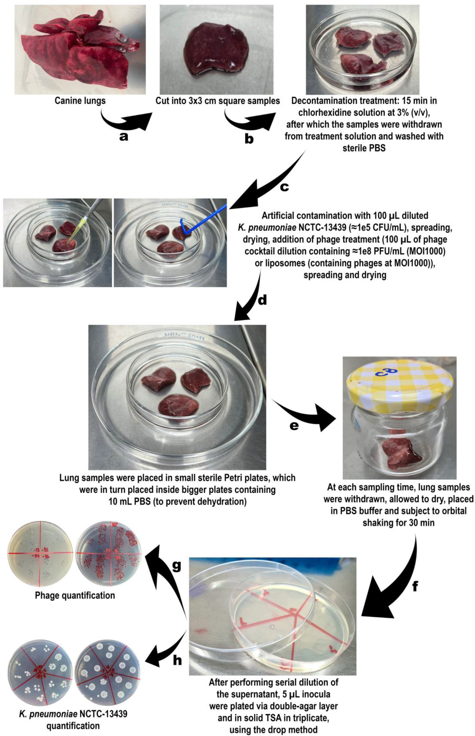
Canine lungs from animals that died naturally were obtained and stored at -20 °C. Before assays, tissue was cut into 3 × 3 cm sections and sterilized in 3% chlorhexidine for 15 min. Twelve groups of samples (three replicates each; 36 samples total) were prepared. After adding PBS and incubating for 1 h at 37 °C, the groups were assigned as follows: (i) tissue control (PBS only); (ii) bacterial control (K. pneumoniae 105 CFU/mL); (iii) free phage treatment (MOI 1000); and (iv) liposome-encapsulated phage treatment (MOI 1000). To maintain moisture, small Petri dishes holding lung samples were placed inside larger dishes containing PBS.
All groups were incubated under identical conditions, and samples were collected hourly for 12 h. Each lung piece was transferred to 10 mL PBS and shaken for 30 min at 37 °C to elute bacteria and phages. Bacterial counts were determined by the drop-plate method on TSA (24 h, 37 °C), and phage titres by the double-layer agar method (18 h, 37 °C). All assays were performed in triplicate and repeated on three separate occasions. A schematic overview of the procedure is shown in Fig. 2.
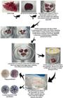
Scheme of the ex vivo phage treatment procedure using the cocktail with phages KpnS01BRG and KpnS02SCE on artificially contaminated canine lung tissue.
Statistical analyses
Data from the ex vivo assays were analyzed using GraphPad Prism 7.04. Normality was assessed by the Kolmogorov-Smirnov test and homoscedasticity by Levene’s test. Differences in bacterial and phage concentrations over time for free phages and liposome-encapsulated phages were evaluated using two-way ANOVA with Bonferroni post hoc tests. Bacterial counts were compared with the corresponding bacterial control, and phage titres were compared with their respective phage controls. Statistical significance was set at p < 0.05.
RESULTS
This study evaluated the structural and functional stabilization of two newly isolated lytic phages against Klebsiella pneumoniae NCTC-13439 using a liposomal formulation aimed at potential lung infection control. Both phages produced clear, distinct plaque morphologies, confirming their virulent nature.
Antibiogram of Klebsiella pneumoniae NCTC-13439
The results of the antibiogram performed to the bacteria used in this study can be seen in Table 2.
Results obtained in the antibiogram performed on the bacteria Klebsiella pneumoniae NCTC-13439.
The strain showed sensitivity to amikacin, gentamicin and tetracycline (Table 2). Amikacin and gentamicin, both aminoglycosides, are important options for treating severe infections [23]. Tetracycline is also an effective antimicrobial for a range of infections [24]. Although Gram-negative bacteria generally restrict antibiotic entry due to their outer membrane, porins, membrane alterations, or shared essential cellular targets can allow susceptibility to some antibiotics typically used against Gram-positive organisms [25].
The strain was resistant to most antibiotics tested, including amoxicillin/clavulanic acid, ampicillin, aztreonam, cefazolin, cefepime, cefoxitin, ceftazidime, clindamycin, chloramphenicol, erythromycin, meropenem, oxacillin and penicillin, indicating that these drugs are not effective for treating this infection. This high level of resistance highlights the need for alternative or combined therapeutic approaches. Ceftriaxone, ciprofloxacin, rifampicin and sulfamethoxazole/trimethoprim showed intermediate responses, suggesting limited and unreliable efficacy.
Liposome characterization via dynamic laser light scattering
DLS analysis provided information on the properties of the liposomes, both with and without encapsulated phages (Table 3).
The mean hydrodynamic diameter increased from 164.48 nm in empty liposomes to 207.76 nm in phage-loaded ones (Table 3), confirming that encapsulation expands vesicle size [26]. The phages themselves showed hydrodynamic diameters of 56.48 nm (KpnS01BRG) and 53.17 nm (KpnS02SCE) (data not shown). The polydispersity index rose from 0.1772 to 0.2712 after encapsulation, indicating greater size heterogeneity [27]. Zeta potential values shifted from -36.29 mV to -30.47 mV, suggesting a modest reduction in negative surface charge, which may influence electrostatic repulsion and interactions with cell surfaces [28,29]. Together, these changes indicate that phage-loaded liposomes become larger, more heterogeneous, and display altered surface charge, modifications that may improve delivery efficiency [30].
Diffusion coefficients were similar in magnitude, but phage-loaded liposomes showed a 21% reduction compared to empty ones (Table 3), consistent with their larger size and with the 16.2% decrease in electrophoretic mobility.
Ex vivo phage treatment experiments in artificially contaminated lung tissue
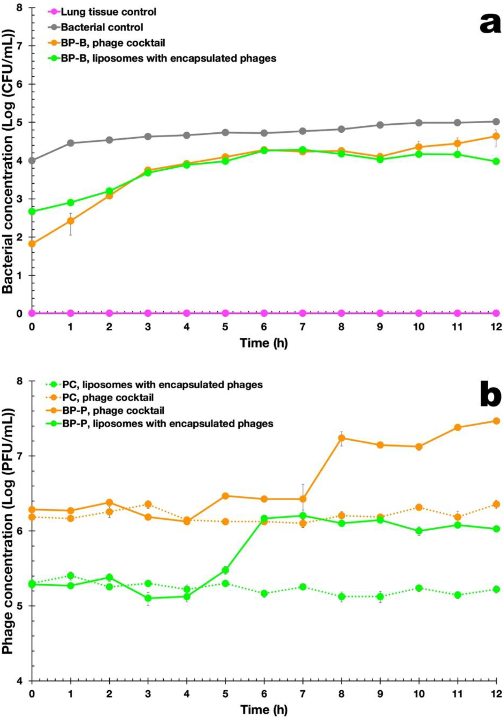
Figure 3 shows the results of the ex vivo inactivation assays performed on canine lung tissue artificially contaminated with K. pneumoniae, using either the free phage cocktail or liposome-encapsulated phages. In Figure 3a, the Tissue Control (magenta) remains at 0 log CFU/mL, confirming successful decontamination of untreated tissue, while the Bacterial Control (gray) shows gradual growth, reaching ~5 log CFU/mL after12 h.
Ex vivo inactivation of K. pneumoniae in contaminated lung tissue by free and liposome-encapsulated phages (MOI 1000, 12 h). (a) Bacterial and (b) phage concentrations.
Treatment with the free phage cocktail (orange) shows an initial rise in bacterial counts up to ~3 h, followed by stabilization around 4.2 log CFU/mL and a temporary decline at 9 h. After this point, bacterial regrowth suggests the emergence of phage resistance.
In contrast, the liposome-encapsulated phage treatment (green) shows a similar early pattern but, from 8 h onward, produces a continuous and sustained bacterial reduction, demonstrating markedly greater efficacy than free phages.
Figure 3b shows the evolution of phage concentration during treatment with the free phage cocktail and liposome-encapsulated phages over 12 h. Phage titers increased more in the free cocktail treatment than in the liposome-encapsulated formulation, indicating active replication in host cells. Encapsulated phages (green line) exhibited a more moderate increase, consistent with a controlled release of virions [30]. Phage controls (PC) remained relatively stable. Although both treatments showed comparable efficacy, encapsulation of phage virions in liposomes could potentially offer additional advantages in terms of phage stability and prolonged release under the specific conditions of this model. These features warrant further investigation in rigorous in vivo trials to determine if such benefits translate to a clinical setting, where factors like host immunity and mucus clearance play a critical role [20, 50].
As expected, disinfected lung tissue showed no bacterial contamination (Fig. 3a). In the bacterial control (BC), bacterial counts increased by 1.02 log CFU/mL over 12 h, consistent with slower growth in solid matrices due to limited nutrient diffusion. Treatment with the free phage cocktail (orange curve, Fig. 3a) reduced bacterial concentration by up to 2.0 log CFU/mL after 1 h, but regrowth occurred after 9 h, indicating the development of bacterial resistance (Fig. 3a). The liposome-encapsulated phage cocktail (green curve, Fig. 3a) showed a similar trend up to 9 h (ANOVA, p = 0.9999 (> 0.05)), followed by a greater reduction by 12 h (ANOVA, p = 0.0024 (< 0.05)), demonstrating higher efficacy than the free phage cocktail. Phage controls (Fig. 3b) remained stable, with only minor variations (±0.2 log PFU/mL; ANOVA, p = 0.9999 (> 0.05)). In the presence of host cells, phage titers increased by 1.1 log PFU/mL (free phage) and 0.8 log PFU/mL (encapsulated phage) after 12 h (ANOVA, p = 0.0001 (< 0.05)), the latter likely due to gradual virion release from liposomes.
When comparing the free phage cocktail versus liposome-encapsulated phages (LEP) in promoting bacterial reduction, the difference is striking, with the LEP promoting a significant reduction in the bacterial load after 9 h and 12 h, as can be seen from inspection of the statistical results displayed in Table 4.
Results from the Bonferroni Post-Hoc statistical tests performed to bacterial counts, comparing the free virion cocktail vs. LEP.
DISCUSSION
Liposomes are microscopic vesicular structures composed of one or more phospholipid bilayers capable of encapsulating and delivering bioactive substances. The preparation method proposed by Alec Bangham in the 1960s [21,22] involves dissolving phospholipids in organic solvent, forming a thin lipid film after solvent evaporation and subsequent hydration in aqueous medium. Developing alternatives to conventional antibiotherapy for controlling infections caused by multidrug-resistant Klebsiella pneumoniae remains a major challenge. In this study, two newly isolated lytic phages, KpnS01BRG (Brazil) and KpnS02SCE (Nigeria), targeting K. pneumoniae NCTC-13439, were encapsulated in liposomes to enhance their stability and suitability for pulmonary applications. Genome analysis confirmed the absence of toxin, virulence, or antibiotic resistance genes [31,32], as well as integrase genes [33], indicating a strictly lytic lifestyle suitable for therapeutic use.
The significant increase in HS (from ~165 nm to ~208 nm after encapsulation of phage virions) and the change in PI (from ~0.177 to ~0.271 nm after encapsulation of phage virions) are robust physical indicators that the virions were encapsulated in the liposomal structures. The maintenance of the phage titre during the extrusion process demonstrates high encapsulation efficiency and liposomal protection, conferring structural and functional stabilization to the phage particles.
The outer membrane of Gram-negative bacteria, including K. pneumoniae, bears a net negative charge due to phospholipids, lipoproteins, and LPS components [34]. Reported Zeta potentials for K. pneumoniae average -30.55 ± 1.56 mV [35]. The liposomal vesicles exhibited comparable negative Zeta potential values, suggesting electrostatic repulsion that may hinder effective interaction between bacterial cells and liposome-encapsulated phages [10].
While the negative Zeta potential (-30.47 mV) of LEP can generate electrostatic repulsion with the negatively charged bacterial surface, we opted for anionic liposomes (soy lecithin) due to their lower alveolar toxicity and better penetration into pulmonary mucus (which is rich in anionic glycoproteins). Cationic liposomes are often trapped in the mucus by electrostatic interactions, preventing them from reaching the bacteria. Modifying the liposomal surface charge to improve interaction with the bacterial cells was not the aim of the present research effort but can certainly be addressed in future experiments targeting in vivo assays. Future optimizations of the LEP discussed herein might use cationic lipids to reverse the surface charge and potentially increase the binding affinity with the bacterial cell wall.
Antibiotic susceptibility testing revealed resistance to all tested drugs except amikacin, gentamicin, and tetracycline, classifying the isolate as XDR (extensively drug-resistant). The diffusion coefficients of liposomes with and without encapsulated phages were of the same order of magnitude (10⁻12 m2·s⁻1), with encapsulated liposomes exhibiting 21% lower diffusion and 16.2% lower electrophoretic mobility, consistent with their slightly larger hydrodynamic size. These findings align with previously reported data for phage and nanoparticle systems [4,10,36,37].
As expected, disinfected lung tissue showed no contamination (Fig. 3a). In the bacterial control (BC), counts increased by 1.02 log CFU/mL over 12 h, consistent with slower growth in solid matrices. Treatment with the free phage cocktail (orange curve) initially reduced bacterial counts by up to 2.0 log CFU/mL after 1 h, but regrowth occurred after 9 h, suggesting bacterial resistance. Liposome-encapsulated phages (green curve) produced similar results up to 9 h (ANOVA, p = 0.9999 (> 0.05)), followed by a significantly greater reduction after 12 h (ANOVA, p = 0.0024 (< 0.05)), demonstrating enhanced efficacy.
Phage controls (Fig. 3b) remained stable throughout the assay (±0.2 log PFU/mL; ANOVA, p = 0.9999 (> 0.05)). In the presence of the host, phage titers increased by 1.1 log PFU/mL (free cocktail) and 0.8 log PFU/mL (encapsulated phages) after 12 h (ANOVA, p = 0.0001 (< 0.05)). The smaller increase observed for encapsulated phages likely reflects the gradual virion release from the liposomes. The maximum bacterial reduction achieved with the liposomal formulation was 1.04 log CFU/mL after 12 h (MOI 1000), while preventing bacterial regrowth (unlike the free phage cocktail). The modest inactivation can be attributed to the electrostatic repulsion between negatively charged bacterial cells and liposomal vesicles. Controlled phage release from liposomes, however, appears to sustain antibacterial activity over time.
Bacterial regrowth following phage treatment is well documented and often linked to variability in bacterial surface receptors. Such resistance can be mitigated through cocktails combining multiple lytic phages with distinct adsorption mechanisms [38-41].
While the quantitative reduction of approximately 1 log CFU/mL observed in our ex vivo canine lung model might appear modest compared to liquid-culture benchmarks, it must be contextualized within the unique constraints of this experimental system. In solid ex vivo models without the aid of neutrophils, 1-log reductions are significant. The reduction should be seen not as a complete cure, but as containment of the critical load. In the absence of immunity in the ex vivo model, the phage virions do all the work alone. In a living lung, this 1-log reduction would allow alveolar macrophages to eliminate the remainder of the infection. The primary limitation is the lack of “immunophage synergy”. In pulmonary infections, the complete resolution of the bacterial load is often contingent upon the cooperative interaction between bacteriophages and the host’s innate immune cells, particularly neutrophils [42,43]. As our ex vivo model lacks an active immune system, the observed reduction represents only the direct bactericidal effect of the phages. In a clinical scenario, even a modest reduction in bacterial density could provide the necessary respite to allow the host’s immune system to effectively clear the remaining pathogens [44]. Furthermore, the ability of the LEP formulation to provide a sustained release and prevent bacterial regrowth (unlike the free phages) is a promising indicator of therapeutic potential for chronic or recalcitrant infections where prolonged activity is required. To enhance efficacy in future developments, increasing the initial Multiplicity of Infection (MOI) or evaluating the synergy between encapsulated phages and sub-inhibitory concentrations of antibiotics (phage-antibiotic synergy, PAS) could be explored.
The ex vivo assays using artificially contaminated canine lung tissue demonstrated that encapsulated phages achieved higher inactivation rates than free phages, particularly beyond 9 h of treatment (ANOVA, p = 0.0001 (< 0.05)). The limited increase in phage titers suggests that the complex tissue matrix may hinder phage diffusion and contact with host cells, restricting amplification [12]. This limitation could potentially be overcome by increasing MOI to enhance initial phage-bacterium encounters.
Overall, the liposomal formulation incorporating both lytic phages represents a promising step toward developing alternative strategies for pulmonary infections caused by K. pneumoniae. Nevertheless, further studies employing intact lungs and naturally infected tissues are essential to validate these findings in in vivo settings and optimize clinical applicability. Assays in live animal models (such as mice or rats) are the essential next step to evaluate the pharmacokinetics and systemic safety of the formulation. In vivo studies in murine models are the next necessary step and are already planned by our research group.
The use of a high multiplicity of infection (MOI 1000) in this study warrants consideration regarding clinical translation. This high dosage was intentionally selected to facilitate “passive” phage therapy (an inundative approach where the initial phage concentration is high enough to ensure bacterial contact and lysis even when the target density is low or the environment is physically complex) [46,47]. In the solid parenchyma of our ex vivo lung model, significant diffusion barriers limit the movement of both free and encapsulated phage virions; thus, a high initial titre was necessary to overcome these physical constraints. Moreover, as this model lacks the “immunophage synergy” provided by host neutrophils in a clinical setting [42], the phages were required to exert their antibacterial effect without the assistance of the innate immune system. While this study serves as a proof-of-concept for the viability and controlled release of liposome-encapsulated phage virions in tissue [45], future in vivo studies should focus on dose-optimization. It is anticipated that the presence of host immunity and the potential for “active” therapy (where phages replicate in situ) may allow for a significant reduction in the required therapeutic dose.
Despite the advantages of the ex vivo pulmonary model in providing a complex three-dimensional architecture for testing phage delivery, several inherent limitations must be considered to avoid overinterpretation. First, the model lacks a functional immune system; in a living host, the “immunophage synergy” between lysing phages and recruited neutrophils is a primary driver of successful bacterial eradication in pneumonia [42,48]. Second, the absence of active mucociliary clearance and natural airflow dynamics means that the distributive forces acting on the liposomal formulations may differ significantly from those in a ventilated, living lung. Furthermore, the lack of continuous blood perfusion and lymphatic drainage prevents the assessment of the systemic clearance of liposomes or the potential for tissue-level regeneration following bacterial insult [49,50]. Consequently, while our results demonstrate the mechanical and structural feasibility of using LEP as a “Trojan Horse” in lung tissue, these findings should be viewed as a foundational proof-of-concept that necessitates further validation in dynamic in vivo pulmonary infection models. Limitations such as the absence of physiological dynamics in the ex vivo model; the need for direct quantification of EE% by ultracentrifugation; the lack of evaluation of prolonged phage release kinetics (beyond 24 h); and the need for validation in live animal models to assess safety (pharmacokinetics), are here duly acknowledged, and will be further addressed in a forthcoming publication with in vivo assays.
The choice of liposomes as a delivery vehicle in this study is based on their unique advantages relative to other emerging phage encapsulation strategies. Unlike polymeric nanoparticles such as poly(lactic-co-glycolic acid) (PLGA), which provide greater structural robustness but can involve the use of harsh organic solvents and slower biodegradation rates, liposomes are composed of phospholipids that mimic the lung’s endogenous surfactant system [45,51]. This biomimicry not only enhances biocompatibility but also facilitates the “Trojan Horse” entry of phages into macrophages, which is critical for treating pathogens like K. pneumoniae that can persist intracellularly [51]. Furthermore, while dry powder formulations for inhalation offer superior long-term stability and ease of administration via portable inhalers [50], they often suffer from significant titre loss during the high-stress spray-drying or freeze-drying processes [52,53]. In contrast, the liposomal encapsulation method employed here maintains high phage viability and protects the virions from antibody neutralization and enzymatic degradation, making it a highly effective (albeit more costly) platform for acute pulmonary interventions [51]. Thus, while dry powders may be more suitable for chronic maintenance therapy, the liposomal approach appears better suited for rapid-response treatments where the preservation of high phage titres and deep tissue penetration are paramount.
CONCLUSIONS
The results of the present work provide a preliminary proof-of-concept suggesting that phage treatment using KpnS01BRG and KpnS02SCE phages encapsulated in liposomal vesicles represents a promising avenue for further study in the control of Klebsiella pneumoniae. However, the clinical applicability of these findings remains to be established through subsequent in vivo safety and efficacy studies. In addition, complementation of the narrow host-range characteristics of the isolated phages with different phages for the same bacteria in the form of a lytic cocktail would allow to surpass and virtually eliminate the drawback of bacterial-acquired phage-resistance.
-
Funding:
This research was funded by São Paulo Research Foundation (FAPESP), grants 2022/10775-9 (Project PsgPhageKill) and 2023/03797-9 (Project RTI). V.M.B. was visiting researcher fellow from FAPESP (2018/05522-9, Project PsaPhageKill). V.M.B. received a research fellowship award from the National Council for Scientific and Technological Development (CNPq) (grant 301978/2022-0).
-
Institutional Review Board Statement: The research developed and described herein was previously exempted from authorization by the Ethics Committee for the Use of Animals of the University of Sorocaba (CEUA-UNISO), through a letter with the approval date of 11/24/2023, given that the animals that donated the lungs died naturally (without inducing death).
-
Informed Consent Statement: Not Aplicable
-
Use of Generative Artificial Intelligence: The authors declare that no artificial intelligence was used in the manuscript.
Acknowledgments:
The authors have no acknowledgments to declare.
Data Availability Statement:
The genome sequences of the phages described in this work have been deposited in GenBank/NCBI under the accession numbers PV081216 (phage KpnS01BRG) and PV081217 (phage KpnS02SCE). Other research data are available upon request for corresponding author.
REFERENCES
-
1 Hatfull GF, Dedrick RM, Schooley RT. Phage therapy for antibiotic-resistant bacterial infections. Annu Rev Med. 2022; 73:197-211. doi: 10.1146/annurev-med-080219-122208.
» https://doi.org/10.1146/annurev-med-080219-122208. - 2 Rodrigo-Troyano A, Sibila O. The respiratory threat posed by multidrug resistant Gram-negative bacteria. Respirology. 2017; 22:1288-99.
-
3 Jansen M, Wahida A, Latz S, Krüttgen A, Häfner H, Buhl EM et al. Enhanced antibacterial effec of the novel T4-like bacteriophage KARL-1 in combination with antibiotics against multi-drug resistant Acinetobacter baumannii. Sci Rep. 2018; 8:14140. doi:10.1038/s41598-018-32344-y.
» https://doi.org/10.1038/s41598-018-32344-y. -
4 Rios AC, Vila MMDC, Lima R, Fiol FS,Tubino M,Teixeira J A, et al Structural and functional stabilization of bacteriophage particles within the aqueous core of a W/O/W multiple emulsion: A potential biotherapeutic system for the inhalational treatment of bacterial pneumonia. Process Biochem, 2018; 64:177-92. doi:10.1016/j.procbio.2017.09.022
» https://doi.org/10.1016/j.procbio.2017.09.022 -
5 Agodi A, Barchitta M, Quattrocchi A, Maugeri A, Aldisio E, Marchese AE et al. Antibiotic trends of Klebsiella pneumoniae and Acinetobacter baumannii resistance indicators in an intensive care unit of Southern Italy, 2008-2013. Antimicrob Resist Infect Control. 2015; 4:43. doi: 10.1186/s13756-015-0087-y
» https://doi.org/10.1186/s13756-015-0087-y -
6 Russo A, Giuliano S, Ceccarelli G, Alessandri F, Giordano A, Brunetti G, et al. Comparison of septic shock due to multidrug-resistant Acinetobacter baumannii or Klebsiella pneumoniae Carbapenemase-Producing K. pneumoniae in Intensive Care Unit Patients. Antimicrob Agents Chemother.2018; 62:e02562-17. doi:10.1128/AAC.02562-17
» https://doi.org/10.1128/AAC.02562-17 -
7 D’Andrea MM, Marmo P, De Angelis LH, Palmieri M, Ciacci N, Di Lallo G et al. φBO1E, a newly discovered lytic bacteriophage targeting carbapenemase-producing Klebsiella pneumoniae of the pandemic Clonal Group 258 clade II lineage. Sci Rep.2017; 7: 2614. doi:10.1038/s41598-017-02788-9
» https://doi.org/10.1038/s41598-017-02788-9 -
8 Haq IU, Chaudhry WN, Akhtar MN, Andleeb S, Qadri I. Bacteriophages and their implications on future biotechnology: a review. Virol J. 2012; 9: 9. doi: 10.1186/1743-422X-9-9
» https://doi.org/10.1186/1743-422X-9-9 -
9 Harada LK, Silva EC, Campos WF, Fiol FS, Vila, M, Dąbrowska, K et al. Biotechnological applications of bacteriophages: State of the art. Microbiol Res. 2018; 212-213:38-58. doi: 10.1016/j.micres.2018.04.007
» https://doi.org/10.1016/j.micres.2018.04.007 -
10 Harada LK, Silva EC, Rossi FPN, Cieza B, Oliveira TJ, Pereira C et al. Characterization and in vitro testing of newly isolated lytic bacteriophages for biocontrol of Pseudomonas aeruginosa. Future Microbiol. 2022; 17:111-41. doi: 10.2217/fmb-2021-0027
» https://doi.org/10.2217/fmb-2021-0027 -
11 Górski A, Międzybrodzki R, Węgrzyn G, Jończyk-Matysiak E, Borysowski J, Weber-Dąbrowska B. Phage therapy: Current status and perspectives. Med Res Rev.2019; 40:459-63. doi:10.1002/med.21593
» https://doi.org/10.1002/med.21593 -
12 Balcão VM, Basu A, Cieza B, Rossi FN, Pereira C, Vila MMDC et al. Pseudomonas-tailed lytic phages: genome mechanical analysis and putative correlation with virion morphogenesis yield. Future Microbiol. 2022; 17:1009-26. doi:10.2217/fmb-2021-0293
» https://doi.org/10.2217/fmb-2021-0293 -
13 Rios AC, Moutinho CG, Pinto FC, Fiol FS, Jozala A, Chaud MV. et al. Alternatives to overcoming bacterial resistances: State-of-the-art. Microbiol Res. 2016; 191:51-80. doi:10.1016/j.micres.2016.04.008
» https://doi.org/10.1016/j.micres.2016.04.008 -
14 Gondil VS, Chhibber S. Bacteriophage and endolysin encapsulation systems: a promising strategy to improve therapeutic outcomes. Front Pharmacol. 2021;12:675440. doi:10.3389/fphar.2021.675440
» https://doi.org/10.3389/fphar.2021.675440 -
15 Rahimzadeh G, Saeedi M, Moosazadeh M, Hashemi SMH, Babaei A, Rezai MS. et al. Encapsulation of bacteriophage cocktail into chitosan for the treatment of bacterial diarrhea. Sci Rep. 2021; 11:15603. doi:10.1038/s41598-021-95132-1
» https://doi.org/10.1038/s41598-021-95132-1 -
16 Rosner D, Clark J. Formulations for bacteriophage therapy and the potential uses of immobilization. Pharmaceuticals. 2021; 14:359. doi:10.3390/ph14040359
» https://doi.org/10.3390/ph14040359 -
17 Singla S, Harjai K, Katare OP, Chhibber S. Bacteriophage-loaded nanostructured lipid carrier: improved pharmacokinetics mediates effective resolution of Klebsiella pneumoniae-induced lobar pneumonia. J Infect Dis. 2015; 212:325-334. doi:10.1093/infdis/jiv029
» https://doi.org/10.1093/infdis/jiv029 -
18 Bassetti M, Vena A, Russo A, Peghin M. Inhaled liposomal antimicrobial delivery in lung infections drugs.2020; 80:1309-18. doi:10.1007/s40265-020-01359-z
» https://doi.org/10.1007/s40265-020-01359-z -
19 Chadha P, Katare OP, Chhibber S. Liposome loaded phage cocktail: Enhanced therapeutic potential in resolving Klebsiella pneumoniae mediated burn wound infections. Burns. 2017; 43:1532-43. doi:10.1016/j.burns.2017.03.029
» https://doi.org/10.1016/j.burns.2017.03.029 -
20 Cinquerrui S, Mancuso F, Vladisavljević GT, Bakker SE, Malik DJ. Nanoencapsulation of bacteriophages in liposomes prepared using microfluidic hydrodynamic flow focusing. Front Microbiol. 2018; 9:2172. doi:10.3389/fmicb.2018.02172
» https://doi.org/10.3389/fmicb.2018.02172 -
21 Bangham A. A Correlation between surface charge and coagulant action of phospholipids. Nature. 1961; 192:1197-8. doi:10.1038/1921197a0
» https://doi.org/10.1038/1921197a0 -
22 Bangham AD, Standish MM, Watkins JC. Diffusion of univalent ions across the lamellae of swollen phospholipids. J Mol Biol.1965; 13: 238-52. doi:10.1016/s0022-2836(65)80093-6
» https://doi.org/10.1016/s0022-2836(65)80093-6 -
23 Frimodt-Møller N, Hansen JU, Plattner M, Huseby DL, Almind SR, Haldimann K et al. Apramycin efficacy against carbapenemand aminoglycoside-resistant Escherichia coli and Klebsiella pneumoniae in murine bloodstream infection models. Int J Antimicrob Agents. 2024; 64:107181.doi: 10.1016/j.ijantimicag.2024.107181
» https://doi.org/10.1016/j.ijantimicag.2024.107181 -
24 Lv ZF, Wang FC, Zheng HL,Wang B, Xie Y, Zhou XJ et al. Meta-analysis: is combination of tetracycline and amoxicillin suitable for Helicobacter pylori infection? World J Gastroenterol.2015; 21:2522-33. doi:10.3748/wjg.v21.i8.2522
» https://doi.org/10.3748/wjg.v21.i8.2522 -
25 Klobucar K, Jardine E, Farha MA, MacKinnon MR, Fragis M, NKonge B et al. Genetic and chemical screening reveals targets and compounds to potentiate Gram-positive antibiotics against Gram-negative bacteria. ACS Infect Dis. 2022; 8:2187-97. doi:10.1021/acsinfecdis.2c00357
» https://doi.org/10.1021/acsinfecdis.2c00357 -
26 Ramli NA, Ali N, Hamzah S, Yatim NI. Physicochemical characteristics of liposome encapsulation of stingless bees’ propolis. Heliyon. 2021; 7:e06649. doi:10.1016/j.heliyon.2021.e06649
» https://doi.org/10.1016/j.heliyon.2021.e06649 -
27 Jończyk E, Kłak M, Międzybrodzki R, Górski A. The influence of external factors on bacteriophages--review. Folia Microbiol (Praha). 2011; 56:191-200. doi:10.1007/s12223-011-0039-8
» https://doi.org/10.1007/s12223-011-0039-8 -
28 Smith MC, Crist RM, Clogston JD, McNeil SE. Zeta potential: a case study of cationic, anionic, and neutral liposomes. Anal Bioanal Chem.2017; 409:5779-87. doi:10.1007/s00216-017-0527-z
» https://doi.org/10.1007/s00216-017-0527-z -
29 Gopi S, Balakrishnan P. Evaluation and clinical comparison studies on liposomal and non-liposomal ascorbic acid (vitamin C) and their enhanced bioavailability. J Liposome Res.2021; 31(4):356-64. doi:10.1080/08982104.2020.1820521
» https://doi.org/10.1080/08982104.2020.1820521 -
30 Liu P, Chen G, Zhang J. A review of liposomes as a drug delivery system: Current status of approved products, regulatory environments, and future perspectives. Molecules.2022; 27:1372. doi:10.3390/molecules27041372
» https://doi.org/10.3390/molecules27041372 -
31 Kakasis A, Panitsa G. Bacteriophage therapy as an alternative treatment for human infections. A comprehensive review. Int J Antimicrob Agents.2019; 53:16-21.doi:10.1016/j.ijantimicag.2018.09.004
» https://doi.org/10.1016/j.ijantimicag.2018.09.004 -
32 Balcão VM, Moreli FC, Silva EC, Belline BG, Martins LF, Rossi FPN et al. Isolation and molecular characterization of a novel lytic bacteriophage that inactivates MDR Klebsiella pneumoniae strains. Pharmaceutics.2022; 14:1421. doi: 10.3390/pharmaceutics14071421
» https://doi.org/10.3390/pharmaceutics14071421 -
33 Hyman P. Phages for phage therapy: Isolation, characterization, and host range breadth. Pharmaceuticals. 2019; 12:35. doi:10.3390/ph12010035
» https://doi.org/10.3390/ph12010035 -
34 Zemb O, Manefield M, Thomas F, Jacquet S. Phage adsorption to bacteria in the light of the electrostatics: A case study using E. coli, T2 and flow cytometry. J Virol Methods.2013; 189:283-9. doi:10.1016/j.jviromet.2013.02.007
» https://doi.org/10.1016/j.jviromet.2013.02.007 -
35 Fontoura I, Raniero L, Castilho ML. Estudo do potencial zeta de cepas de Klebsiella pneumoniae correlacionada a permeabilidade de antimicrobianos [Study of the zeta potential of Klebsiella pneumoniae strains correlated to antimicrobial permeability]. Brazilian J Infect Dis.2023; 27:17. doi:10.1016/j.bjid.2023.102843
» https://doi.org/10.1016/j.bjid.2023.102843 -
36 Katayama K, Nomura H, Ogata H, Eitoku T. Diffusion coefficients for nanoparticles under flow and stop-flow conditions. Phys Chem Chem Phys. 2009; 11:10494-9. doi:10.1039/B911535H
» https://doi.org/10.1039/B911535H -
37 Glasser CA, Vila MMDC, Pereira JC, Chaud MV, Oliveira Junior JM, Tubino M et al. Development of a water-in-oil-in-water multiple emulsion system integrating biomimetic aqueous-core lipid nanodroplets for protein entity stabilization. Part II: process and product characteriz. 2016; 42(12):1990-2000. doi:10.1080/03639045.2016.1188109
» https://doi.org/10.1080/03639045.2016.1188109 -
38 Forti F, Roach DR, Cafora M, Pasini ME. Design of a broad-range bacteriophage cocktail that reduces Pseudomonas aeruginosa biofilms and treats acute infections in two animal models. Antimicrob Agents Chemother. 2018; 62:e02573-17. doi:10.1128/AAC.02573-17
» https://doi.org/10.1128/AAC.02573-17 -
39 Costa P, Pereira C, Gomes ATPC, Almeida A. Efficiency of single phage suspensions and phage cocktail in the inactivation of Escherichia coli and Salmonella Typhimurium: An in vitro preliminary study. Microorganisms. 2019; 7(4):94. doi: 10.3390/microorganisms7040094
» https://doi.org/10.3390/microorganisms7040094 -
40 Pinheiro LAM, Pereira C, Barreal ME, Gallego PP, Balcão VM, Almeida A. Use of phage ϕ6 to inactivate Pseudomonas syringae pv. actinidiae in kiwifruit plants: in vitro and ex vivo experiments. Appl Microbiol Biotechnol.2019; 104:1319-30. doi: 10.1007/s00253-019-10301-7
» https://doi.org/10.1007/s00253-019-10301-7 -
41 Pinheiro LAM, Pereira C, Frazão C, Balcão VM, Almeida A. Efficiency of phage φ6 for biocontrol of Pseudomonas syringae pv. syringae: An in vitro preliminary study. Microorganisms. 2019; 7:286. doi:10.3390/microorganisms7090286
» https://doi.org/10.3390/microorganisms7090286 -
42 Roach DR, Leung CY, Henry M, Morello E, Singh D, Di Santo JP et al. Synergy between the host immune system and bacteriophage is essential for successful phage therapy against an acute respiratory pathogen. cell host microbe. 2017; 22(1):38-47.e4. doi: 10.1016/j.chom.2017.06.018.
» https://doi.org/10.1016/j.chom.2017.06.018. -
43 Weissfuss C, Hoffmann K, Behrendt U, Bürkle M, Debarbieux L.; Ricard J.-D et al. immunophage synergy: supporting innate immunity with phages to combat bacterial pneumonia. ERJ Open Research 2025; 11 (suppl 15): TP304. doi: 10.1183/23120541.LSC-2025.TP304.
» https://doi.org/10.1183/23120541.LSC-2025.TP304. -
44 Weissfuss C, Hoffmann K, Behrendt U, Bürkle M, Twamley SG, Korf IHE et al. Neutrophils, not macrophages, aid phage-mediated control of pulmonary Pseudomonas aeruginosa infection. Front Immunol. 2025; 16:1681461. doi: 10.3389/fimmu.2025.1681461.
» https://doi.org/10.3389/fimmu.2025.1681461. -
45 Yue Y, Xu Z, Soteyome T, Premarathna M, Yin X, Liu J. Phage encapsulation and delivery technology: a strategy for treating drug-resistant pathogenic microorganisms. Pharmaceuticals (Basel). 2025; 18(11):1688. doi: 10.3390/ph18111688.
» https://doi.org/10.3390/ph18111688. -
46 Abedon ST. Lysis from without. Bacteriophage. 2011;1(1):46-9. doi: 10.4161/bact.1.1.13980.
» https://doi.org/10.4161/bact.1.1.13980. -
47 Abedon ST. Active bacteriophage biocontrol and therapy on sub-millimeter scales towards removal of unwanted bacteria from foods and microbiomes. AIMS Microbiol. 2017;3(3):649-88. doi: 10.3934/microbiol.2017.3.649.
» https://doi.org/10.3934/microbiol.2017.3.649. -
48 Nang S C, Lin Y.-W, Fabijan A P, Chang RYK, Rao GG, Iredell J et al. Pharmacokinetics/pharmacodynamics of phage therapy: a major hurdle to clinical translation. Clinical Microbiology and Infection. 2023; 29 (6): P702-9. doi: 10.1016/j.cmi.2023.01.021.
» https://doi.org/10.1016/j.cmi.2023.01.021. -
49 Yaqub N, Wayne G, Birchall M, Song W. Recent advances in human respiratory epithelium models for drug discovery. Biotechnol Adv. 2022; 54:107832. doi: 10.1016/j.biotechadv.2021.107832.
» https://doi.org/10.1016/j.biotechadv.2021.107832. -
50 Bodier-Montagutelli E, Morello E, L'Hostis G, Guillon A, Dalloneau E, Respaud R, et al. Inhaled phage therapy: a promising and challenging approach to treat bacterial respiratory infections. Expert Opin Drug Deliv. 2017;14(8):959-72. doi: 10.1080/17425247.2017.1252329.
» https://doi.org/10.1080/17425247.2017.1252329. -
51 Singla S, Harjai K, Katare OP, Chhibber S. Encapsulation of bacteriophage in liposome accentuates its entry in to macrophage and shields it from neutralizing antibodies. PLoS One. 2016;11(4):e0153777. doi: 10.1371/journal.pone.0153777.
» https://doi.org/10.1371/journal.pone.0153777. -
52 Pathak V, Chan HK, Zhou QT. Formulation of bacteriophage for inhalation to treat multidrug-resistant pulmonary infections. Kona. 2025;42:200-12. doi: 10.14356/kona.2025016.
» https://doi.org/10.14356/kona.2025016. -
53 Chang RYK, Chow MYT, Wang Y, Liu C, Hong Q, Morales S et al. The effects of different doses of inhaled bacteriophage therapy for Pseudomonas aeruginosa pulmonary infections in mice. Clin Microbiol Infect. 2022; 28(7):983-9. doi: 10.1016/j.cmi.2022.01.006.
» https://doi.org/10.1016/j.cmi.2022.01.006.
-
Editor-in-Chief:
Paulo Vitor Farago
-
Associate Editor:
Paulo Vitor Farago

 Legend: (a) lungs cut into ~3 × 3 cm samples; (b) samples decontaminated in 3% chlorhexidine and rinsed with sterile PBS; (c) samples artificially contaminated with K. pneumoniae NCTC-13439, dried, and treated with phages (MOI 1000); (d) samples kept in a moist environment during treatment; (e) at set intervals, samples transferred to PBS under orbital shaking to elute bacteria and phages; (f) eluates serially diluted; (g) phage titres determined by the double-layer agar method; (h) bacterial counts determined by the drop-plate method on TSA.