Enhancing the antifertility performance by cashew nut shell extract (Anacardium occidentale L.) on Rattus norvegicus Berkenhout 1769
Abstract
Various plants can be used as safe and effective antifertility agents, one of which is cashew (Anacardium occidentale L.). Cashew nut shells contain phenolic compounds Cashew Nut Shell Liquid (CNSL) or lacquer oil, which are effective antifertility agents in female rats, but there has been no research on male rats. The purpose of the study of antifertility agents of cashew nut shell extract in male rats includes analysis of the body weight, histological structure of the testes, testosterone levels, and assessing the microscopic quality of spermatozoa. Experimental research in the laboratory with a Completely Randomized Design. The test animals were Rattus norvegicus male Wistar strain aged two months, weighing 170-190 gr, totaling 24 divided into four treatment groups, each with six replications. The treatments given were 0.5% CMCNa (Control/T0), cashew nut shell extract 250 mg/kg body weight (Treatment 1/T1), 500 mg/kg body weight (T2), and 750 mg/kg body weight (T3). The treatment period was 25 days and every seven days body weight was measured. Testicular preparations were made using the paraffin method and Hematoxylin Eosin staining. Testosterone levels were tested using the Enzyme-linked Immunosorbent Assay method. Microscopic spermatozoa quality tests included motility, morphology, and number of spermatozoa. Analysis of the histological structure of the testes was analyzed descriptively qualitatively by comparing between treatments. One-way ANOVA at the 5% level was used to measure rat body weight, testosterone levels, motility, morphology and number of spermatozoa. The results showed that cashew nut shell extract had no effect on rat body weight (p>0.001). Repeated one-way measures to find out if the rat's mean body weight varied amongst the four treatments, an ANOVA was conducted. The rat's body weight did not differ statistically significantly between the treatments, according to an ANOVA (F statistic is 0.457 and the corresponding p value is 0.649). The rats body weight did not differ statistically significantly across the four treatments, according to the results of Bonferroni's test for multiple comparisons. The extract affected the histology of the seminiferous tubules in T3, namely atrophy, irregular shape, vacuolization and membrane fluidity. Between the interstitial tissue and the seminiferous tubules of the testes of rats in groups T1, T2 and T3 there was a gap or empty space called the phenomenon of tissue stretching (compliance). The extract significantly affected testosterone levels (p<0.001) and microscopic quality of spermatozoa, namely motility (p<0.001), morphology (p<0.001) and number of spermatozoa (p<0.001) which decreased with increasing extract dose. The results of the study concluded that the extract did not significantly affect the weight of rat, there were differences in the structure of the seminiferous tubules in T3, the extract significantly affected testosterone hormone levels and microscopic quality of spermatozoa (motility, normal morphology and number of spermatozoa) which decreased with increasing extract dose. The results of this study can be a reference for natural antifertility compounds.
Keywords:
cashew nut shell; testosterone; motility; mortality; sperm count
Resumo
Várias plantas podem ser usadas como agentes antifertilidade seguros e eficazes, entre elas o cajueiro (Anacardium occidentale L.). A casca da castanha de caju contém compostos fenólicos, como o líquido da casca da castanha (CNSL) ou óleo de laca, que é um agente antifertilidade eficaz em ratas, mas não há investigação em ratos machos. O objetivo da investigação sobre os agentes antifertilidade do extrato da casca da castanha de caju em animais machos inclui a análise da estrutura histológica dos testículos, os níveis de testosterona e a avaliação da qualidade microscópica dos espermatozoides. A investigação experimental foi realizada em laboratório com um Delineamento Inteiramente Casualizado. Os animais testados eram machos da linhagem Rattus norvegicus Wistar com dois meses de idade, pesando 170-190 gramas, totalizando 24 animais. Os tratamentos receberam CMCNa 0,5% (Controle/C), extrato da casca da castanha de caju 250 mg/kg de peso corporal (Tratamento 1/T1), 500 mg/kg de peso corporal (T2), e 750 mg/kg de peso corporal (T3). O período de tratamento foi de 25 dias, com medição do peso corporal a cada sete dias. As preparações testiculares foram efetuadas utilizando o método de parafina e coloração com Hematoxilina-Eosina. Os níveis de testosterona foram testados utilizando o método ELISA (Enzym Link Immunosorbent Assay). Os testes de qualidade microscópica dos espermatozoides incluem a motilidade, a morfologia e o número de espermatozoides. A estrutura histológica dos testículos foi analisada de forma descritiva e qualitativa, comparando os tratamentos. Foi utilizada uma ANOVA unidirecional ao nível de 5% para medir o peso corporal do rato, os níveis de testosterona, a motilidade, a morfologia e a contagem de espermatozoides. Os resultados mostraram que o extrato da casca da castanha de caju não teve nenhum efeito no peso corporal dos animais (p > 0,001). A ANOVA indicou ausência de diferença estatisticamente significativa entre os tratamentos (F = 0,457; p = 0,649), o que foi confirmado pelo teste de comparações múltiplas de Bonferroni. No entanto, observou-se que o extrato afetou a histologia dos túbulos seminíferos em T3, como atrofia, forma irregular e tamanho reduzido. O tecido intersticial T1, T2 e T3 registrou conformidade. O extrato afetou significativamente os níveis de testosterona e a qualidade microscópica dos espermatozoides (motilidade, morfologia normal e contagem de espermatozoides), os quais diminuíram com o aumento da dose do extrato. Esses resultados sugerem que o extrato possui potencial como composto antifertilidade natural.
Palavras-chave:
casca de castanha de caju; testosterona; motilidade; mortalidade; contagem de espermatozoides
1. Introduction
Rapid population growth is a problem faced by developing countries, including Indonesia. The results of the 2020 Population Census show that Indonesia's population is 270.20 million people (Badan Pusat Statistik, 2021). Economic growth and social welfare are things that are influenced by rapid population growth, so a way is needed to regulate population growth. In addition, there are not many reliable contraceptives available for men, so women are responsible for population growth (Louwagie et al., 2023). Contraceptive methods are a way to prevent pregnancy (Ochala, 2021). Many contraceptive methods are used to control fertility, but they cannot be used long term because they cause side effects, such as nausea, headaches, abdominal cramps, breast tenderness, increased vaginal discharge, and decreased libido (Hlongwa et al., 2021). Therefore, it is necessary to find a safe and effective contraceptive agent.
So far, various efforts have been made by experts to obtain highly efficacious contraceptive agents. One of these efforts is the use of plants as safe and effective antifertility agents. Research on plants or plant extracts as antifertility agents in male and female animals has been widely conducted. Handayani and Gofur (2024) researching the decoction of pulutan leaves (Urena lobata I) can reduce testosterone levels in male mice, so it has the potential as an antifertility. Oroxylum indicum Vent. bark as an antifertility in female mice (Das et al., 2024). Research by Dawane et al. (2024) found that administering Entada rheedii Spreng seed extract to female rats increased embryo development failure and acted as an antiimplantation and abortifacient. Another study on the antifertility of ethyl alcohol extract of Cynodon dactylon in female rats has the potential as an estrogenic and antiprogestogenic compound, this extract also causes vaginal cornification, increased uterine weight and uterine proliferation (Malpani et al., 2020). Obembe et al. (2023) studied Buchholzia coriacea seeds which showed antifertility properties by interfering with testicular enzymes and inflammatory cytokines. Another plant that has the potential as an antifertility is cashew (Anacardium occidentale L).
Anacardium occidentale L has high economic and medicinal value. Dare et al. (2011) has extensively studied the content and potential uses. Cashew nut shell extract contains cashew nut shell liquid (CNSL) or lacquer oil. Natural CNSL is a complex mixture of phenolic compounds, it mainly consists of anacardic acid (70%), cardanol (5%), and cardol (18%), and methyl cardol (7%) (Bhatia et al., 2024). The extraction process of anacardic acid undergoes decarboxylation to become cardanol (Patel et al., 2019; Simpen, 2018). Research on the potential of compounds in cashew nut shells has been widely carried out. Prasad et al. (2007) examined that anacardic acid has antifertility and antiimplantation activity in female mice. CNSL has the potential to be anticancer (Schultz et al., 2010), inhibit the activity of various enzymes (Zhuang et al., 2010) and anti-inflammatory (Sahoo et al., 2024 ). Cashew nut shell extract affects the levels of the hormone progesterone, decreasing as the dose of the extract used increases (Harlita et al., 2015). Research by Harlita and Puspitasari (2019) showed that nut shell extract has the activity of inhibiting CYP19 aromatase, an enzyme that converts testosterone into estradiol. This inhibition causes reduced fertility in female rats.
In relation to performance as an antifertility agent in male animals, theoretically there are two important measurements to be studied, namely the microscopic quality of spermatozoa and testosterone hormone levels. Sperm quality can be measured based on: semen volume, spermatozoa count, spermatozoa density, spermatozoa movement (motility), spermatozoa speed, spermatozoa shape (morphology), and spermatozoa viability (Chung et al., 2022). Quality spermatozoa have high viability, normal morphology, normal number, and progressive motility (Walters et al., 2004). The testes consist of germinal epithelium containing seminiferous tubules and interstitial tissue containing Leydig cells. Disorders of these two tissues affect the testosterone hormone produced (Nassar and Leslie, 2018; O’Donnell et al., 2015). Nevertheless, nothing is known about the potential of cashew nut shell extract as an antifertility agent in male animals. Based on the description above, this study aims to determine the antifertility effects of cashew nut shell extract on male rats Rattus norvegicus Berkenhout 1769.
2. Materials and Methods
2.1. Cashew nut shell preparation
Cashew nut shells were obtained from Wonogiri, Central Java, Indonesia. Cashew nut shells were sliced into small pieces, dried in an oven at 50 °C for 48 hours, then made into powder. Cashew nut shell powder of 500 grams was extracted using the maceration method with 500 mL of 96% ethanol solvent. The solution was then stirred with an Ace homogenizer for 30 minutes (2x15 minutes) and then left for 24 hours at room temp after which it was filtered. The extracted filtrate was evaporated using a vacuum rotary evaporator at a temperature of 58-60 °C until no ethanol droplets were visible to obtain a viscous extract. The extract was stored for in vivo testing (Hidayat and Wulandari, 2021).
2.2. Animal test
Experimental laboratory study with a Completely Randomized Design using 24 male Wistar rats (Rattus norvegicus Berkenhout 1769) aged 2 months, weighing 170-150 g, obtained from the Integrated Research and Community Service Unit 4, Universitas Gadjah Mada, Indonesia. The test animals were acclimatized for seven days. The rats were fed with standard pellets and drank ad libitum. In this study, there were four treatment groups, namely the administration of 0.5% CMCNa (Control/T0), cashew nut shell extract 250 mg/kg body weight (Treatment1/T1), 500 mg/kg body weight (T2), and 750 mg/kg body weight (T3). Changes in dosage based on cashew nut shell extract toxicity testing on test animals, where a dose of 500 mg/kg body weight affects test animals (Harlita et al., 2016). Below and above this dose is the range of the dose that was used.
Each treatment consisted of 6 male rats. The extract was given orally as much as 2 mL/200 g body weight. The duration of treatment was 25 days because it is included in one cycle of rat spermatogenesis and the body weight was weighed every seven days. At the end of treatment, rats were anaesthetised and dissected. Each test animal was anesthetized using ketamine at a dose of 0.1 mL and then placed on a surgical board to have its testes removed for histology preparation. All experimental procedures were analyzed and previously approved by the Health Ethics Commission of Dr. Moewardi Hospital, Surakarta, Indonesia No. 1.017/VIII/HREC/2020.
Histological preparations of the testes were made using the paraffin method. The paraffin method procedure according to Chaudhuri (2023) is as follows: (1) Fixation: The testes were placed in saline solution (NaCl 0,9%) then cleaned of fat and then placed in neutral buffered formalin 10% fixative solution for 4 hours; (2) Dehydration: Soak in 70% alcohol (1 hour), 80% alcohol (30 minutes), 90% alcohol (30 minutes), and ethanol (30 minutes); (3) Clearing: Soak in xylol overnight until transparent tissue is obtained; (4) Infiltration: Infiltration was performed in an oven at 55-60 °C by placing the organ in xylol paraffin at a ratio of 3:1 (30 min) - xylol paraffin at a ratio of 1:1 (30 min) - xylol paraffin at a ratio of 1:3 (30 min); pure paraffin I (1 h); liquid paraffin II (30 min); (5) Embedding: The tissue is embedded in an embedding box that has been filled with liquid paraffin, orienting the location of the tissue so that when sliced with a microtome knife it is located in the center of the incision, leave it until the tissue freezes; (6) Sectioning: The paraffin block containing the tissue is cut with a rotary microtome with a thickness of 4-5 µm. The incision is placed on a glass object and then placed on a hotplate at 40-50 °C until the paraffin dries; (7) Staining using Hematoxilin and Eosin: Starting with a multistage dehydration process of distilled water, 30% alcohol, 40% alcohol, 50% alcohol, 60% alcohol, 70% alcohol, 80% alcohol, 90% alcohol, and ethanol for two minutes each. The preparation was put into hematoxilin for 7 seconds and then washed with running water (15 minutes). Dehydration with 70% alcohol (2 minutes), Eosin Y (2 minutes) then rinse with 70% alcohol (2 minutes), 80% alcohol, 90% alcohol, and ethanol for 1 minute each and then put in xylol solution (15 minutes); (8) Mounting with Canadian balsam covered with a cover glass.
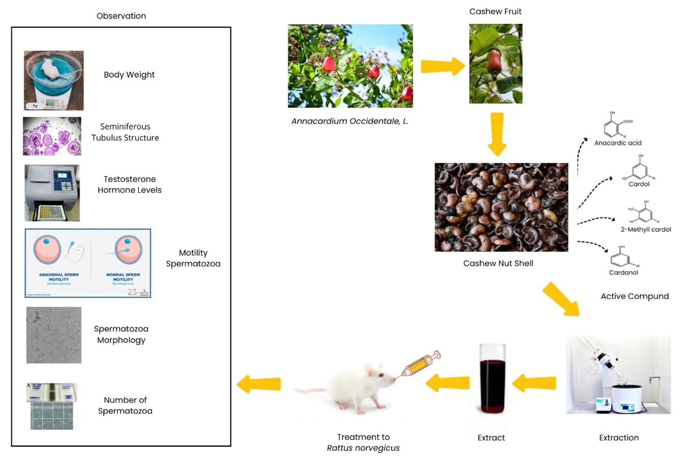
The parameters used in this study were the body weight of mice measured 5 times every 7 days for 25 days of treatment. Histological structure was performed using testis preparations with the paraffin method and hematoxylin eosin staining and then observed under an electric microscope at 200x magnification. Testosterone hormone levels were measured using the ELISA method. Rat blood was taken from the orbital vein for analysis of testosterone levels. Rat blood was taken as 1 mL on days 0, 7, 14, and 21 of treatment. Spermatozoa were taken from the cauda epididymis and used to analyse the microscopic structure of spermatozoa, namely motility, morphology, and the number of spermatozoa. The research methodology scheme can be seen in Figure 1.
2.3. Testicular histology
The testes were fixed with neutral buffered formalin 10%, then histological preparations were made using the paraffin method and hematoxylin-eosin staining (Chaudhuri, 2023). Paraffin blocks containing tissue were cut with a rotary microtome with a thickness of 4-5 µm. The testis preparations were observed under a microscope with a magnification of 200 µm. The number of seminiferous tubules observed in each testis preparation was 50-100. In this study, ordinal gradation was not carried out but only observed the histological structure of the seminiferous tubules such as the density of spermatogenesis cells, the distance between seminiferous tubules, and the presence or absence of spermatozoa, by comparing the control and treatment.
2.4. Testosterone hormone
Blood for testosterone analysis was taken from the orbital sinus at the end of treatment, then tested using the Enzyme Linked Immunosorbent Assay (ELISA) method, using a rat testosterone kit (REF, EIA-1559 produced by DRG Instruments GmbH Germany). Measurement of testosterone levels using the ELISA Kit was repeated twice for each treatment to minimize variability in results. Quantitative data in the form of testosterone levels obtained from the ELISA Reader results were converted using the Curve Expert program and analyzed quantitatively with one way anova.
2.5. Spermatozoa analysis
The testes, epididymis, ductus deferens were dissected, washed with physiological solution at 38 °C. To obtain sperm, the flotation technique is used. The cauda epididymis is dissected with fine scissors to allow the sperm to swim out for 10 to 15 minutes. The sperm suspension was gently aspirated into a plastic transfer pipette (inner diameter, 2 mm; Samco, San Fernando, CA) and placed in a 5 mL tube for motility testing, morphology, and number of spermatozoa (Bezerra et al., 2023; Varisli et al., 2013).
2.5.1. Spermatozoa motility
Motility is seen from the percentage of spermatozoa that move progressively forward. Progressive motility is normal sperm movement characterized by rapid and straight sperm movement (Chung et al., 2022; Luthfi and Noor, 2021). A pipette was used to extract 10 µL of sperm suspension from the available samples, which was then introduced into the counting chamber (Luthfi and Noor, 2021). Subjectively determined per field of view with a 10×40 magnification light microscope. In this study only progressive motility was observed.
Sperm motility was evaluated following the filling of both counting chambers. A light microscope was used to view the sperm motility at 200x or 400x magnification. The center of the hemocytometer counting grid area should be systematically scanned, beginning with the square in the upper left corner of the 25 large squares, moving on to the next three squares to the right, the squares in the row below, and so forth. Only sperm that were intact that is, had a head and a tail were evaluated. To reduce variance, around 100 sperm were allocated to each counting chamber (Luthfi and Noor, 2021).
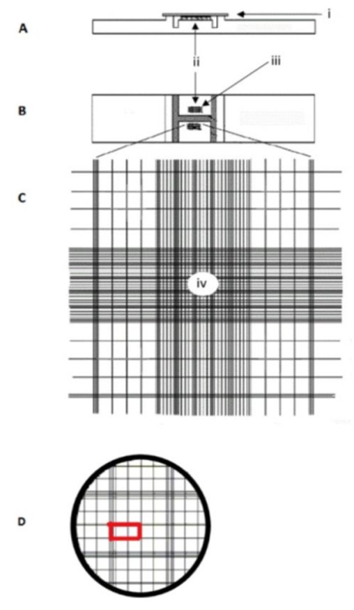
Sperm motility follows the criteria set by WHO (Chung et al., 2022) and is denoted by the letters a, b, c (from fastest to slowest) as follows: (a) progressive motility; ≥ 25 μm/s where 25 μm is equal to the length of 2 squares (smallest box) on the haemocytometer (see red rectangle in Figure 2). (b) non progressive motility; < 5 μm/s. (c) no motility.
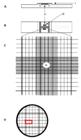
Haemocytometer. A = Side view; B = Top view; C = One of the two haemocytometer counting chambers; D = Insert of iv. Legend: i. Cover glass; ii. Counting chamber; iii. Counting grid area; iv. One of the 25 larger squares (Chung et al., 2022; Luthfi and Noor, 2021).
The percentage of motility is determined by the Formula 1 according to Chung et al. (2022):
2.5.2. Spermatozoa morphology
Using a pipette, 10 µL of sperm suspension were extracted from the available sperm samples and placed into the counting chamber (Luthfi and Noor, 2021). One drop of spermatozoa suspension and one drop of 1% eosin and 10% nigrosin dyes were placed in a glass slide. Observed with a light microscope magnification of 40×10, as many as 100 spermatozoa per preparation with 5 replication, the results are expressed in percent (%). In this study, observations were made five times to minimize assessment bias (Inveresk Research, 2000) (Research, Sciences, and Sequani 2000). Giemsa's dye was used to stain the slides, which were then examined at x400 magnification. To reduce variance, each counting chamber was allotted about 100 sperm. Sperm motility is calculated using the specified criteria (Adamkovicova et al., 2016; Luthfi and Noor, 2021).
Spermatozoa have normal morphology if the head is curved like a hook, straight neck, and a single free-tipped tail. Abnormal morphology, if the head size is small or too large; two heads with one neck or one tail, broken or branched neck; branched tail; curled, and broken. Head and tail deformities were the two categories used to categorize the morphological abnormalities. Mid-piece sperm abnormalities were included in the evaluation of the sperm tail (Adamkovicova et al., 2016; Inveresk Research, 2000; Seed et al., 1996).
2.5.3. Number of spermatozoa
A total of 10 µL of sperm suspension was taken using a pipette from the available sperm samples to be inserted into the counting chamber (Luthfi and Noor, 2021). The number of spermatozoa was determined using a Thoma hemocytometer (Varisli et al., 2013). Spermatozoa diluted with physiological NaCl solution are placed into a hemocytometer. The counted spermatozoa were located at the center and edge of the chamber (top and left side). The average number of spermatozoa (n) was obtained from the total sum of spermatozoa in each chamber divided by 4. The length of each chamber was 1 mm, and the height was 0.1 mm, so the chamber volume was equal to 0.1 mm3 or 1.0 mm2 ×0.1 mm or 1.0 × 10⁴ mL.
The Formula 2 calculated the number of spermatozoa according to Meyers (2000):
2.6. Data analysis
Testosterone level reading data were converted using the Curve Expert program. All qualitative data were analyzed descriptively and quantitative data were analyzed statistically. The histological structure of the testes was analyzed descriptively qualitatively by comparing between treatments. Rat weight, testosterone levels, motility, morphology and spermatozoa count were analyzed using one way anova at a significance level of 5%, if there was a difference, further testing was carried out using the Duncan Multiple Range Test (DMRT).
3. Results and Discussion
The results of research that has been conducted on the effects of administering cashew nut shell extract to male rats on body weight, testosterone hormone levels, and spermatozoa quality (motility, morphology, and number of spermatozoa) are explained below.
3.1. Body weight
The physical condition of the results of body weight measurements can be seen in Table 1. The weight gain found during the five weeks of treatment in all treatment groups, but did not differ significantly (F count 0.732 > F table 0.432). The Control Group (T0), Treatment 1 (T1) and Treatment 2 (T2) showed an average weight gain with a graph that continued to increase, but in Treatment 3 (T3) although there was weight gain, it was not as much as groups T0, T1 and T2. The administration of a single dose of oral cashew nut shell extract had no effect on the growth and development of body weight of male white rats during the five weeks of observation after administration of the test substance. The occurrence of a decrease in body weight in a day that did not reach 5% showed the influence of behavior on test animals, which is common due to treatment. Changes in body weight serve as an indication of the general health status of animals (Koriem et al., 2019).
Based on Table 1, the results of the average measurement of the weight of rat in the treatment groups T0, T1, T2 and T3 increased every week. The difference in the average measurement of the weight of rat at the end of treatment (week 5) and the beginning of treatment (week 1) at T0 = 18.5 gr, T1 = 18.84 gr, T2 = 15.67 gr, and T3 = 12.34 gr. The average weight of T3 rat experienced a smaller increase in weight compared to other treatments. The earliest and most obvious signs of the harmful effects of the test samples are actual changes in body weight. Experimental animals given high doses in toxicity tests typically lose weight as a result of their diminished appetite (Sireeratawong et al., 2010).
One-way repeated measures ANOVA was performed to determine whether the mean body weight of rat differed between the four treatments. The results of the one-way repeated measures ANOVA can be seen in Table 2.
Repeated one-way measures to find out if the rat's mean body weight varied amongst the four treatments, an ANOVA was conducted. The rat's body weight did not differ statistically significantly between the treatments, according to an ANOVA (F statistic is 0.457 and the corresponding p value is 0.649). The rats body weight did not differ statistically significantly across the four treatments, according to the results of Bonferroni's test for multiple comparisons.
3.2. Histological structure of testes
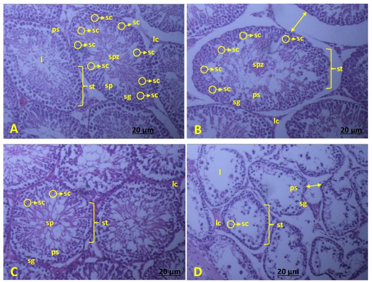
The histological structure of the testes in each treatment group can be seen in Figure 3. The comparison between the testes tissue structures of each treatment group appears different, both in the interstitial tissue and in the seminiferous tubules. Group C shows a normal picture of the seminiferous tubules and interstitial tissue, all spermatogenic cells, namely spermatogonia, primary spermatocytes, spermatids and spermatozoa were found. Leydig cells were found in the interstitial tissue. Leydig cells play a role in producing the hormone testosterone (Figure 3A). In the seminiferous tubules of groups T1 and T2, the spermatogenic cells found were spermatogonia, primary spermatocytes, spermatids and spermatozoa. In T3, only spermatogonia were found in the basal part and a small number of primary spermatocytes. Between the interstitial tissue and the seminiferous tubules of the testes of rats in groups T1, T2 and T3, there is a gap or empty space, which is called the tissue stretching phenomenon (Compliance) (Figures 3B, 3C and 3D). This testicular tissue stretching phenomenon indicates the presence of fluid that is rapidly released from the circulation into the interstitial tissue due to an abnormal condition, called edema (Hall, 1991).
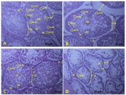
Histological structure of testis. Hematoxylin Eosin (HE) staining. A = T0. Control; B = T1. Extract 250mg/kg body weight; C = T2. Extract 500 mg/kg body weight; D = T3. Extract 750 mg/kg body weight. lc = Leydig cells; st = seminiferous tubules; sg = Spermatogonium; ps = Primary spermatocyte; sp = Spermatid; spz = Spermatozoa; sc = Sertoli cells; l = Lumen; } = one of seminiferous tubules; ↔ = compliance.
Based on Figure 3. the structure of the seminiferous tubules T0 and T1 are oval in shape and large. T2 is round and smaller than T0 and T1. T3 is irregular in shape and smaller than T0, T1, and T2. The seminiferous tubules' basal cells are smaller in T3 compared to T0, T1, and T2. This happens because of testosterone's inability to reach the tubules for these cells to mature. Another option is brought on by the polarity of the metabolites of the ethanolic extract, which tend to remove water from the cells, reducing their size. FSH and testosterone are inhibited in sperm that have been injured in the lumen of the seminiferous tubules of the T3 rat testis. The tension that develops around the tubules is probably also connected to the suppression of testosterone binding to ABP. Therefore, the diameter is lower since the strain shown in T3 is caused by vacuolization and membrane fluidity. This is because cashew nut shell extract contains phenolic compounds (Bhatia et al., 2024).
Damage after phenol activation to cells through the membrane. Activation will cause membrane leakage followed by collagen formation if the cell has a nucleus. Conversely, the membrane will become denser and undergo structural changes due to aggregation in cells that do not have a nucleus. The leaking membrane causes fluid to escape from the cell into the tissue, resulting in “pitting oedema” or “nonpitting oedema” if it is with the cell mass or tissue. Ethanolic extract of cashew nut shell at a dose of 750 mg/kg BW (T3) contains phenolic compounds that are cytotoxic. Cytotoxic occurs due to the nature of polarity and phenol bonds that are more than polyphenols. Cytotoxic causes inflammation accompanied by udem. The resulting udem includes pitting udem and nonpitting udem. Cytotoxic to tissues causes focal atrophy and diffuse atrophy. Metabolites of cashew nut shell extract can draw fluid out of normal cells because they are polar phenolic compounds. According to Leopold and Kriedemann (1983), phenols are compounds that at low concentrations can spur growth, but at high concentrations can inhibit growth because they inhibit protein formation and amino acid transport. So phenol compounds in seminiferous tubules, in small amounts act as estrogen so that it has estrogenic potential but in excessive amounts cause cell death so it is cytotoxic.
The condition of the T3 seminiferous tubules shows atrophy with no spermatogenic cells (spermatids and spermatozoa) that have degenerated, but Leydig cells are still found. This can be seen by comparing the lumen of the seminiferous tubules in each treatment. The atrophy of seminiferous tubules in Figure 3D is called focal atrophy. Focal atrophy affects spermatids and spermatozoa (Greaves and Faccini, 1984). The empty spaces between the spermatogenic cells show the effect of cashew nut shell extract. Testicular atrophy is mainly caused by the loss of spermatozoa cells and a decrease in seminiferous tubular diameter (Albadri et al., 2013). Disorders of the seminiferous tubules and Leydig cells will affect the testosterone hormone produced (Nassar and Leslie, 2018; O’Donnell et al., 2015).
By comparing Figures 3A, 3B, 3C and 3D, in the 3D image, cell death is seen at the spermatids and spermatozoa (from the lumen towards the basal), so that neither of these cells was found. This suggests that the metabolites of cashew nut shell ethanolic extract can enter through the basal membrane of the tubules. In each treatment when compared to the control, the number of sertoli cells decreased, especially in T3, where very few sertoli cells were found. According to O’Donnell et al. (2015), sertoli cells play a role in feeding the developing spermatocytes. so that the reduced number of sertoli cells causes the process of spermatogenesis to be inhibited. this is evidenced in T3 no spermatozoa were found in the seminiferous tubules. Extract metabolites that enter the tubules are phenolic compounds because they act like estrogen. This is evidenced by the presence of spermatogonia cells and primary spermatocytes in Figure 3D. The phenols are inhibitors of protein formation and amino acid transport; low concentrations spur growth, but high concentrations inhibit growth. The presence of phenol compounds in seminiferous tubules is the same; in small amounts, they act as estrogen so that it has estrogenic potential, but in excessive amounts, they cause cell death, so they are cytotoxic.
3.3. Testosterone hormone levels
The average results of testosterone level measurements with cashew nut shell extract treatment can be seen in Table 3. Cashew nut shell extract has a significant effect on testosterone levels. There is a decrease in testosterone levels along with the increase in the dose of cashew nut shell extract. The average testosterone level at T0 was 170.43 ng/mL, T1 was 143.97 ng/mL, T2 was 108.33 ng/mL and T3 showed the lowest hormone level of 71.11 ng/mL at T3.
The data in Table 3 and Figure 3 show that treating cashew nut shells with ethanolic extract significantly affects testosterone levels, reducing testosterone levels. If testosterone levels are linked to morphometric changes in testicular tissue, then T3 has the lowest testosterone levels. In Figure 3, when treatment doses increase, fewer Sertoli cells are present, which results in fewer Androgen-Binding Proteins (ABP). The testes' Sertoli cells produce a protein called Androgen-Binding Protein (ABP), which binds to testosterone and carries it to the epididymis through the seminiferous tubule fluid. ABP functions to regulate testosterone in the male reproductive system and shares structural similarities with Sex Hormone Binding Globulin (SHBG) (Munell et al., 2002).
The phenolic chemicals in cashew seed extract have estrogenic properties that influence hypothalamic cell receptors, causing the pituitary to release Interstitial Cell Stimulating Hormone (ICSH) in response to the Hypothalamus-Pituitary-Gonad (HPG) axis (Tammasse and Tamrin, 2023). Additionally, ICSH promotes the creation of testosterone; as a result of its positive activation, testosterone levels rise, but then fall as a result of cell damage. Damage to Leydig cells, which results in alterations in the testes' histological structure across all therapies, causes the drop in testosterone levels. Leydig cells release testosterone, hence increasing the number of Leydig cells can increase the hormone's secretion (Widjaja et al., 2023). Male fertility and spermatogenesis depend heavily on testosterone (Smith and Walker, 2014).
In the experimental group, along with the increase in the dose of extract, there was a reduction in the number of leydig cells, thereby reducing testosterone secretion. This is in accordance with the opinion Chen et al. (2020) testosterone can increase if leydig cell activation is stimulated by Interstitial Cell Stimulating Hormone (ICSH), but it is also related to the availability of its precursor, namely cholesterol. Damaged spermatozoa in the lumen of the semiferrous tubules of the Rattus novergicus T3 testis (Figure 3D) showed inhibition of FSH and testosterone, so that testosterone levels decreased. The inhibition of testosterone binding to Androgen Binding Protein is likely also related to the strain that occurs around the tubules. This is in accordance with the opinion of Pusparanee et al. (2016) that the increase in testosterone levels is due to the layer of germ epithelial cells becoming thicker, the lumen of the seminiferous tubules being dense with sperm and increasing the number of sertoli cells which ultimately produce many spermatid and spermatozoa cells which enter the lumen.
In relation to the hypothalamus-pituitary-gonad axis, the phenolic compounds of the extract that are estrogenic affect the receptors of hypothalamus cells to stimulate the pituitary to produce ICSH. Furthermore, ICSH stimulates testosterone production, but because there is damage to the testicular structure, testosterone hormone levels are reduced. According to Smith and Walker (2014) testosterone is the main androgen in the testes that regulates spermatogenesis. Testosterone is produced by Leydig cells in response to stimulation with luteinizing hormone (LH) and acts as a paracrine factor that diffuses into the seminiferous tubules.
Cashew nut shell extract contains estrogenic phenolic chemicals so they affect steroidogenetic. Because they force primordial cells to undergo apoptosis, estrogen and progesterone in the male reproductive system can prevent spermatogenesis. When rat are given estrogen constantly, their testes will rapidly become aspermic because meiosis division is imperfect even while mitotic activity persists. FSH or testosterone can be used to restore sperm development (Pramaningtyas et al., 2022). Androgen is not necessary for the spermatogonia to mature into spermatids, but it is necessary for the spermatids to mature into spermatozoa. Therefore, low testosterone levels hinder the maturation of spermatozoa (Susilawati, 2011).
3.4. Microscopic quality of spermatozoa
Fertility is influenced by the microscopic condition of spermatozoa, including motility, morphology, and number of spermatozoa. Normal spermatozoa have good quality so that they are able to perform capacitation, acrosome reaction and penetrate the zona pellucida wall during fertilization so that the success of fertilization increases (WHO, 2021) (Chung et al. 2022). Microscopic quality testing of spermatozoa includes spermatozoa motility, spermatozoa morphology, and spermatozoa count. The average microscopic quality of spermatozoa can be seen in Table 4.
Motility is the ability of spermatozoa to proliferate in a liquid environment is known as motility. For spermatozoa to break through the protective cells enclosing the ovum, sperm motility is crucial (Sujoko et al., 2019). Numerous factors, such as age, medications, and chemicals, affect sperm motility (Syntin and Robaire, 2001). The quantity of spermatozoa that move gradually in order to reach the female reproductive system for fertilization is one measure of sperm motility, which is one of the criteria for assessing spermatozoa quality (van Der Horst et al., 2018).
According to Table 4, spermatozoa motility significantly decreased in all treatments when cashew nut shell extract was administered (P <0.05). Hormones and the metabolic activity of spermatozoa both affect sperm motility. In the epididymis, testosterone attaches itself to receptor proteins. In cells, receptor proteins carry out signal transduction functions. Chemical cues cause target cells to react after initially activating receptors (Champbell, 2004). Male white rat's testosterone levels may be able to trigger reactions in the epididymis' spermatozoa. The hormone testosterone and other elements, such as nutrients and enzymes released by the epididymal epithelium, affect the maturation of spermatozoa in the epididymis from non-motile to motile spermatozoa (Susetyarini et al., 2019).
The study found that the phenolic chemicals in cashew nut shell extract interfered with the spermatogenesis process, which resulted in a decrease in spermatozoa motility. According to Bhatia et al. (2024), the phenolic chemicals found in cashew nut skin extract include anacardic acid (70%), cardanol (5%), cardol (18%), and methyl cardol (7%). Spermatozoa motility may be decreased if the epididymal spermatozoa maturation process is disturbed (Rotimi et al., 2024).
The maturation process is a process of change that includes structural changes in the head and tail of spermatozoa. This process can cause changes in the form of increased motility of more aggressive spermatozoa. Sperm morphometry is combined with measurements of spermatozoa motility and spermatozoa function tests such as vitality, DNA fragmentation and acrosome integrity, as explained by Lee et al. (2015).
The results of the analysis of variance on the morphology of spermatozoa in the treatment of cashew nut shell extract, had a significant effect when compared to the control (P <0.05). The characteristics of normal spermatozoa are that they have a head shape like a fishing hook and a long, straight tail. Abnormal spermatozoa have an irregular head shape, can be shaped like a banana, or irregular (amorphous), or too bent, and the tail is not straight or even has no tail, or only has a tail without a head (Halimah et al., 2020).
The number of normal spermatozoa morphology in treated rats was less when compared to controls. Administration of cashew nut shell extract changed spermatozoa morphology and decreased spermatozoa fertility. Cashew nut shell extract affected sertoli cells in the seminiferous tubules, causing failure in spermatogenesis. Damage to the seminiferous tubules caused abnormalities in early development. In addition, cashew nut skin extract affected steroid hormones involved in the reproductive process. Decreased sperm count and quality were associated with decreased testosterone levels and oxidative damage caused by suppression of antioxidant enzyme activity (Adamkovicova et al., 2016). In the treatment, there was a decrease in testosterone hormone levels compared to the control and this will affect the number of normal morphology of spermatozoa. This is supported by the opinion (Ghavi et al., 2017) that testosterone, FSH and LH secretion affect the formation of spermatozoa. Normal spermatozoa morphology, if there is a disturbance in the production of spermatogenesis hormones, it will affect the morphology of spermatozoa.
In Table 4, the administration of cashew nut shell extract significantly affected the normal number of spermatozoa in all treatments compared to the control (P <0.05). The number of spermatozoa (million/mL) in T3 was 12.62, lower than T2 which was 24.25, lower than T1 which was 36.60, when compared to the control of 47.60. The decrease in the number of spermatozoa in each treatment was because the cashew nut shell extract caused disruption to spermatogenesis and disruption to the testosterone hormone. This can be seen in the histological structure in each treatment, especially in T3 there was atrophy in the seminiferous tubules, irregular lumen shape and disruption to the spermatogenesis stage (Figure 3D). According to O’Donnell et al. (2015)) testosterone is a very important hormone in maintaining the survival of spermatozoa while they are stored in the epididymis. Decreased sperm count and sperm quality associated with low serum testosterone concentrations is a cause of reproductive dysfunction leading to male infertility (Illiano et al., 2020). Zhao et al. (2020) stated normal levels of LH, FSH and testosterone are essential for maintaining normal morphology and progressive motility of spermatozoa.
There is a physiological reaction (endogenous ROS) in the testes, particularly in the seminiferous tubules. This response is primarily the consequence of normal cell metabolism, with a minor portion coming from external exposure (exogenous ROS). On the other hand, excessive ROS generation causes cell damage if the normal physiological process is disturbed Consequently, the number of spermatozoa is impacted by cellular damage and spermatozoa cell death (Pasupathi et al., 2009).
4. Conclusions
The results of the study on Enhancing the Antifertility Performance by Cashew Nut Shell (Anacardium occidentale L) Extract on Rattus norvegicus Berkenhout 1769 can be concluded that there is no significant difference in the weight of the rats. The effect of the extract on the histological structure of the testes in T3 occurs atrophy of the seminiferous tubules, irregular shape, vacuolization and membrane fluidity. Interstitial tissue T1, T2 and T3 experience compliance. The administration of the extract has a significant effect on testosterone hormone levels, and the microscopic quality of spermatozoa (motility, normal morphology and number of spermatozoa) which decreases with increasing extract dose. Further research is needed on the isolation of active compounds from cashew nut shell and studies on the recovery of test animals after administration of cashew nut shell extract.
Acknowledgements
The author would like to thank the Institute for Research and Community Service (LPPM) Universitas Sebelas Maret for providing funds for the excellent research scheme with Contract Number 452/UN27.21/PN/2020.
Data Availability Statement
The research data analyzed in this study are not publicly available by any means. The entire data set that supports the results of this study was published in the article itself. Data is available at: DOI of the dataset or link where it is published.
References
-
ADAMKOVICOVA, M., TOMAN, R., MARTINIAKOVA, M., OMELKA, R., BABOSOVA, R., KRAJCOVICOVA, V., GROSSKOPF, B. and MASSANYI, P., 2016. Sperm motility and morphology changes in rats exposed to cadmium and diazinon. Reproductive Biology and Endocrinology, vol. 14, no. 1, pp. 42. http://doi.org/10.1186/s12958-016-0177-6 PMid:27503218.
» http://doi.org/10.1186/s12958-016-0177-6 -
ALBADRI, C.T., ANI, I.M.A. and HIBA, H.M.A., 2013. Alcohol consumption and its effect on testicular structure and on sperm count and motility in parent mice and their offspring. International Medical Journal Malaysia, vol. 12, no. 1, pp. 43-48. http://doi.org/10.31436/imjm.v12i1.2001
» http://doi.org/10.31436/imjm.v12i1.2001 -
BADAN PUSAT STATISTIK, 2021 [viewed 7 January 2025]. Hasil sensus penduduk (sp2020) pada september 2020 mencatat jumlah penduduk sebesar 270,20 juta jiwa [online]. Available from: https://www.bps.go.id/id/pressrelease/2021/01/21/1854/hasil-sensus-penduduk--sp2020--pada-september-2020-mencatat-jumlah-penduduk-sebesar-270-20-juta-jiwa-.html
» https://www.bps.go.id/id/pressrelease/2021/01/21/1854/hasil-sensus-penduduk--sp2020--pada-september-2020-mencatat-jumlah-penduduk-sebesar-270-20-juta-jiwa-.html -
BEZERRA, L.G.P., SILVA, A.M., DANTAS, M.R.T., SANTOS, R.P., MOREIRA, S.S.J., PEREIRA, A.G., OLIVEIRA, M.F., COMIZZOLI, P. and SILVA, A.R., 2023. Development of assays for the characterization of sperm motility parameters, viability, and membrane integrity in the epididymis and vas deferens of the greater rhea (Rhea americana). Animal Reproduction, vol. 20, no. 4, e20230113. http://doi.org/10.1590/1984-3143-ar2023-0113 PMid:38213765.
» http://doi.org/10.1590/1984-3143-ar2023-0113 -
BHATIA, B., AMARNATH, N., RASTOGI, S.K. and LOCHAB, B., 2024. Isolation of cardanol fractions from cashew nutshell liquid (cnsl): a sustainable approach. Sustainable Chemistry, vol. 5, no. 2, pp. 68-80. http://doi.org/10.3390/suschem5020006
» http://doi.org/10.3390/suschem5020006 - CHAMPBELL, R.M., 2004. Biology Jakarta: Erlangga.
-
CHAUDHURI, P., 2023. Basic histological techniques (microtechniques) for staining of animal tissue. Uttar Pradesh Journal of Zoology, vol. 44, no. 24, pp. 225-230. http://doi.org/10.56557/upjoz/2023/v44i243830
» http://doi.org/10.56557/upjoz/2023/v44i243830 -
CHEN, P., ZIRKIN, B.R. and CHEN, H., 2020. Stem leydig cells in the adult testis: characterization, regulation and potential applications. Endocrine Reviews, vol. 41, no. 1, pp. 22-32. http://doi.org/10.1210/endrev/bnz013 PMid:31673697.
» http://doi.org/10.1210/endrev/bnz013 -
CHUNG, E., ARAFA, M., BOITRELLE, F., KANDIL, H., HENKEL, R., SALEH, R., SHAH, R., VOGIATZI, P., ZINI, A. and AGARWAL, A., 2022. The new 6th edition of the WHO Laboratory manual for the examination and processing of human semen: is it a step toward better standard operating procedure? Asian Journal of Andrology, vol. 24, no. 2, pp. 123-124. http://doi.org/10.4103/aja2021118
» http://doi.org/10.4103/aja2021118 -
DARE, S.S., HAMMAN, W.O., MUSA, S., GOJI, A.D.T., OYEWALE, A.A., ABBA, S. and EZEKIEL, I., 2011 [viewed 7 January 2025]. Effects of aqueous extract of Anacardium occidentale (cashew) leaf on pregnancy outcome of wistar rats. International Journal of Animal and Veterinary Advances [online], vol. 3, no. 2, pp. 77-82. Available from: https://maxwellsci.com/print/ijava/v3-77-82.pdf
» https://maxwellsci.com/print/ijava/v3-77-82.pdf -
DAS, B., MAHAPATRA, M., GURUNG, B., DEY, A., NONGALLEIMA, K., DAS, S., DAS TALUKDAR, A., CHOWDHURY, A., CHOUDHURY, M.D. and DEB, L., 2024. Antifertility activity of Oroxylum indicum vent. stem bark on female wistar rats. Indian Journal of Traditional Knowledge, vol. 23, no. 2, pp. 189-199. http://doi.org/10.56042/ijtk.v23i2.8722
» http://doi.org/10.56042/ijtk.v23i2.8722 -
DAWANE, J., THAKUR, L. and SURYAWANSHI, S., 2024. Evaluation of antifertility potential of Entada rheedii spreng seed kernel extract in female wistar rats. Journal of Natural Remedies, vol. 24, no. 7, pp. 1517-1523. http://doi.org/10.18311/jnr/2024/35409
» http://doi.org/10.18311/jnr/2024/35409 -
GHAVI, F., MOSALANEJAD, L., ABDOLLAHIFARD, S. and JAHROMI, M.G., 2017. Male infertility and its impact on women’s sexual behaviors: need attention to psychological problem as a psychological rehabilitation. Iranian Rehabilitation Journal, vol. 15, no. 2, pp. 87-94. http://doi.org/10.18869/nrip.irj.15.2.87
» http://doi.org/10.18869/nrip.irj.15.2.87 - GREAVES, P., and FACCINI, J. M., 1984. Rat histopathology: a glossary for use in toxicity and carcinogenicity studies Amsterdam: Elsevier.
-
HALIMAH, K., RAHAYU, S.R. and YUNIASTUTI, A., 2020 [viewed 7 January 2025]. The effect of noise to morphology of rats (mus musculus) spermatozoa. Public Health Perspective Journal [online], vol. 5, no. 3, pp. 1-2. Available from: http://journal.unnes.ac.id/sju/index.php/phpj
» http://journal.unnes.ac.id/sju/index.php/phpj - HALL, J.E., 1991. Guyton and hall textbook of medical physiology. Amsterdam: Elsevier Health Sciences.
-
HANDAYANI, N. and GOFUR, A., 2024. Effect of Pulutan (Urena lobata l) leaves decoction on the hormone testosterone levels of Balb C mice (Mus musculus). In: International Conference on Life Sciences and Technology (ICoLiST 2023). 24-25 August 2023, Malang. Malang: BIO Web of Conferences. http://doi.org/10.1051/bioconf/202411701048
» http://doi.org/10.1051/bioconf/202411701048 -
HARLITA, H. and PUSPITASARI, D., 2019 [viewed 7 January 2025]. Pengaruh ekstrak kulit biji mete (anacardium occidentale l) terhadap siklus estrus tikus albino (rattus norvegicus berkehout 1769). Seminar Nasional Pendidikan Sains [online], vol. 1, pp. 2–6. Available from: https://core.ac.uk/reader/289792060
» https://core.ac.uk/reader/289792060 -
HARLITA, R.M.P., PROBOSARI, R.M. and ARIYANTO, J., 2015. Perubahan histologis uterus tikus putih (Rattus norvegicus) galur wistar: aktifitas antifertilitas ekstrak kulit biji mete (Anacardium occidentale L.). Bioedukasi: Jurnal Pendidikan Biologi, vol. 8, no. 2, pp. 1-4. http://doi.org/10.20961/bioedukasi-uns.v8i2.3860
» http://doi.org/10.20961/bioedukasi-uns.v8i2.3860 -
HARLITA, N.H., SATUTI, N.H.N., SAGI, M. and ASTUTI, P., 2016. Acute toxicity of cashew nut shell extract (Anacardium occidentale L.) in albino rat (Rattus norvegicus Berkenhout 1769). Pakistan Journal of Biological Sciences, vol. 19, no. 2, pp. 89-94. http://doi.org/10.3923/pjbs.2016.89.94 PMid:29023045.
» http://doi.org/10.3923/pjbs.2016.89.94 -
HIDAYAT, R. and WULANDARI, P., 2021. Methods of extraction: maceration, percolation and decoction. Eureka Herba Indonesia, vol. 2, no. 1, pp. 73-79. http://doi.org/10.37275/ehi.v2i1.15
» http://doi.org/10.37275/ehi.v2i1.15 -
HLONGWA, M., KALINDA, C., PELTZER, K. and HLONGWANA, K., 2021. Factors associated with modern contraceptive use: a comparative analysis between younger and older women in Umlazi Township, KwaZulu-Natal, South Africa. Women’s Health, vol. 17, pp. 17455065211060641. http://doi.org/10.1177/17455065211060641 PMid:34798800.
» http://doi.org/10.1177/17455065211060641 -
ILLIANO, E., TRAMA, F., ZUCCHI, A., IANNITTI, R.G., FIORETTI, B. and COSTANTINI, E., 2020. Resveratrol‐based multivitamin supplement increases sperm concentration and motility in idiopathic male infertility: a pilot clinical study. Journal of Clinical Medicine, vol. 9, no. 12, pp. 1-10. http://doi.org/10.3390/jcm9124017 PMid:33322606.
» http://doi.org/10.3390/jcm9124017 -
KORIEM, K.M.M., ARBID, M.S. and EL-ATTAR, M.A., 2019. Acute and subacute toxicity of ammi visnaga on rats. Interdisciplinary Toxicology, vol. 12, no. 1, pp. 26-35. http://doi.org/10.2478/intox-2019-0004 PMid:32189984.
» http://doi.org/10.2478/intox-2019-0004 -
LEE, K.H., CHEN, H.P., LEUNG, C.M., CHEN, H.L., TSAI, S.S. and HSU, P.C., 2015. Effects of indium chloride exposure on sperm morphology and DNA integrity in rats. Journal of Food and Drug Analysis, vol. 23, no. 1, pp. 152-160. http://doi.org/10.1016/j.jfda.2014.10.001 PMid:28911439.
» http://doi.org/10.1016/j.jfda.2014.10.001 - LEOPOLD, A.C. and KRIEDEMANN, P.E., 1983. Plant growth and development 2nd ed. New Delhi: Tata McGraw-Hill Publishing Company Ltd.
-
LOUWAGIE, E.J., QUINN, G.F.L., POND, K.L. and HANSEN, K.A., 2023. Male contraception: narrative review of ongoing research. Basic and Clinical Andrology, vol. 33, no. 1, pp. 30. http://doi.org/10.1186/s12610-023-00204-z PMid:37940863.
» http://doi.org/10.1186/s12610-023-00204-z -
LUTHFI, M.J. and NOOR, M.M., 2021. A simple technique for rapid assessment of rat (Rattus norvegicus) sperm motility. Biology, Medicine, & Natural Product Chemistry, vol. 9, no. 2, pp. 105-107. http://doi.org/10.14421/biomedich.2020.92.105-107
» http://doi.org/10.14421/biomedich.2020.92.105-107 -
MALPANI, A., MAHURKAR, N. and ASWAR, U., 2020. Phytochemical analysis and antifertility potential of cynodon dactylon in female wistar rats: a herbal approach towards contraception. Chinese Herbal Medicines, vol. 12, no. 3, pp. 281-288. http://doi.org/10.1016/j.chmed.2020.06.001 PMid:36119013.
» http://doi.org/10.1016/j.chmed.2020.06.001 - MEYERS, T.R., 2000. Fish pathology section Juneau: Alaska Department of Fish and Game, Commercial Fisheries Division.
-
MUNELL, F., SUAREZ-QUIAN, C.A., SELVA, D.M., TIRADO, O.M. and REVENTOS, J., 2002. Androgen-binding protein and reproduction: where do we stand? Journal of Andrology, vol. 23, no. 5, pp. 598-609. http://doi.org/10.1002/j.1939-4640.2002.tb02296.x PMid:12185088.
» http://doi.org/10.1002/j.1939-4640.2002.tb02296.x - NASSAR, G.N. and LESLIE, S.W., 2018. Physiology, testosterone. Treasure Island: StatPearls Publishing. PMid: 30252384.
- O’DONNELL, L., STANTON, P. and KRETSER, D.M., 2015. Endocrinology of the male reproductive system and spermatogenesis. South Dartmouth: MDText.com. PMid: 25905260.
-
OBEMBE, O.O., OJETOLA, A.A., ATERE, T.G., ABAYOMI, T.A., DARE, B.J., ADEYEMI, D.H. and OYEYIPO, I.P., 2023. Buchholzia coriacea seed induce antifertility by interfering with steroidogenic enzymes and inflammatory cytokines in rat testis. Journal of Reproductive Immunology, vol. 157, pp. 103923. http://doi.org/10.1016/j.jri.2023.103923 PMid:36868158.
» http://doi.org/10.1016/j.jri.2023.103923 -
OCHALA, E., JAN, S.A.G. and MAT, S., 2021 [viewed 7 January 2025]. A literature review on contraceptive practices, barriers and measures to improve use among postpartum women. African Journal of Health, Nursing and Midwifery [online], vol. 4, no. 1, pp. 1–23. Available from: https://abjournals.org/ajhnm/papers/volume-4/issue-1/a-literature-review-on-contraceptive-practices-barriers-and-measures-to-improve-use-among-postpartum-women/
» https://abjournals.org/ajhnm/papers/volume-4/issue-1/a-literature-review-on-contraceptive-practices-barriers-and-measures-to-improve-use-among-postpartum-women/ -
PASUPATHI, P., SARAVANAN, G., CHINNASWAMY, P. and BAKTHAVATHSALAM, G., 2009. Glutathione, glutathione-dependent enzymes and antioxidant status in gastric carcinoma patients. Journal of Applied Biomedicine, vol. 7, no. 2, pp. 101-109. http://doi.org/10.32725/jab.2009.010
» http://doi.org/10.32725/jab.2009.010 -
PATEL, K., PANCHAL, N. and INGLE, P., 2019 [viewed 7 January 2025]. Review of extraction techniques extraction methods: microwave, ultrasonic, pressurized fluid, soxhlet extraction, Etc. International Journal of Advanced Research in Chemical Science [online], vol. 6, no. 3, pp. 6-21. Avaliable from: https://www.arcjournals.org/international-journal-of-advanced-research-in-chemical-science/volume-6-issue-3/2
» https://www.arcjournals.org/international-journal-of-advanced-research-in-chemical-science/volume-6-issue-3/2 -
PRAMANINGTYAS, M.D., ISLAMIANA, D. and ADNAN, M.L., 2022 [viewed 7 January 2025]. Apoptosis pada spermatogenesis. Jurnal Kedokteran Syiah Kuala [online], vol. 22, no. 3, pp. 147-157. Available from: https://jurnal.usk.ac.id/JKS/article/view/21375
» https://jurnal.usk.ac.id/JKS/article/view/21375 -
PRASAD, F., PINGALE, S.S., RAGHUNATH, D.P. and SHAIKH, S.S., 2007 [viewed 7 January 2025]. In vivo anti-fertility and anti-implantation activity of anacardic acid methyl ester in rats. Pharmacologyonline [online], vol. 3, pp. 485-490. Available from: https://pharmacologyonline.silae.it/files/archives/2007/vol3/43_Prasad.pdf
» https://pharmacologyonline.silae.it/files/archives/2007/vol3/43_Prasad.pdf -
PUSPARANEE, H., LEE, H.W., HALIMAH, A.S. and MAHANEM, M.N., 2016 [viewed 7 January 2025]. Effects of Gynura procumbens on sperm quality and testosterone level in streptozotocin-induced type 1 diabetic rats. International Journal of Pharmacognosy and Phytochemical Research [online], vol. 8, no. 1, pp. 22-30. Available from: https://impactfactor.org/PDF/IJPPR/8/IJPPR,Vol8,Issue1,Article5.pdf
» https://impactfactor.org/PDF/IJPPR/8/IJPPR,Vol8,Issue1,Article5.pdf - INVERESK RESEARCH. Huntingdon Life Sciences. Sequani. Glaxo Wellcome, 2000. Industrial Reproductive Toxicology (IRDG) Computer Assisted Sperm Analysis (CASA) group rat sperm morphological assessment edition. London: Glaxo Wellcome, 15 p.
-
ROTIMI, D.E., IYOBHEBHE, M., OLUWAYEMI, E.T., OLAJIDE, O.P., AKINSANOLA, B.A., EVBUOMWAN, I.O., ASALEYE, R.M. and OJO, O.A., 2024. Energy metabolism and spermatogenesis. Heliyon, vol. 10, no. 19, e38591. http://doi.org/10.1016/j.heliyon.2024.e38591 PMid:39397940.
» http://doi.org/10.1016/j.heliyon.2024.e38591 -
SAHOO, A., GHOSH, A., DEY, A., MAJI, S., DAS, S., SULTANA, I., DE, S.K. and CHATTOPADHYAY, S., 2024. Assessment of oxidative liver injury caused by cashew nut shell liquid (cnsl) via interacting with bcl2 gene in male wistar rats and basic in-silico approaches. Proceedings of the Zoological Society, vol. 77, pp. 136-146. http://doi.org/10.1007/s12595-024-00516-6
» http://doi.org/10.1007/s12595-024-00516-6 -
SAHOO, A., GHOSH, A., DEY, A., MAJI, S., DAS, S., SULTANA, I., DE, S.K. and CHATTOPADHYAY, S., 2024. Assessment of oxidative liver injury caused by cashew nut shell liquid (cnsl) via interacting with bcl2 gene in male wistar rats and basic in-silico approaches. Proceedings of the Zoological Society, vol. 77, pp. 136-146. http://doi.org/10.1007/s12595-024-00516-6
» http://doi.org/10.1007/s12595-024-00516-6 -
SCHULTZ, D.J., WICKRAMASINGHE, N.S., IVANOVA, M.M., ISAACS, S.M., DOUGHERTY, S.M., IMBERT-FERNANDEZ, Y., CUNNINGHAM, A.R., CHEN, C. and KLINGE, C.M., 2010. Anacardic acid inhibits estrogen receptor α-DNA binding and reduces target gene transcription and breast cancer cell proliferation. Molecular Cancer Therapeutics, vol. 9, no. 3, pp. 594-605. http://doi.org/10.1158/1535-7163.MCT-09-0978 PMid:20197399.
» http://doi.org/10.1158/1535-7163.MCT-09-0978 -
SEED, J., CHAPIN, R.E., CLEGG, E.D., DOSTAL, L.A., FOOTE, R.H., HURTT, M.E., KLINEFELTER, G.R., MAKRIS, S.L., PERREAULT, S.D., SCHRADER, S., SEYLER, D., SPRANDO, R., TREINEN, K.A., VEERAMACHANENI, D.N. and WISE, L.D., 1996. Methods for assessing sperm motility, morphology, and counts in the rat, rabbit, and dog: a consensus report. Reproductive Toxicology (Elmsford, N.Y.), vol. 10, no. 3, pp. 237-244. http://doi.org/10.1016/0890-6238(96)00028-7 PMid:8738562.
» http://doi.org/10.1016/0890-6238(96)00028-7 -
SIMPEN, I., 2018 [viewed 7 January 2025]. Isolasi cashew nut shell liquid dari kulit biji jambu mete (anacardium occidentale l) dan kajian beberapa sifat fisiko-kimianya. Jurnal Kimia [online], vol. 2, no. 2, pp. 71-76. Available from: https://ojs.unud.ac.id/index.php/jchem/article/view/2715
» https://ojs.unud.ac.id/index.php/jchem/article/view/2715 - SIREERATAWONG, S., PIYABHAN, P., SINGHALAK, T., WONGKRAJANG, Y., TEMSIRIRIRKKUL, R., PUNSRIRAT, J., RUANGWISES, N., SARAYA, S., LERDVUTHISOPON, N. and JAIJOY, K., 2010. Toxicity evaluation of sappan wood extract in rats. Journal of the Medical Association of Thailand, vol. 93, suppl. 7, pp. S50. PMid:21294398.
-
SMITH, L.B. and WALKER, W.H., 2014. The regulation of spermatogenesis by androgens. Seminars in Cell & Developmental Biology, vol. 30, pp. 2-13. http://doi.org/10.1016/j.semcdb.2014.02.012 PMid:24598768.
» http://doi.org/10.1016/j.semcdb.2014.02.012 -
SUJOKO, H., SETIADI, M.A. and BOEDIONO, A., 2019 [viewed 7 January 2025]. Seleksi spermatozoa domba garut dengan metode sentrifugasi gradien densitas percoll. Jurnal Veteriner [online], vol. 10, no. 3, pp. 125-132. Available from: https://ojs.unud.ac.id/index.php/jvet/article/view/3356
» https://ojs.unud.ac.id/index.php/jvet/article/view/3356 -
SUSETYARINI, E., COREBIMA, A.D., AMIN, M. and SUSILAWATI, T., 2019. Hubungan testosteron dengan motilitas spermatozoa tikus putih jantan (Ratus norwegicus) setelah diberi senyawa aktif daun beluntas untuk penyusun buku antifertilitas. Belantika Pendidikan, vol. 2, no. 2, pp. 52-57. http://doi.org/10.47213/bp.v2i2.31
» http://doi.org/10.47213/bp.v2i2.31 - SUSILAWATI, T., 2011. Spermatologi Malang: UB Press.
-
SYNTIN, P. and ROBAIRE, B., 2001. Sperm structural and motility changes during aging in the brown norway rat. Journal of Andrology, vol. 22, no. 2, pp. 235-244. http://doi.org/10.1002/j.1939-4640.2001.tb02176.x PMid:11229797.
» http://doi.org/10.1002/j.1939-4640.2001.tb02176.x -
TAMMASSE, I.F.U. and TAMRIN, F., 2023. Different of hypothalamic-pituitary-gonadal axis in male and female. Reproductive Medicine International, vol. 6, no. 1, pp. 1-6. http://doi.org/10.23937/2643-4555/1710023
» http://doi.org/10.23937/2643-4555/1710023 -
VAN DER HORST, G., SKOSANA, B., LEGENDRE, A., OYEYIPO, P. and DU PLESSIS, S.S., 2018. Cut-off values for normal sperm morphology and toxicology for automated analysis of rat sperm morphology and morphometry. Biotechnic & Histochemistry, vol. 93, no. 1, pp. 49-58. http://doi.org/10.1080/10520295.2017.1380842 PMid:29319353.
» http://doi.org/10.1080/10520295.2017.1380842 - VARISLI, O., AGCA, C. and AGCA, Y., 2013. Short-term storage of rat sperm in the presence of various extenders. Journal of the American Association for Laboratory Animal Science; JAALAS, vol. 52, no. 6, pp. 732-737. PMid:24351761.
-
WALTERS, A.H., EYESTONE, W.E., SAACKE, R.G., PEARSON, R.E. and GWAZDAUSKAS, F.C., 2004. Sperm morphology and preparation method affect bovine embryonic development. Journal of Andrology, vol. 25, no. 4, pp. 554-563. http://doi.org/10.1002/j.1939-4640.2004.tb02826.x PMid:15223844.
» http://doi.org/10.1002/j.1939-4640.2004.tb02826.x - WORLD HEALTH ORGANIZATION – WHO, 2021. The examination and processing of human semen 6th ed. Geneva: WHO.
-
WIDJAJA, G., IBRAHIM, N.J., HADI, S.J., CABABAT, F., TURKI JALIL, A., AL-CHARAK, A.G., YASIN, G., SULTAN, M.Q. and FAKRI MUSTAFA, Y., 2023. The effect of eucalyptus globulus hydroalcoholic extract on lh, fsh and testosterone concentrations and sperm morphology. Archives of Razi Institute, vol. 78, no. 1, pp. 115-123. http://doi.org/10.22092/ARI.2022.359470.2426 PMid:37312694.
» http://doi.org/10.22092/ARI.2022.359470.2426 -
ZHAO, W., JING, J., SHAO, Y., ZENG, R., WANG, C., YAO, B. and HANG, D., 2020. Circulating sex hormone levels in relation to male sperm quality. BMC Urology, vol. 20, no. 1, pp. 101. http://doi.org/10.1186/s12894-020-00674-7 PMid:32680497.
» http://doi.org/10.1186/s12894-020-00674-7 -
ZHUANG, J.X., HU, Y.H., YANG, M.H., LIU, F.J., QIU, L., ZHOU, X.W. and CHEN, Q.X., 2010. Irreversible competitive inhibitory kinetics of cardol triene on mushroom tyrosinase. Journal of Agricultural and Food Chemistry, vol. 58, no. 24, pp. 12993-12998. http://doi.org/10.1021/jf103723k PMid:21121650.
» http://doi.org/10.1021/jf103723k
Edited by
-
Editor:
Takako Matsumura Tundisi