A series of complications following extrusion of root canal sealer: a rare case report
Abstract
Aim In this study, we aimed to draw attention to the mismanagement of a rare complication that develops during root canal treatment. The report follows the guidelines of the Case Report Guide (CARE) and focuses on steps to eliminate the complication that has occurred.
Methods The widespread acute infection of a 51-year-old female patient who applied to our clinic with an extraoral abscess was first brought under control. Afterward, during the root canal treatment performed on the lower third molar tooth, which was the source of the infection, endodontic paste residues that penetrated the mandibular canal were surgically removed, and the patient was followed up.
Results It was understood that the source of the patient’s widespread infection was the medicament-impregnated cotton that the endodontic treatment physician placed into the tooth extraction socket for analgesia. It was observed that the patient’s paresthesia, which occurred due to nerve damage, did not regress during long-term follow-up.
Conclusion This case report shows that while it is essential to avoid complications in dentistry, the knowledge and ability to manage them are truly remarkable.
Keywords
Mandibular nerve injuries; Foreign bodies; Root canal therapy
Introduction
Nerve damage is a typical complication observed during oral and maxillofacial dentistry operations1. Procedures that may cause nerve damage include local anesthesia, impacted tooth extraction, orthognathic surgery, maxillofacial traumas and treatments, implant surgery, pre-prosthetic surgical procedures, temporomandibular joint surgery/arthroscopy, and endodontic treatments2.
Although nerve injuries due to endodontic reasons are not very common, injuries have been reported in the literature as a result of mechanical trauma during excessive shaping of the protruding root canal, pressure resulting from overfilling at the apical area, the toxic effect of root canal irrigation solutions on tissues, and injuries caused by filling material invasion into the inferior alveolar canal due to overfilling of the canal through the apical foramen in the mandible3-4.
Case
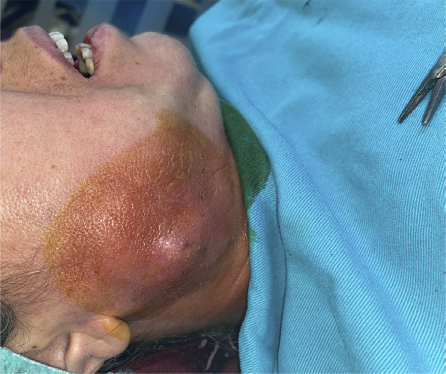
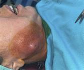
A 51-year-old patient without any systemic diseases and routine medication use presented to our clinic with a complaint of significant swelling and pain in the face (Fig. 1). According to the medical history obtained from the patient, root canal treatment was applied to the lower right third molar tooth in a private dental clinic. When the patient experienced pain, the dentist extracted the tooth and curetted the area. In the following two weeks, the patient reported using 1000 mg oral amoxicillin + clavulanic acid along with non-steroidal anti-inflammatory drugs. Over the last three days, the patient complained of extraoral swelling and pain in the buccal region of the lower right jaw.
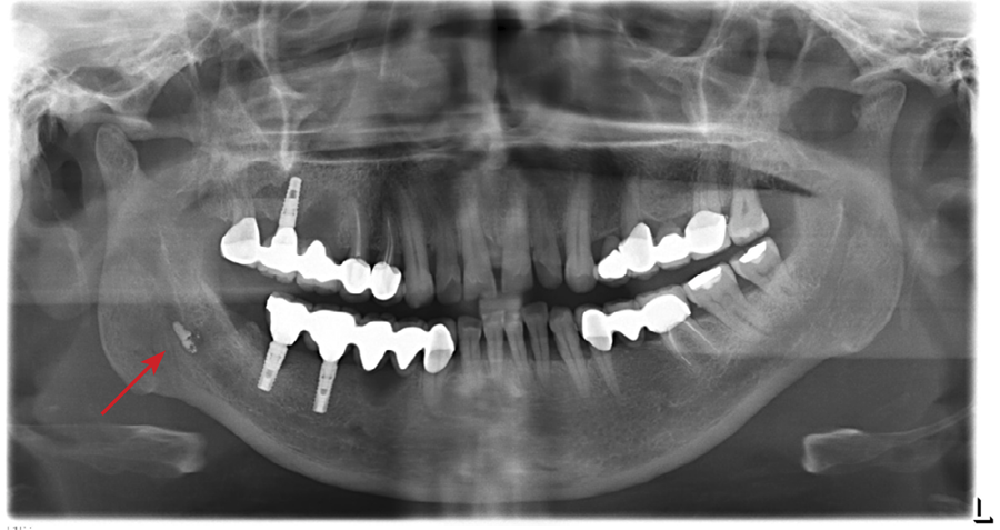
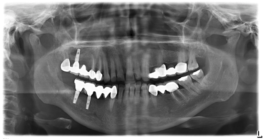
When the patient presented to our clinic, trismus was evident. Limited intraoral examination revealed that the epithelialization of the extraction site had not occurred, but there was no apparent pus drainage. Additionally, the patient was found to have paresthesia in a wide area affecting the right lower jaw, cheek, and lip regions. Evaluation of the panoramic radiograph showed that the root canal filling paste used during the canal treatment of the lower right third molar tooth had overflowed into the mandibular canal, and it was considered that this buccal abscess could be related to this situation (Fig. 2).
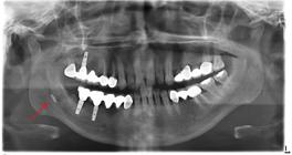
The patient was advised to perform extraoral warm compresses along with intravenous 1-gram cefazolin therapy. Extraoral drainage started on the second follow-up day, and a Penrose drain was sutured at the drainage site. The patient received regular medical dressing changes for five days, with 1 gram of cefazolin and 0.5% metronidazole therapy. At the end of the five days, a decrease in pus drainage and an improvement in mouth opening were observed. A surgical procedure under general anesthesia was planned for the patient.
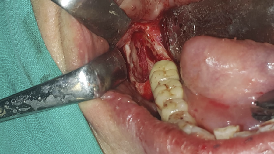
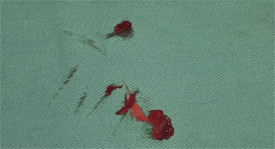
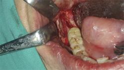
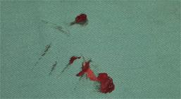
During the operation, a full-thickness flap was raised, extending towards the ramus from the region of the second molar tooth. Afterward, the extraction socket of the third molar tooth was explored. During socket curettage, a cotton-like foreign body was found. Access to the mandibular canal was achieved through bone reduction made from the socket and buccal cortex. Carefully removing the root canal filling paste from the canal, ensuring the integrity of the nerve, was done with attention (Fig. 3 ). A collagen sponge was placed in the area and sutured. The cotton-like foreign body removed during the operation and the debris removed from the canal were sent for pathological evaluation (Fig. 4).
After the operation, a panoramic radiograph taken immediately revealed that the material in the canal had been largely removed, with only a tiny piece still present.
The pathology report regarding the materials removed during the operation confirmed the preliminary diagnosis. It indicated that the foreign body was consistent with cotton, and the material removed was identified as root canal filling paste. The discussion with the patient revealed that the dentist who performed the canal treatment and extraction had placed an analgesic agent, analgesic-impregnated cotton, in the area when the patient’s symptoms worsened.
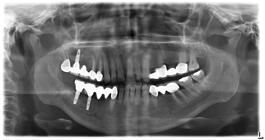
At the 6-month follow-up, it was learned that the patient did not experience a recurrence of pain and swelling symptoms, but the preoperative paresthesia persisted. In the panoramic radiograph, it was observed that the unrecoverable remnants of the paste had shrunk in size (Fig. 5).
Discussion
According to published research, the most frequent causes of endodontic nerve injuries are root canal overshaping-related mechanical trauma, overfilling-related pressure at the apex, root canal irrigation solutions’ toxic effects, and filling material extrusion from the apical foramen into the mandibular inferior alveolar canal3-5.
When root canal filling materials come into touch with neural tissues, they can cause toxic consequences such as anesthesia, hypoesthesia, paresthesia, or dysesthesia, which can be permanent6.
Root canal sealants are another thing that might cause paresthesia. Chemical toxicity is the primary cause of paresthesia brought on by sealers. In endodontic treatments, sealers based on zinc oxide and eugenol are most frequently used. Eugenol’s neurotoxic effects, which have been shown to produce paresthesia or anesthesia when in direct contact with the inferior alveolar nerve, are the main reason for toxicity. According to a study by Trowbridge et al.7, intradental nerve activity is inhibited by freshly mixed zinc oxide eugenol.
Sealers based on calcium hydroxide (Ca[OH]2) are also frequently used. Clinical studies have validated the irreversible nerve damage caused by Ca[OH]2-based sealers, as Boiesen and Bodrin8 reported in their experimental model9. Ahlgren et al.10, reporting on a case of mechanical compression causing paresthesia, was caused by the extrusion of a large amount of Ca(OH)2-based sealer into the periapical area close to the inferior alveolar nerve.
Numerous past clinical reports suggest that surgically removing the root canal filling material from the mandibular canal is a successful treatment that resolves the problem completely11-13. On the other hand, it has been noted that routine radiographic follow-up without surgical intervention is appropriate in certain clinical cases where the patient has no complaints14,15.
In the cases reported by Garde et al.16 and Köseoğlu et al.11, the preference was surgical removal of the canal sealer to decompress the nerve. However, rather than surgically removing the sealer, Gonzáles-Martín et al.13 and Poveda et al.17 conducted periodic follow-up visits. Typically, a patient’s symptoms and preferences guide the selection of treatment options. A patient should be monitored clinically and radiologically if they refuse surgical treatment, as some patients may do.
Considering that the canal filling paste led to an extraoral abscess in the patient, we preferred surgical removal. During the operation, we concluded that the medicated cotton we removed from the cavity was the reason for the infection. Although the surgery was successful and did not result in any iatrogenic nerve injury, we learned at the 18-month follow-up that the pre-existing paresthesia in the patient persisted without any improvement.
In conclusion, the information presented in this clinical case emphasizes the importance of careful root canal preparation and avoiding excessive root canal filling paste use. Additionally, being aware of the neurotoxic effects of materials used as root canal filling paste or pulpal medicaments is crucial for preventing complications or effectively managing the situation when complications arise. Knowledge of emergency interventions in case of such complications is also essential.
Acknowledgments
The authors would like to thank Ertan Ali Delibaşı at the Department of Oral and Maxillofacial Surgery, Dental Faculty, Gazi University, Turkey for invaluable support and supervision.
References
-
1 Küçükkurt S, Tükel HC, Murat Ö. [Nerve injuries: causes, diagnosis, and treatment]. Atatürk Üniv Dis Hek Fak Derg. 2019;29(1):150-9. Turkish. doi: 10.17567/ataunidfd.523097.
» https://doi.org/10.17567/ataunidfd.523097 -
2 Er N, Canakci FG, Capar G, Alkana A. [Peripheral nerve injuries and their treatment in dentistry]. Atatürk Üniv Dis Hek Fak Derg.2016;26(1). Turkish. doi:10.17567/dfd.33334.
» https://doi.org/10.17567/dfd.33334 -
3 Knowles KI, Jergenson MA, Howard JH. Paresthesia associated with endodontic treatment of mandibular premolars. J Endod. 2003 Nov;29(11):768-70. doi: 10.1097/00004770-200311000-00019.
» https://doi.org/10.1097/00004770-200311000-00019 -
4 Fanibunda K, Whitworth J, Steele J. The management of thermomechanically compacted gutta percha extrusion in the inferior dental canal. Br Dent J. 1998 Apr;184(7):330-2. doi: 10.1038/sj.bdj.4809618.
» https://doi.org/10.1038/sj.bdj.4809618 -
5 Andrabi SM, Alam S, Zia A, Khan MH, Kumar A. Mental nerve paresthesia secondary to initiation of endodontic therapy: a case report. Restor Dent Endod. 2014 Aug;39(3):215-9. doi: 10.5395/rde.2014.39.3.215.
» https://doi.org/10.5395/rde.2014.39.3.215 -
6 Brodin P, Rød A, Aars H, Orstavik D. Neurotoxic effects of root filling materials on rat phrenic nerve in vitro. J Dent Res. 1982 Aug;61(8):1020-3. doi: 10.1177/00220345820610080501.
» https://doi.org/10.1177/00220345820610080501 -
7 Trowbridge H, Edwall L, Panopoulos P. Effect of zinc oxide-eugenol and calcium hydroxide on intradental nerve activity. J Endod. 1982 Sep;8(9):403-6. doi: 10.1016/S0099-2399(82)80094-0.
» https://doi.org/10.1016/S0099-2399(82)80094-0 -
8 Boiesen J, Brodin P. Neurotoxic effect of two root canal sealers with calcium hydroxide on rat phrenic nerve in vitro. Endod Dent Traumatol. 1991 Dec;7(6):242-5. doi: 10.1111/j.1600-9657.1991.tb00210.x.
» https://doi.org/10.1111/j.1600-9657.1991.tb00210.x -
9 Yatsuhashi T, Nakagawa K, Matsumoto M, Kasahara M, Igarashi T, Ichinohe T, et al. Inferior alveolar nerve paresthesia relieved by microscopic endodontic treatment. Bull Tokyo Dent Coll. 2003 Nov;44(4):209-12. doi: 10.2209/tdcpublication.44.209.
» https://doi.org/10.2209/tdcpublication.44.209 -
10 Ahlgren FK, Johannessen AC, Hellem S. Displaced calcium hydroxide paste causing inferior alveolar nerve paraesthesia: report of a case. Oral Surg Oral Med Oral Pathol Oral Radiol Endod. 2003 Dec;96(6):734-7. doi: 10.1016/j.tripleo.2003.08.018.
» https://doi.org/10.1016/j.tripleo.2003.08.018 -
11 Köseoglu BG, Tanrikulu S, Sübay RK, Sencer S. Anesthesia following overfilling of a root canal sealer into the mandibular canal: a case report. Oral Surg Oral Med Oral Pathol Oral Radiol Endod. 2006 Jun;101(6):803-6. doi: 10.1016/j.tripleo.2005.07.015.
» https://doi.org/10.1016/j.tripleo.2005.07.015 -
12 Scolozzi P, Lombardi T, Jaques B. Successful inferior alveolar nerve decompression for dysesthesia following endodontic treatment: report of 4 cases treated by mandibular sagittal osteotomy. Oral Surg Oral Med Oral Pathol Oral Radiol Endod. 2004 May;97(5):625-31. doi: 10.1016/S1079210404000502.
» https://doi.org/10.1016/S1079210404000502 -
13 Gonzáles-Martín M, Torres-Lagares D, Gutiérrez-Pérez JL, Segura-Egea JJ. Inferior alveolar nerve paresthesia after overfilling of endodontic sealer into the mandibular canal. J Endod. 2010 Aug;36(8):1419-21. doi: 10.1016/j.joen.2010.03.008.
» https://doi.org/10.1016/j.joen.2010.03.008 -
14 Scarano A, Di Carlo F, Quaranta A, Piattelli A. Injury of the inferior alveolar nerve after overfilling of the root canal with endodontic cement: a case report. Oral Surg Oral Med Oral Pathol Oral Radiol Endod. 2007 Jul;104(1):e56-9. doi: 10.1016/j.tripleo.2007.01.036.
» https://doi.org/10.1016/j.tripleo.2007.01.036 -
15 Batur YB, Ersev H. Five-year follow-up of a root canal filling material in the maxillary sinus: a case report. Oral Surg Oral Med Oral Pathol Oral Radiol Endod. 2008 Oct;106(4):e54-6. doi: 10.1016/j.tripleo.2008.06.008.
» https://doi.org/10.1016/j.tripleo.2008.06.008 - 16 Garde JB, Jawale BA, Bendgude V, Dadhe D, Kulkarni A. Inferior alveolar neuralgia resulting from foreign body within the inferior alveolar canal: a rare incidence. Int J Dent Case Reports 2012;2:113-6.
-
17 Poveda R, Bagán JV, Fernández JM, Sanchis JM. Mental nerve paresthesia associated with endodontic paste within the mandibular canal: report of a case. Oral Surg Oral Med Oral Pathol Oral Radiol Endod. 2006 Nov;102(5):e46-9. doi: 10.1016/j.tripleo.2006.03.022.
» https://doi.org/10.1016/j.tripleo.2006.03.022
-
Data Availability:
Datasets related to this article will be available upon request to the corresponding author.
Edited by
-
Editor:
Dr. Altair A. Del Bel Cury
Datasets related to this article will be available upon request to the corresponding author.