 Transposition flap for desmotomy reconstruction in dogs with patellar luxation
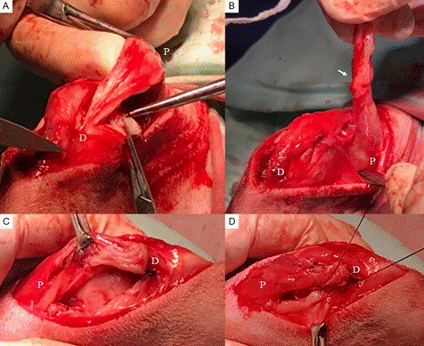
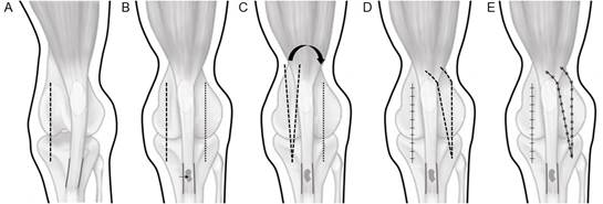
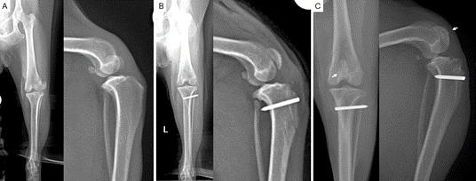
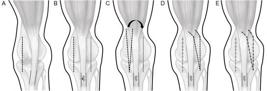
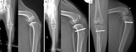
This study described a transposition flap that can be used as an adjunctive procedure for medial or lateral patellar luxations and to evaluate its use in dogs retrospectively. Dogs with grade 3 and grade 4 patellar luxation were retrospectively enrolled in this study. Five dogs had medial luxation, and three had lateral luxation. The transposition flap was used in 11 hind limbs with patellar luxation. After deepening the trochlear groove (chondroplasty or wedge recession trochleoplasty), transposition of the tibial tuberosity, and desmotomy, a triangular flap was created with the stretched parapatellar soft tissue in the opposite direction to the patellar luxation. The base of the flap was located approximately 1 cm proximal to the patella and the apex near the tibial tuberosity. After the flap was elevated by cutting through the apex, it was transposed to the desmotomy area, and its apex was sutured to the distal aspect of the desmotomy. The edges of the flap were then sutured to the medial and lateral edges of the desmotomy. All dogs had a good outcome with stabilized patellae and complete functional recovery at least 2 months after surgery. In conclusion, the transposition flap provide soft-tissue coverage to the desmotomy area and can be considered a complementary procedure for the stabilization of patellar luxation. However, the results should be interpreted as preliminary due to the limited sample size, short-term outcomes, and the descriptive nature of the analysis.
Key words:
dog; surgery; patella; soft tissue
 Thumbnail
 Thumbnail
 Thumbnail