 Fowl adenovirus serotype 1 detection in broilers: gizzard erosion pattern characterization and confirmation by FISH and PCR
ABSTRACT:
Adenoviral gizzard erosions have been affecting domestic and wild birds worldwide, highlighting the need for new diagnostic approaches. Therefore, this study aimed to identify and score gizzard lesions caused by fowl adenovirus serotype 1 (FAdV-1) in chickens of different ages from commercial farms located in two states of the South and Central-West regions of Brazil, as well as to apply the fluorescent in situ hybridization (FISH) technique as a diagnostic tool. A total of 56 samples were collected from broilers aged between 1 and 45 days, showing poor performance and suspected gizzard erosion or ulceration. Samples were analyzed macroscopically and histologically, and the presence of the agent was confirmed by polymerase chain reaction (PCR) and FISH techniques. Macroscopically, only 5.35% of the gizzards presented lesions, classified as grades 1 and 2, corresponding to Group 1 (66.67%) and Group 2 (33.33%), respectively. The lesions were focal to multifocal, whitish, and resembled the detachment of the koilin membrane, measuring approximately 0.4 cm in diameter. Histologically, 92.85% of the analyzed samples showed alterations: 9.61% presented only koilin membrane rarefaction (grade 1), 50% exhibited lymphocytic and granulocytic ventriculitis with fragmentation or erosion of the koilin layer (grade 2), and 40.38% showed fibrosis associated with ventriculitis and erosion of the koilin layer. Intranuclear inclusion bodies were also observed in 26.78% of the samples. Positive signals for FAdV-1 were detected in 57.14% and 30.35% of the samples analyzed by FISH and PCR, respectively. Broilers aged one to two weeks exhibited acute lesions, while those aged three weeks or older presented chronic lesions. The chronicity of the lesions, associated with lower viral load in the tissues of broilers older than three weeks, negatively influenced the PCR results. In conclusion, different degrees of gizzard erosion were identified in chickens of various ages in Brazil. The FISH technique can be used as a diagnostic method for the detection of FAdV-1 in gizzard tissues.
INDEX TERMS:
Avian pathology; broilers; diagnosis; FISH; gizzard erosion; histopathology
RESUMO:
As erosões da moela de origem adenoviral têm afetado aves domésticas e silvestres em todo o mundo, indicando a necessidade de investigar novas técnicas de detecção. Diante disso, este estudo teve como objetivo identificar e pontuar lesões ventriculares causadas por adenovírus aviário sorotipo 1 (FAdV-1) em aves de diferentes faixas etárias provenientes de granjas de frangos localizadas em dois estados das regiões Sul e Centro-Oeste do Brasil, além de aplicar a técnica de hibridização fluorescente in situ (FISH) como ferramenta diagnóstica. Foram coletadas 56 amostras de aves com idades entre 1 e 45 dias, apresentando baixo desempenho zootécnico e suspeita de erosão ou ulceração da moela. As amostras foram submetidas à análise macroscópica e histológica, e a presença do agente foi confirmada pelas técnicas de reação em cadeia da polimerase (PCR) e FISH. Macroscopicamente, apenas 5,35% das moelas apresentaram lesões, classificadas como graus 1 e 2, pertencentes ao Grupo 1 (66,67%) e ao Grupo 2 (33,33%), respectivamente. As lesões eram focais a multifocais, de coloração esbranquiçada, semelhantes ao descolamento da membrana de coilina, medindo aproximadamente 0,4 cm de diâmetro. Histologicamente, 92,85% das amostras apresentaram alterações: 9,61% mostraram apenas rarefação da membrana de coilina (grau 1), 50% exibiram ventriculite linfocítica e granulocítica associada à fragmentação ou erosão da camada de coilina (grau 2), e 40,38% apresentaram fibrose associada à ventriculite e erosão da camada de coilina. Corpúsculos de inclusão intranucleares foram observados em 26,78% das amostras. Sinais positivos para FAdV-1 foram detectados em 57,14% e 30,35% das amostras analisadas pelas técnicas de FISH e PCR, respectivamente. Aves com uma a duas semanas de idade apresentaram lesões agudas, enquanto aquelas com três semanas ou mais apresentaram lesões crônicas. A cronificação das lesões, associada à menor carga viral nos tecidos de aves com mais de três semanas de idade, influenciou negativamente o desempenho da PCR. Em conclusão, foram identificados diferentes graus de erosão da moela em frangos de distintas idades no Brasil. A técnica de FISH pode ser utilizada como um método diagnóstico para detecção de FAdV-1 em tecidos ventriculares.
TERMOS DE INDEXAÇÃO:
Patologia aviária; frangos de corte; diagnostico; FISH; erosão de moela; histopatologia
Introduction
Fowl adenoviruses (FAdV) belong to the genus Aviadenovirus of the Adenoviridae family. According to the current binomial nomenclature of species names adopted by the International Committee on Taxonomy of Viruses (ICTV), FAdVs are classified into five species (ICTV 2024), with their former designations shown in parentheses: Aviadenovirus ventriculi (FAdV-A), Aviadenovirus quintum (FAdV-B), Aviadenovirus hydropericardii (FAdV-C), Aviadenovirus gallinae (FAdV-D), and Aviadenovirus hepatitidis (FAdV-E), comprising a total of 12 recognized serotypes (FAdV-1, -2, -3, -4, -5, -6, -7, -8a, -8b, -9, -10, and -11) that infect a wide range of domestic and wild birds worldwide (Fitzgerald 2020, Hess 2020). FAdV transmission occurs both vertically and horizontally and the virus is considered an opportunistic pathogen that usually causes subclinical infections (Chen et al. 2020). However, some FAdV species and their respective serotypes have been identified as primary agents in the poultry industry, such as A. ventriculi (FAdV-A, serotype FAdV-1), which causes gizzard erosion with intranuclear inclusion bodies; some strains of A. hydropericardii (FAdV-C, serotype FAdV-4), which are associated with hepatitis-hydropericardium syndrome; and strains of A. gallinae (FAdV-D, serotypes FAdV-2, -3, -9 and -11) and A. hepatitidis (FAdV-E, serotypes FAdV-6, -7, -8a and -8b), which are involved in inclusion body hepatitis (Hess 2020). The virus is commonly detected by a combination of viral isolation and histopathology, which is occasionally associated with electron microscopy and/or polymerase chain reaction (PCR) (Ojkic et al. 2008). In addition, many laboratories use serotyping by virus neutralization, immunohistochemistry (IHC), and in situ hybridization (ISH) (Fitzgerald 2020). To note, however, fluorescence in situ hybridization (FISH) has not been reported as a method for FAdV diagnosis in veterinary medicine. This technique relies on fluorescence-labeled probes that hybridize to specific nucleic acid sequences within intact cells, enabling direct visualization of viral genomes (Moter & Gobel 2000).
Gizzard erosion of adenoviral origin was first reported in the 1990s, when inclusion bodies were observed in mucosal epithelial cells (Tanimura et al. 1993). In 2001, FAdV-1 was isolated and identified as the cause of gizzard condemnation in Japanese slaughterhouses (Ono et al. 2001). Since then, outbreaks have occurred worldwide, including in Poland, Korea, Hungary, Germany, Belgium, and Iran (Domanska-Blicharz et al. 2011, Kecskeméti et al. 2012, Lim et al. 2012, Schade et al. 2012, Garmyn et al. 2018, Mirzazadeh et al. 2019).
Some Brazilian states have reported the presence of FAdV in domestic chickens: the first report of hepatitis-pericardial syndrome in commercial flocks from a farm was described in the state of São Paulo, southwestern Brazil (Mettifogo et al. 2014); in the state of Minas Gerais, also located in the southwestern region, the virus was detected in both family and industrial poultry farming, with high dissemination rates in both scenarios (Pereira et al. 2014). A study detected the molecular sequences of FAdV and characterized the serotypes that affect commercial farms in Brazil, identifying the FAdV-8a, FAdV-8b, and FAdV-11 agents (De la Torre et al. 2018). However, data on FAdV-1 as a causative agent of gizzard erosion in Brazil are scarce, especially regarding the country’s southern region, where poultry farming is extensive.
Therefore, this study aimed to characterize the pathological findings associated with FAdV-1 infection in broilers of different age groups from southern and central-western Brazil, with suspected gizzard erosion/ulceration and reduced performance. Macroscopic and histological lesions were scored, and the presence of the virus was confirmed by PCR. In addition, considering the need for improved diagnostic approaches, this study also sought to apply the FISH technique.
Materials and Methods
Ethical approval. This study was approved by the Ethics Committee on Animal Use of the Palotina Sector of the “Universidade Federal do Paraná” (UFPR), under protocol CEUA/Palotina 04/2020.
Samples. Gizzards from 56 low-performance broiler chickens (Gallus gallus domesticus), aged 1 to 45 days, from 21 different batches were evaluated, with up to five broilers from the same batch. They were grouped by age as follows: Group 1 (one week old), Group 2 (two to three weeks old), and Group 3 (three weeks old to slaughter). The broilers were from different flocks and farms located in the western and southern regions of the Paraná state (southern Brazil) and Mato Grosso do Sul (central-western Brazil), respectively. These farms comply with biosecurity protocols and have a history of good animal zootechnical indexes, with a recent drop in performance.
Macroscopy. The samples were classified and scored according to the degree of koilin layer damage as follows: score 0 (no damage), score 1 (mild damage: less than one-third of the koilin layer/mucosa affected), score 2 (moderate damage: one-third to half of the koilin layer/mucosa affected), and score 3 (severe damage: more than half of the koilin layer/mucosa affected) (Nakamura et al. 2002).
Histological technique. After collection and fixation, the samples were cleaved, stored in embedding cassettes, and immersed in alcohol and xylene at different concentrations (Luna 1968). They were then embedded in paraffin, and the resulting blocks were sectioned to a thickness of 3-5 µm sections using a Leica® model Rm2245 microtome. Finally, they were mounted on glass slides as usual and stained with haematoxylin and eosin (HE) for optical microscopic analysis.
Histological analysis. The gizzard erosions were analyzed for the detection of adenovirus presence. Similarly to the macroscopic analysis, the evaluated changes were classified as follows: score 0 (no histological changes), score 1 (acute necrosis of the koilin layer with cell debris), score 2 (acute necrosis of the koilin layer with the presence of lymphohistiocytic and granulocytic inflammatory infiltrate in the lamina propria), and score 3 (acute necrosis/ulceration of the koilin layer with cell debris, associated with the presence of lymphohistiocytic and granulocytic inflammatory infiltrate and fibrosis in the lamina propria). The presence or absence of inclusion bodies in the cells of the mucosal surface epithelium was also evaluated.
DNA extraction and PCR. Ultrapure autoclaved water was used as a negative control in all reactions. As a positive control, an autogenous vaccine gently provided by the INATA Laboratory (Uberlândia, state of Minas Gerais, Brazil) was used. The reference vaccine strain employed was the prototype of FAdV-1, CELO strain. Viral DNA was extracted using the Nucleospin® Tissue kit (Macherey-Nagel, Germany), according to the manufacturer’s instructions. For PCR, the FAdV-1A (5’TTCGAGATCAAGGCCAAGT3’) was used as a sense primer and the FAdV-1B (5’GGTCGAAGTTGCGTAGGAAG3’) as an antisense primer; they had an expected fragment size of 178 bp (Niczyporuk et al. 2010). The reactions occurred with a final volume of 25 µL, which contained 3.75 µL of 10x buffer, 1 µL of MgCl2, 2 µL of 1x dNTP, 1 µL of each primer, 0.4 µL of Taq DNA Polymerase (Platinum® Taq DNA Polymerase-Invitrogen), 11.85 µL of ultrapure autoclaved water, and 4 µL of genomic DNA. The samples were subjected to the following conditions: initial denaturation at 95 °C for 2 minutes, denaturation at 94 °C for 1 minute, primer annealing at 58 °C for 1 minute, chain extension at 72 °C for 2 minutes, and final extension at 72 °C for 7 minutes. A total of 50 replication cycles were performed on the MyGenie96 Thermal Block thermal cycler (Bioneer, Korea). The samples amplified for visualization were run through a 2% agarose gel electrophoresis in TBE buffer pH 8.4 (89 mM Tris; 89 mM boric acid; 2 mM EDTA) at 150 V and 80 mA. Subsequently, they were stained with ethidium bromide (0.5 mg/mL) and visualized in a transilluminator under ultraviolet light.
DNA probe. We adapted the methodology adopted by Grafl et al. (2012) - who originally used the long fibre gene FAdV-1 (5-CGGGGTCGCAGCAGCTGCAGCTCGCGAGCGGAGAACTCG-3) for ISH - to a commercially synthesized probe (Invitrogen®). The probe was labeled with the fluorochrome Alexa Fluor 555 (Thermo Fisher®) and purified by high-performance liquid chromatography (HPLC).
FISH technique. FISH was performed on all gizzard fragments from broilers with macroscopic and histological lesions suggestive of FAdV-1 infection. To this end, the 3-5-μm tissue sections were prepared on silanized slides; they were then deparaffinized with xylene (two washes of 10 minutes each) and rehydrated with absolute, 90%, 80%, and 70% alcohol (one wash of 5 minutes each). Finally, they were left in distilled water for 5 minutes. After the slides were dried at room temperature, they were positioned in incubation trays for immunohistochemistry. We used 495 µL of the hybridization buffer (100 mM Tris, pH 7.2, 0.9 M NaCl, 0.1% NADS) for probe dilution, which was performed with nuclease-free pipettes. The buffer was transferred to 1.5-mL microtubes, to which 5 µL of the probe was added to achieve a final concentration of 2 ng of probe per µL of solution. Subsequently, each slide received 100 µL of this solution and was then incubated at 45 °C overnight. After this period, the slides were washed three times with hybridization buffer and washing solution (100 mM Tris, pH 7.2, 0.9 M NaCl), both preheated to 45 °C. The final step consisted of mounting the slides using a mounting medium for fluorescence microscopy (PorLong Diamond Antifade Mountant, Thermo Fisher®). They were immediately analyzed in a fluorescence microscope (Olympus, FSX 100, Japan) with an appropriate filter for the fluorochrome (460-495 nm) and magnification from 40x to 60x. The FISH slides with positive staining were classified according to the agent’s presence intensity in the tissue as follows: weak (up to 10% of the tissue), mild (11% to 30%), moderate (31% to 60%), accentuated (61% to 90%), and severe (91% to 100%).
Results
Macroscopy
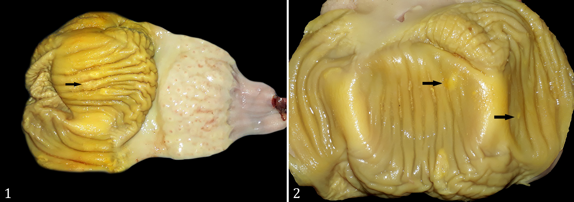
Erosions observed on the koilin layer that suggested FAdV-1 infection were found in only three gizzards (5.35%, 3/56). Of this total, two belonged to Group 1 and were classified as score 1; the other was from Group 2 and was classified as score 2 (Table 1). These erosions are characterized by focal to multifocal pale areas measuring approximately 0.4 cm in diameter, with discoloration and detachment of the koilin layer (Fig. 1-2).
Macroscopic image of gizzard erosion in Gallus domesticus. (1) Presence of focal areas measuring approximately 0.4 cm in diameter, with erosion of the koilinia membrane. (2) Multifocal areas with moderate discoloration of the koilinia membrane are observed.
Histology
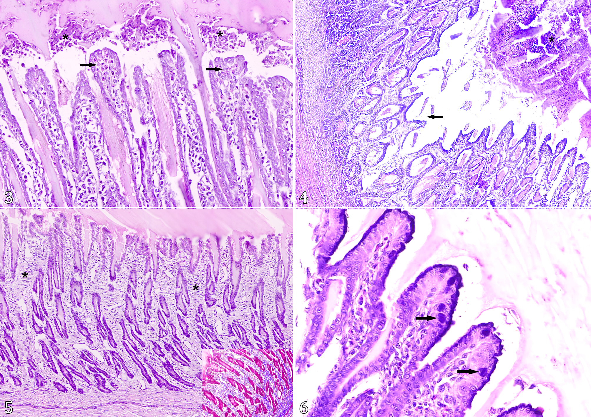
Histological alterations were observed in 92.85% (52/56) of the samples, of which 9.61% (5/52) presented acute necrosis of the koilin layer (score 1) with no intranuclear inclusion bodies; half of the samples (26/52) had acute necrosis of the koilin layer with lymphohistiocytic and granulocytic inflammatory infiltrate in the lamina propria and sometimes in the koilin layer (Fig. 3) (score 2), of which 50% (13/26) had intranuclear inclusion bodies; finally, 40.38% (21/52) presented acute necrosis/ulceration of the koilin layer, as well as lymphohistiocytic and granulocytic inflammatory infiltrate (Fig. 4) and associated fibrosis (Fig. 5) (score 3), which can be evidenced by Masson’s trichrome staining (Fig. 5, inset). Intranuclear inclusion bodies were identified in 9.52% of them (2/21). When present, the bodies were basophilic and located in the nucleus of the mucosal surface epithelium cells. They had an approximate size of 11 µm and did not exceed the amount of one to six bodies per high magnification field (Fig. 6). We identified no lesions in 7.14% (4/56) of the samples, all belonging to Group 3. The histopathological classification according to the score of the lesion and the presence or absence of inclusion bodies for each group is detailed in Table 1.
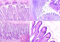
Histology of gizzard erosion in Gallus domesticus. Koilin. (3) Lymphohistiocytic and granulocytic infiltrate is observed in the lamina propria (arrow). Associated with this, acute necrosis of the koilin layer is noted, with granulocytic infiltrate and cellular debris (asterisk). HE, obj. 8.5x. (4) Detachment of the mucosal koilin layer (erosion) (arrow) can be observed, with the presence of lymphohistiocytic and granulocytic inflammatory infiltrate (asterisk). HE, obj. 8.5x. (5) Fibrosis is observed throughout the lamina propria. HE, obj. 8.5x. Inset: Connective tissue within the lamina propria is stained blue. Masson’s trichrome stain, obj. 4x. (6) Presence of intranuclear inclusion bodies in the cells of the superficial mucosal epithelium measuring up to 11 µm. HE, obj. 40x.
PCR
Of all the gizzards evaluated, 30.35% (17/56) obtained amplification for FAdV-1, of which 47.06% (8/17) belonged to Group 1, 41.18% (7/17) to Group 2, and 11.76% (2/17) to Group 3, as shown in Table 1.
FISH
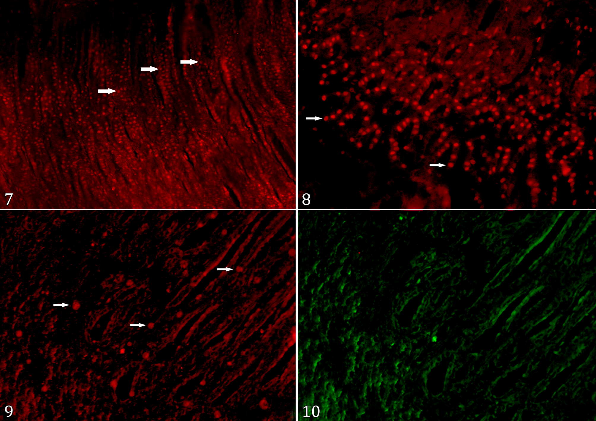
Of the 56 evaluated gizzards by the FISH technique, 57.14% (32/56) were positive for FAdV-1. In those, a bright, strong, oval-shaped signal was identified in the nucleus of the cells of mucosa (Fig. 7-9), which disappeared after changing the light filter (Fig. 10). The 32 positive cases were classified into Group 1 (40.62%, 13/32); Group 2 (15.62%, 5/32), and Group 3 (43.75%, 14/32) (Table 1). Of the 24 FISH-negative samples, only one (4.17%), belonging to Group 3, presented no histological alterations. As for the other ones, 8.33% (2/24), belonging to Group 1, presented histological lesions classified as score 1; 45.83% (11/24) presented histological lesions classified as score 2, of which 45.45% (5/11) was from Group 1 and 54.54% (6/11) from Group 2; finally, 41.67% (10/24) presented histological lesions classified as score 3, all belonging to Group 3.
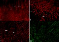
Photomicrograph showing positive fluorescence for fowl adenovirus serotype 1 (FAdV-1) in broiler gizzard (Gallus gallus domesticus) using fluorescent in situ hybridization (FISH) technique (460-495 nm). (7) Accentuated intranuclear fluorescence in the gizzard epithelial cells. FISH, obj. 10x. (8) Moderate intranuclear fluorescence in the gizzard epithelial cells. FISH, obj. 20x. (9) Mild intranuclear fluorescence in the gizzard epithelial cells. FISH, obj. 20x. (10) Counterstain in the histological section shown in Figure 9. FISH, obj. 20x.
Discussion
The diagnosis of FAdV-1 is made through macroscopic examination, histopathology, PCR for detection of viral DNA, ISH, and/or viral isolation (golden test) (Latimer et al. 1997, Gjevre et al. 2013, Grafl et al. 2013). The evaluation of the FISH technique in this study was motivated by the need for complementary diagnostic methods for FAdV-1, especially due to the difficulty of performing viral isolation for diagnosis. Conventional approaches such as histopathology and PCR, although widely used, have important limitations. Histopathology indicates the presence of lesions consistent with adenoviral infection. However, it is not specific, whereas PCR is highly sensitive and specific but requires DNA extraction and does not allow direct localization of the virus within tissue lesions. In our study, 30.35% of ventricles tested positive in PCR. Similar results were found in Iran, where 22.91% of ventricles were PCR-positive for FAdV-1 (Mirzazadeh et al. 2019).
When comparing the results of PCR and FISH techniques, there was agreement in 39.28% (22/56) of the cases, both for positive and negative results, of which 40.90% (9/22) belonged to Group 1, 18.18% (4/22) to Group 2, and 40.9% (9/22) to Group 3. However, there was divergence in 60.71% (34/56) of the cases, in which the sample obtained positive results from only one technique; 32.35% (11/34) of them belong to Group 1, 20.59% (7/34) to Group 2, and 47.06% (16/34) to Group 3.
In our research, the localization of FISH labeling agrees with that seen in ISH in broiler gizzards (Grafl et al. 2012, 2013). The marking observed indicates that the presence of the agent stood out in relation to the fluorescence of the tissue. Confirmation was performed by changing the filter from green to red, after which the positive markings disappeared and were no longer observable in the sections, as also seen by Piccoli (2023). Although this technique is highly sensitive and specific, Moter & Gobel (2000) state that some false-positive results may occur, such as in tissues containing elastin, collagen, or erythrocytes and eosinophils, due to the autofluorescence of these components. However, given the intranuclear labeling of the agent, there was no difficulty visualizing the positive marking for FAdV-1 in our study.
There was no FAdV-1 staining in FISH in 42.85% of the samples, but most of them presented histological lesions. According to Neves & Guedes (2012), false negatives may occur in cases of insufficient probe penetration into the microorganism due to the structure of the cell wall or low concentration of target DNA for annealing.
However, in the present study, Group 3 showed the highest percentage of discrepancy between FISH and PCR techniques. In contrast, Piccoli et al. (2023) reported a positive correlation between FISH and PCR in 83% of liver samples from birds infected with chlamydia. This difference may be partially attributed to the chronicity of lesions observed in older broiler chickens, a condition generally associated with a lower viral load, which reduces the amount of viral DNA available for PCR amplification. Furthermore, most studies investigating the detection of FAdV-1 by PCR include a prior viral isolation step using embryonated eggs or susceptible cell cultures derived from chicken liver, kidney, or fibroblasts, which allows viral replication at high titers and increases the sensitivity of the assay (Hess 2020, Leland & Ginocchio 2007). In the present study, PCR was performed directly on gizzard tissue, without prior viral isolation. It is important to emphasize that the gizzard is predominantly composed of muscle tissue, while the lesions associated with FAdV and viral replication are mainly located in the mucosal epithelium. Since no mucosal scrapings or targeted sampling of the epithelial layer were performed, the dilution of viral DNA in muscle-rich tissue may have further reduced the sensitivity of the PCR. This, along with the lower viral load associated with the older age of the broilers, may also have limited the sensitivity of the PCR compared to FISH.
In this context, FISH proved to be a valuable tool, allowing in situ visualization of viral genomes in preserved tissues, enabling not only detection but also a clear correlation between viral presence and histopathological changes. The validation of the technique in this study was supported by three main findings: a strong agreement between FISH-positive cases and PCR-positive cases (except in Group 3), confirming its reliability; the specific nuclear localization of the fluorescent signal in mucosal epithelial cells, consistent with adenoviral replication sites, which corroborates its specificity; and the identification of additional FISH-positive cases in PCR-negative samples, suggesting greater sensitivity of this method in formalin-fixed, paraffin-embedded tissues, where DNA integrity may be compromised. Although viral isolation was not performed and sensitivity and specificity could not be calculated, FISH allowed specific in situ detection of FAdV-1 and its correlation with lesions, supporting its use as a valid complementary diagnostic tool in broilers.
The macroscopic changes observed are similar in broilers, both experimentally and naturally infected with FAdV-1. A study in Italy evaluated 35 gizzards from broilers aged 42 to 63 days, obtained from 12 outbreaks over 11 years; erosion areas were found to vary in size and to have a brownish to blackish color (Manarolla et al. 2009). FAdV-1 was diagnosed in Belgium at two distinct broiler farms, causing koilin layer erosion in 27- and 35-day-old broilers, with lesions in 60% (6/10) and 80% (8/10) of the analyzed gizzards, respectively (Garmyn et al. 2018). In our study, we observed a few gizzards of one to three-week-old broilers with discoloration and detachment of the koilin layer. There was no macroscopic alteration in the other broilers, although 92.85% presented histological lesions. These findings may be related to the low viral load in the tissue since there is an experimentally detected coincidence between the maximum viral load in the gizzard and the onset of pronounced macroscopic and microscopic lesions, along with a decline in lesions at older ages (Grafl et al. 2013). In these cases, we highlight the importance of performing histological evaluation in tandem with other diagnostic tools, since macroscopic evaluation alone may be inconclusive for FAdV-1 adenoviral-associated lesions diagnosis, as seen in this study. Moreover, other factors may be associated with gizzard erosion, including congenital characteristics, ingested fiber structure, nutritional deficiencies, toxic substances, and microbial colonization (Gjevre et al. 2013).
Changes in the histology of ventricles affected by FAdV-1 have been reported in several studies (Tanimura et al. 1993, Abe et al. 2001, Schade et al. 2012, Matczuk et al. 2017). According to these studies, in such cases, there is koilin layer necrosis, histiocytic inflammatory infiltrate in the lamina propria, submucosa, or muscle, and occasionally intranuclear inclusion bodies in glandular epithelial cells. These findings corroborate the alterations found in our study. The lesions classified as score 1 - in which only the koilin layer presents alterations - were visualized only in broilers from Group 1, although there were also gizzards with class 2 alterations in the same group. In this case, we believe that environmental factors such as heat stress may have contributed to lesion aggravation, given that housings’ high population density and excessive heat are frequent in Brazil and cause corticosterone release. Regardless of the group, the inflammatory infiltrate, when present, remained in the lamina propria and rarely affected the submucosa and muscle regions. The same applies to the inclusion bodies, which occurred in low quantity; these results corroborate the Iranian study in which, in a natural outbreak, there were few or no inclusion bodies (Mirzazadeh et al. 2021). In experimentally infected broilers, inclusion bodies were detected up to two weeks after infection (Grafl et al. 2013). In our study, broilers from Group 3 presented inclusion bodies, suggesting that the infection may have occurred later and been milder in them. Additionally, these gizzards had histological lesions with a score 3, presenting fibrosis in the lamina propria. Fibrosis was also noted in 51- and 55-day-old broilers in a study in Japan, although limited to the base of the ventricle ulcers in more advanced cases (Ono et al. 2001).
In our study, no histological lesion was observed in 7.14% (4/56) of the broilers older than three weeks (Group 3), suggesting that their immunity was higher. However, of the four, three broilers were positive in FISH, whereas all were negative in PCR, confirming the presence of positive broilers with no anatomopathological lesions and highlighting their relevance as carriers. Further studies are needed to verify the influence of these carrier broilers on FAdV-1 dissemination.
Conclusions
The results demonstrated the presence of fowl adenovirus serotype 1 (FAdV-1) on commercial broiler farms in southern and central-western Brazil, causing gizzard erosion. Both polymerase chain reaction (PCR) and fluorescent in situ hybridization (FISH) - until then, not employed as a diagnostic method - were efficient at detecting and confirming the presence of the virus.
FISH proved to be a reliable diagnostic tool for FAdV-1. Its validation was supported by agreement with PCR results, specific nuclear localization consistent with adenoviral replication, and the detection of additional positive cases missed by PCR.
These findings indicate that FISH combines sensitivity and morphological correlation, making it a valuable complementary method for the diagnosis of FAdV-1 in poultry.
To our knowledge, this is the first study performed in broiler gizzards addressing the detection and scoring of FAdV-1 lesions in these Brazilian regions, as well as employing FISH as a diagnostic method.
Acknowledgments
The authors gratefully acknowledge the “Centro de Assessoria de Publicação Acadêmica” (Academic Publishing Advisory Center - CAPA; www.capa.ufpr.br) of the “Universidade Federal do Paraná” (UFPR) for assistance with English language translation and developmental editing. They also thank “Coordenação de Aperfeiçoamento de Pessoal de Nível Superior” (CAPES) for the scholarship support. In addition, the authors acknowledge Juliana C. Goulart, Arthur C. Cheng, Carolina Fontana, and Bianca Barroti for their valuable technical assistance with slide preparation, including tissue processing and final staining procedures.
References
-
Abe T, Nakamura K, Tojo T, Yuasa N. Gizzard erosion in broiler chicks by group I avian adenovirus. Avian Dis 2001; https://doi.org/10.2307/1593034
» https://doi.org/10.2307/1593034 -
Chen L, Yin L, Peng P, Zhou Q, Du Y, Zhang Y, Xue C, Cao Y. Isolation and characterization of a novel fowl adenovirus serotype 8a strain from China. Virol Sin 2020; https://doi.org/10.1007/s12250-019-00172-7
» https://doi.org/10.1007/s12250-019-00172-7 -
De la Torre D, Nuñes LFN, Parra SHS, Astolfi-Ferreira CS, Ferreira AJP. Molecular characterization of fowl adenovirus group I in commercial broiler chickens in Brazil. Virus Dis 2018; https://doi.org/10.1007/s13337-018-0430-z
» https://doi.org/10.1007/s13337-018-0430-z -
Domanska-Blicharz K, Tomczyk G, Smietanka K, Kozaczynski W, Minta Z. Molecular characterization of fowl adenoviruses isolated from chickens with gizzard erosions. Poult Sci 2011; https://doi.org/10.3382/ps.2010-01214
» https://doi.org/10.3382/ps.2010-01214 - Fitzgerald SD. Adenovirus infections. In: Swayne DE. Diseases of Poultry. Vol. 1. 14th ed. Hoboken: Wiley-Blackwell; 2020. p.321.
-
Garmyn A, Bosseler L, Braeckmans D, Van Erum J, Verlinden M. Adenoviral gizzard erosions in two Belgian broiler farms. Avian Dis 2018; https://doi.org/10.1637/11826-030918-Case.1
» https://doi.org/10.1637/11826-030918-Case.1 -
Gjevre A-G, Kaldhusdal M, Eriksen GS. Gizzard erosion and ulceration syndrome in chickens and turkeys: a review of causal or predisposing factors. Avian Pathol 2013; https://doi.org/10.1080/03079457.2013.817665
» https://doi.org/10.1080/03079457.2013.817665 -
Grafl B, Aigner F, Liebhart D, Marek A, Prokofieva I, Bachmeier J, Hess M. Vertical transmission and clinical signs in broiler breeders and broilers experiencing adenoviral gizzard erosion. Avian Pathol 2012; https://doi.org/10.1080/03079457.2012.740614
» https://doi.org/10.1080/03079457.2012.740614 -
Grafl B, Liebhart D, Gunes A, Wernsdorf P, Aigner F, Bachmeier J, Hess M. Quantity of virulent fowl adenovirus serotype 1 correlates with clinical signs, macroscopical and pathohistological lesions in gizzards following experimental induction of gizzard erosion in broilers. Vet Res 2013; https://doi.org/10.1186/1297-9716-44-38
» https://doi.org/10.1186/1297-9716-44-38 - Hess M. Aviadenovirus infections. In: Swayne DE. Diseases of Poultry. Vol. 1. 14th ed. Hoboken: Wiley-Blackwell; 2020. p.322-331.
-
ICTV. Taxonomy Browser. Virus Taxonomy Database Search Engine. International Committee on Taxonomy of Viruses. 2024. Accessed Dec 17, 2025. https://ictv.global/taxonomy
» https://ictv.global/taxonomy - Kecskeméti S, Bistyák A, Matiz K, Glávits R, Kaján GL, Benko M. Megfigyelések csirkék adenovírus okozta zúzógyomorfekélyével kapcsolatban. Magy Allatorv Lapja 2012;134(3):145-149.
-
Latimer KS, Niagro FD, Williams OC, Ramis A, Goodwin MA, Ritchie BW, Campagnoli RP. Diagnosis of avian adenovirus infections using DNA in situ hybridization. Avian Dis 1997; https://doi.org/10.2307/1592329
» https://doi.org/10.2307/1592329 -
Leland DS, Ginocchio CC. Role of cell culture for virus detection in the age of technology. Clin Microbiol Rev 2007; https://doi.org/10.1128/cmr.00002-06
» https://doi.org/10.1128/cmr.00002-06 -
Lim T-H, Kim B-Y, Kim M-S, Jang J-H, Lee D-H, Kwon Y-K, Lee J-B, Park S-Y, Choi I-S, Song C-S. Outbreak of gizzard erosion associated with fowl adenovirus infection in Korea. Poult Sci 2012; https://doi.org/10.3382/ps.2011-02050
» https://doi.org/10.3382/ps.2011-02050 - Luna LG. Routine staining procedures. In: Luna LG. Manual of Histologic Staining Methods of the Armed Forces Institute of Pathology. 3rd ed. New York: McGraw-Hill; 1968. p.32-46.
-
Manarolla G, Pisoni G, Moroni P, Gallazi D, Sironi G, Rampin T. Adenoviral gizzard erosions in Italian chicken flocks. Vet Rec 2009; https://doi.org/10.1136/vr.164.24.754
» https://doi.org/10.1136/vr.164.24.754 -
Matczuk AK, Niczkyporuk JS, Kuczkowski M, Woźniakowski G, Nowak M, Wieliczko A. Whole genome sequencing of fowl aviadenovirus A - a causative agent of gizzard erosion and ulceration, in adult laying hens. Infect Genet Evol 2017; https://doi.org/10.1016/j.meegid.2016.12.008
» https://doi.org/10.1016/j.meegid.2016.12.008 -
Mettifogo E, Nuñes LFN, Parra SHS, Astolfi-Ferreira CS, Ferreira AJP. Fowl adenovirus group I as a causal agent of inclusion body hepatitis/hydropericardium syndrome (IBH/HPS) outbreak in Brazilian broiler flocks. Pesq Vet Bras 2014; https://doi.org/10.1590/S0100-736X2014000800004
» https://doi.org/10.1590/S0100-736X2014000800004 -
Mirzazadeh A, Asasi K, Schachner A, Mosleh N, Liebhart D, Hess M, Grafl B. Gizzard erosion associated with fowl adenovirus infection in slaughtered broiler chickens in Iran. Avian Dis 2019; https://doi.org/10.1637/aviandiseases-D-19-00069
» https://doi.org/10.1637/aviandiseases-D-19-00069 -
Mirzazadeh A, Grafl B, Abbasnia M, Emadi-Jamali S, Abdi-Hachesoo B, Schachner A, Hess M. Reduced performance due to adenoviral gizzard erosion in 16-day-old commercial broiler chickens in Iran, confirmed experimentally. Front Vet Sci 2021; https://doi.org/10.3389/fvets.2021.635186
» https://doi.org/10.3389/fvets.2021.635186 -
Moter A, Gobel UB. Fluorescence in situ hybridization (FISH) for direct visualization of microorganisms. J Microbiol Methods 2000; https://doi.org/10.1016/S0167-7012(00)00152-4
» https://doi.org/10.1016/S0167-7012(00)00152-4 -
Nakamura K, Ohyama T, Yamada M, Abe T, Tanaka H, Mase M. Experimental gizzard erosions in specific-pathogen-free chicks by serotype 1 group I avian adenoviruses from broilers. Avian Dis 2002; https://doi.org/10.1637/0005-2086(2002)046[0893:EGEISP]2.0.CO;2
» https://doi.org/10.1637/0005-2086(2002)046[0893:EGEISP]2.0.CO;2 -
Neves SMN, Guedes RMC. Hibridização in situ fluorescente: princípios básicos e perspectivas para o diagnóstico de doenças infecciosas em medicina veterinária. Arq Inst Biol 2012; https://shre.ink/AQYn
» https://shre.ink/AQYn -
Niczyporuk JS, Samorek-Salamonowicz E, Czekaj H. Incidence and detection of aviadenoviruses of serotypes 1 and 5 in poultry by PCR and duplex PCR. Bull Vet Inst Pulawy 2010; https://shre.ink/AQ4e
» https://shre.ink/AQ4e -
Ojkic D, Martin E, Swinton J, Vaillancourt J-P, Boulianne M, Gomis S. Genotyping of Canadian isolates of fowl adenoviruses. Avian Pathol 2008; https://doi.org/10.1080/03079450701805324
» https://doi.org/10.1080/03079450701805324 -
Ono M, Okuda Y, Yazawa S, Shibata I, Tanimura N, Kimura K, Haritani M, Mase M, Sato S. Epizootic outbreaks of gizzard erosion associated with adenovirus infection in chickens. Avian Dis 2001; https://doi.org/10.2307/1593040
» https://doi.org/10.2307/1593040 -
Pereira CG, Marin SY, Santos BM, Resende JS, Resende M, Gomes AM, Martins NRS. Occurrence of aviadenovirus in chickens from the poultry industry of Minas Gerais. Arq Bras Med Vet Zootec 2014; https://doi.org/10.1590/1678-41625899
» https://doi.org/10.1590/1678-41625899 -
Piccoli RJ, Gruchouskei L, Beninca ALV, Berón MM, Cheng AC, Andrade JA, Faccin M, Grzegozevski AP, Silva GCR, França JC, Fernandes NLM, Carvalho AL, Viott AM. Detection of Chlamydia sp. by fluorescence in situ hybridization (FISH) in histologic sections of the liver from exotic and native avian species. Braz J Vet Pathol 2023; https://bjvp.org.br/bjvp/article/view/419/40
» https://bjvp.org.br/bjvp/article/view/419/40 -
Schade B, Schmitt F, Bohm B, Alex M, Fux R, Cattoli G, Terregino C, Monne I, Currie RJW, Olias P. Adenoviral gizzard erosion in broiler chickens in Germany. Avian Dis 2012; https://doi.org/10.1637/10330-082312-Case.1
» https://doi.org/10.1637/10330-082312-Case.1 -
Tanimura N, Nakamura K, Imai K, Maeda M, Gobo T, Nitta S, Ishihara T, Amano H. Necrotizing pancreatitis and gizzard erosion associated with adenovirus infection in chickens. Avian Dis 1993; https://doi.org/10.2307/1591697
» https://doi.org/10.2307/1591697
-
Data availability statement
The data used in this study are available and can be accessed upon request from the corresponding author
Edited by
-
Editor-in-Chief
Fabiano José Ferreira de Sant’Ana
The data used in this study are available and can be accessed upon request from the corresponding author