 Robotic kidney autotransplantation with extracorporeal arterial and venous reconstruction for complex ureteral stricture: a technical note
ABSTRACT
Introduction: Kidney autotransplantation is an organ-preserving option for selected patients with complex ureteral or renovascular pathology not amenable to in situ reconstruction. Conventional open autotransplantation, however, is associated with substantial morbidity. Robotic kidney autotransplantation (RKAT) has emerged as a minimally invasive alternative, but detailed technical reports of complex cases requiring vascular reconstruction remain limited.
Technical Description: We report the technique of RKAT in a 32-year-old woman with a radiation-induced high-grade ureteral stricture refractory to endoscopic management and not amenable to standard ureteral reconstruction. The procedure consisted of robotic nephrectomy, extracorporeal back-table reconstruction of dual renal arteries using a fish-mouth technique, extension of the short right renal vein with a bovine pericardial conduit, robotic implantation to the right external iliac vessels, and neocystoureterostomy. A detailed operative description is accompanied by a narrated surgical video.
Initial Experience: Console times for nephrectomy and implantation were 99 and 228 minutes, respectively. First warm ischemia time was 3 minutes, cold ischemia time was 3 hours, and second warm ischemia time was 36 minutes. The postoperative course was uncomplicated, and the patient was discharged on postoperative day 3 with preserved renal function and satisfactory graft perfusion.
Conclusion: Robotic kidney autotransplantation with extracorporeal vascular reconstruction is a feasible minimally invasive option for carefully selected patients with complex ureteral pathology. This report provides technical insight into the operative strategy and reconstructive steps involved in RKAT.
Keywords:
Robotics; Kidney Autotransplantation; Ureteral Stricture; Surgical Technique;
Transplantation
RESUMO
Introdução: O autotransplante renal é uma opção preservadora de órgãos para pacientes selecionados com patologias ureterais ou renovasculares complexas que não são adequadas à reconstrução in situ. O autotransplante aberto convencional, no entanto, está associado a morbidade substancial. O autotransplante renal robótico (ATRR) surgiu como uma alternativa minimamente invasiva, mas relatos técnicos detalhados de casos complexos que exigem reconstrução vascular ainda são limitados.
Descrição Técnica: Relatamos a técnica de ATRR em uma mulher de 32 anos com estenose ureteral de alto grau induzida por radiação, refratária ao manejo endoscópico e não adequada à reconstrução ureteral padrão. O procedimento consistiu em nefrectomia robótica, reconstrução extracorpórea em bancada de artérias renais duplas usando técnica de boca de peixe, alongamento da veia renal direita curta com um conduto de pericárdico bovino, reimplante robótico nos vasos ilíacos externos direitos e neocistoureterostomia. Uma descrição cirúrgica detalhada é acompanhada por um vídeo cirúrgico narrado.
Experiência Inicial: Os tempos de console para nefrectomia e implantação foram, respectivamente, 99 e 228 minutos. O primeiro tempo de isquemia quente foi de três minutos, o de isquemia fria, de três horas, e o de segunda isquemia quente, de 36 minutos. A evolução pós-operatória foi simples, e a paciente recebeu alta no terceiro dia pós-operatório, com função renal preservada e perfusão satisfatória do enxerto.
Conclusão: O autotransplante renal robótico com reconstrução vascular extracorpórea é uma opção viável minimamente invasiva para pacientes com patologia ureteral complexa cuidadosamente selecionados. Este relato fornece uma visão técnica sobre a estratégia e os passos reconstrutivos envolvidos no ATRR.
Palavras-chave:
Robótica; Autotransplante de Rim; Estenose ureteral; Técnica cirúrgica; Transplante
INTRODUCTION
The first kidney autotransplantation (KAT) was performed in the 1960s as a treatment for unilateral renal artery stenosis. Since then, it has evolved into a valuable organ-salvaging procedure for managing complex renovascular, ureteral, and select malignant pathologies that are not amenable to in-situ reconstruction1-5. Despite its clinical utility, the widespread adoption of KAT has been limited, primarily due to the technical expertise required and the significant morbidity associated with traditional open approaches. Conventional KAT typically involves a midline pubo-xiphoid incision or a combination of separate flank and pelvic incisions for nephrectomy and reimplantation, with reported morbidity rates ranging from 42% to 46%6.
In 2000, laparoscopic-assisted KAT was introduced, in which nephrectomy was performed laparoscopically while transplantation proceeded via a conventional open incision, often through a midline or retroperitoneal (Gibson) approach7-8. Although this hybrid technique reduced the invasiveness of the nephrectomy portion, the procedure still entailed substantial surgical trauma and recovery time. More recently, robotic kidney autotransplantation (RKAT) has emerged as a minimally invasive alternative, requiring only a single incision to perform both nephrectomy and reimplantation5. RKAT is typically performed in three stages: nephrectomy, back-table preparation, and reimplantation. During the preparation phase, the kidney is preserved either intracorporeally or extracorporeally to allow cold ischemia and vascular reconstruction. Ex-vivo preparation may also include procedures such as venous or arterial graft extension, treatment of lithiasis, ureteral stent placement, or tumor excision, depending on the underlying pathology.
Although RKAT remains a relatively novel technique, early evidence, primarily in the form of case reports and small case series demonstrates its safety and feasibility in well-selected patient cohort5,9-13. This technical note presents an early-stage application of robotic kidney autotransplantation with extracorporeal vascular reconstruction, emphasizing procedural detail, safety considerations, and initial postoperative experience.
Surgical Description and Technique Aspects
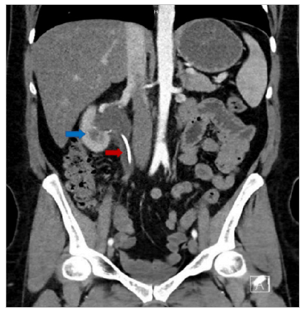
A 32-year-old woman with a history of pelvic radiation for cervical cancer presented three years later with right flank pain, pyelonephritis, and microscopic hematuria. Computed tomography demonstrated a 6mm right mid-pole stone and hydroureteronephrosis. She was initially treated for pyelonephritis and subsequently underwent cystoscopy with stent placement for a radiation-induced distal ureteral stricture. Repeat imaging confirmed a long-segment, high-grade ureteral stricture with persistent hydroureteronephrosis (Figure 1). Despite multiple endoscopic interventions, including balloon dilation and repeat stenting, the stricture remained refractory. Reconstructive urology determined that the lesion was not suitable for standard ureteral reconstruction, including Boari flap or psoas hitch, because of its length and proximal extent. Given the patient’s young age, salvageable kidney, and need for definitive treatment, the case was discussed in a multidisciplinary setting and RKAT was selected as a kidney-preserving strategy.
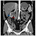
Coronal contrast-enhanced CT scan of the abdomen and pelvis demonstrating right-sided moderate to severe hydronephrosis (blue arrow), and dilation of the proximal right ureter (red arrow).
Positioning: (pre-operative preparations and requirements)
The operation was performed using the DaVinci Xi surgical system (Intuitive Surgical Inc., Sunnyvale, CA, USA). A narrated video of the RKAT operation can be accessed through the following link: https://www.youtube.com/watch?v=ckbeS1G4TV8 . After induction of general anesthesia, the patient was positioned in the left lateral decubitus position with the right side up for the nephrectomy portion. All bony prominences were padded, and intravenous antibiotics were administered within 30 minutes before incision.
Operative technique for right nephrectomy
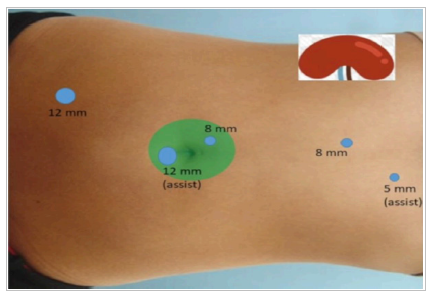
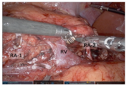
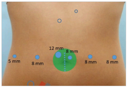
Port placement for the nephrectomy is shown in Figure 2. Initial access was obtained through a 7-cm periumbilical incision, and a GelPort was inserted, through which an 8-mm camera port and a 12-mm assistant port were introduced. Two additional robotic ports (an 8-mm port in the right subcostal area and a 12-mm port in the right lower abdomen along the mid-clavicular line) were placed, and the robot was docked. An additional 5-mm laparoscopic port was placed in the epigastric midline area, through which a locking laparoscopic grasper was inserted and used to retract the right lobe of the liver to provide exposure. The right colon was mobilized along the avascular plane, exposing the duodenum. The duodenum was then partially kocherized, exposing the inferior vena cava (IVC) and the right renal vein. The dilated right ureter was identified, circumferentially controlled, and mobilized down to the area of the stricture. The renal vessels were then dissected, and circumferential control was obtained of the right renal vein and the two renal arteries (Figure 3). The kidney was mobilized by dissecting it off the adrenal gland and taking down the posterior attachments until it was completely freed and only attached through the vessels and ureter. The ureter was incised above the stricture, and the indwelling stent was removed. The video shows the technique for using a robotic stapler to divide the renal artery, followed by the renal vein, completing the nephrectomy. The patient was administered 40 mg of Furosemide, 12.5 g of mannitol, and 2,000 units of heparin prior to dividing the vessels, as per institutional guidelines for living donor nephrectomy. The kidney was removed through the GelPort and placed in an ice basin for back-table flushing and reconstruction.
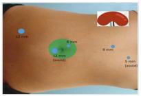
Picture of the port placement for right nephrectomy. Two 8 mm ports are inserted in the left subcostal and periumbilical areas. A 12 mm port is inserted in the left lower abdomen and another assist 12mm port in the periumbilical area. An extra 5 mm port is placed in the epigastric region for assisting with liver retraction.
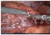
View of right renal vein and two renal arteries just before removal of the native kidney. RA-1 = first renal artery, RA-2 = second renal artery, RV = renal vein.
Back-table preparation and vascular reconstruction:
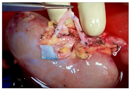
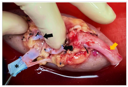
After removal, the kidney is placed in an ice basin. The staple lines on the vessels are excised, and the kidney is flushed with preservative solution (Histidine-Tryptophan-Ketoglutarate solution-HTK). In this case, there were two renal arteries, which were reconstructed on the back table by suturing the medial walls in a fish-mouth technique (Figure 4). The renal vein was also quite short, as this was the right kidney, and the decision was made to extend its length using a conduit fashioned from bovine pericardium (Figure 5). The orientation of the vessels was then marked, and the kidney was wrapped in a piece of gauze to facilitate positioning inside the body.
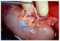
Back table picture of the kidney showing the two renal arteries after fish-mouth reconstruction.
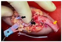
Back table picture of the kidney showing the reconstructed renal vein with tabularized bovine pericardium extension graft (black *). The two renal arteries (black arrows) and the short ureter (yellow arrow) can be seen as well.
Implantation:
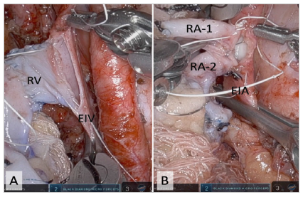
The patient was positioned supine with approximately 10 degrees of Trendelenburg. An 8-mm camera port and a 12-mm assistant port were placed in the peri-umbilical GelPort, and three additional 8-mm ports were placed transversely across the abdomen, along with a lateral 5-mm assistant port. Port placement can be seen in Figure 6. As the first step of the implantation operation, the peritoneum overlying the right external iliac vessels was incised, starting approximately 2 cm inferior to the deep inguinal ring and extending medially and laterally creating flaps superiorly and inferiorly. Approximately 200 cc of fluid was instilled into the urinary bladder for orientation. The external iliac artery and vein were dissected circumferentially using a combination of monopolar cautery and a vessel sealer device, proximally from just distal to the bifurcation of the common iliac artery to just before the level of the crossing circumflex iliac vein. This ensured adequate length for an unhindered vascular anastomosis. When ready for implantation, the external iliac vein was clamped proximally and distally using Scanlan® Reliance 25 mm straight bulldog clamps, and the kidney was placed intraperitoneally through the GelPort with the vessel orientation clearly marked. The vascular anastomosis technique is demonstrated in the accompanying video. An anterior venotomy was created in the external iliac vein using robotic scissors, and an end-to-side anastomosis was performed between the renal vein and the external iliac vein using 6-0 Gore-Tex suture (Figure 7).
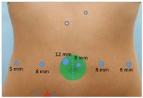
Picture of the port placement for the implantation phase. This includes an 8 mm port and a 12 mm assistant port in the GelPort. Additionally, two 8 mm ports are placed in the left abdomen in a straight line and an 8 mm port, and a 5 mm port are placed on the right abdomen.
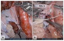
Views of A) venous and B) arterial anastomoses being completed. RA = renal artery, RV = renal vein, EIV = external iliac vein, EIA = external iliac artery.
Upon completion of the venous anastomosis, a third bulldog clamp was applied to the renal vein, and the clamps on the external iliac vein were removed and applied on the external iliac artery. An arteriotomy was made on the external iliac artery, and the renal anastomosis was completed using the same technique as for the vein anastomosis (Figure 7). After completion of the arterial anastomosis, the kidney was reperfused by removing the clamp on the renal vein, followed by the clamps on the artery. The gauze around the kidney was carefully removed, and duplex ultrasound was performed to ensure adequate flow. The kidney was then retroperitonealized against the anterolateral abdominal wall, and the peritoneal flaps were loosely approximated around the kidney using robotic clips. A small peritoneal window was left medially to protect against lymphocele formation.
Lastly, the bladder anastomosis was performed by distending the bladder and making a small opening in its anterolateral aspect. A neo-cysto-ureterostomy was performed using absorbable suture (5-0 Vicryl or 5-0 PDS), beginning at the ‘heel’ of the ureter and proceeding toward the toe end (continuous suture). A ureteral stent was placed across the anastomosis prior to its completion. In this patient, the ureter was relatively short but came together without excessive tension. A single drain was left adjacent to the ureter anastomosis, with the end brought out through a lateral port site. The decision to leave a drain is made on a case-by-case basis; most patients do not require one. The robot was then undocked and the ports removed. The periumbilical fascial incision was closed with #1 PDS suture, and the skin approximated using absorbable monofilament suture.
Operative (console) times for nephrectomy and implantation were 99 minutes and 228 minutes, respectively. The first warm ischemia time (stapling of renal artery to placement of kidney on ice) was 3 minutes. The cold ischemia time (time from when kidney placed on ice after extraction to when taken off ice for implantation) was 3 hours and included time for extracorporeal vascular reconstruction. The second warm ischemia time (from removal of the kidney from ice to reperfusion) was 36 minutes; 5 minutes for positioning, 18 minutes for venous anastomosis, and 13 minutes for arterial anastomosis.
Risk and Safety Assessment
Robotic kidney autotransplantation should be reserved for carefully selected patients with a salvageable kidney and pathology not amenable to simpler reconstructive alternatives. In the present case, standard ureteral reconstruction was not feasible because of the length and proximal extent of the stricture. Extracorporeal graft preparation was chosen to permit controlled back-table arterial reconstruction and extension of the short right renal vein before implantation. After reperfusion, duplex ultrasound was used to confirm satisfactory graft flow, and the graft was retroperitonealized with a small peritoneal window left to reduce the risk of postoperative lymphocele formation. A decellularized bovine pericardial patch (PhotoFix®, CryoLife Inc.) was selected for reconstruction because it offered a biologic option for vascular reconstruction of the short right renal vein. The conventional approach of using donor iliac vein to reconstruct the right renal vein in deceased donor kidney transplantation was not feasible in this case because this was an autotransplant, and the patient would not be receiving immunosuppressive therapy. Alternative biologic options included autologous femoral vein or internal jugular vein, although these would have increased procedural morbidity, or cryopreserved vein, which requires additional preparation time. A prosthetic graft, such as PTFE or Gore-Tex, could also have been considered; however, these materials are generally more thrombogenic than biologic alternatives. The patient was not anticoagulated for implantation and was maintained postoperatively only on low-dose antiplatelet therapy with aspirin 81 mg daily for 3 months. Postoperative systemic anticoagulation was not used because bovine pericardium has been associated with a low risk of thrombotic complications in vascular reconstruction and does not routinely require long-term anticoagulation. [15]. Given the complexity of robotic nephrectomy, vascular reconstruction, and implantation, this approach should be undertaken by surgeons and centers with appropriate experience.
Initial Experience
The postoperative course was uncomplicated. Duplex ultrasound on postoperative day 1 demonstrated good graft perfusion. The patient was discharged on postoperative day 3 with preserved renal function; serum creatinine at discharge was 1.15 mg/dL. The Foley catheter and drain were removed on postoperative day 3, and the ureteral stent was removed later in the outpatient setting. At 6 months and 1 year postoperative follow-up, the patient remained free of complications, with stable renal function and no evidence of vascular or ureteral compromise on follow-up imaging.
DISCUSSION
Kidney autotransplantation remains an important kidney-sparing option for selected patients with complex ureteral or renovascular pathology when endoscopic or in situ reconstruction is not feasible. Robotic kidney autotransplantation has extended the minimally invasive application of this procedure, although published experience remains limited. In this context, the present case contributes practical technical detail rather than comparative outcome evidence.
A key technical lesson from this case is that extracorporeal RKAT may be advantageous when vascular reconstruction is anticipated. Comparative data suggest that intracorporeal RKAT is associated with shorter cold and total ischemia times, whereas longer-term renal functional outcomes appear similar between the two approaches12. When examined against the ERUS RAKT Working Group benchmarks reported by Breda et al.12, our intraoperative metrics fall within or favorably alongside the published ranges. Our first warm ischemia time of 3 minutes is identical to the reported eRAKAT median of 3 minutes (IQR 2-4) and the iRAKAT median of 3 minutes (IQR 2.7-3.2). Our cold ischemia time of 180 minutes is longer than the eRAKAT median of 151 minutes (IQR 125-199), reflecting the additional back-table time required for both fish-mouth reconstruction of dual renal arteries and bovine pericardial extension of the short right renal vein. As expected, our cold ischemia time is substantially longer than the iRAKAT median of 27.5 minutes (IQR 20-55), consistent with the well-described trade-off of the extracorporeal approach. Our rewarming (second warm) ischemia time of 36 minutes was shorter than both the eRAKAT median (44 minutes, IQR 43-52) and the iRAKAT median (46.5 minutes, IQR 22-61). Our operative time of 327 minutes compares favorably with the eRAKAT operative-time median of 360 minutes (IQR 330-400) and falls within the iRAKAT range (median 307 minutes, IQR 287-418). Although a single case cannot establish performance benchmarks, these comparisons suggest that the operative metrics achieved here are within the range expected of experienced robotic transplant programs, even in the setting of complex dual-vessel reconstruction. These findings suggest that the choice between intracorporeal and extracorporeal approaches should be individualized according to anatomy, anticipated reconstructive complexity, and surgical expertise12-14.
This report is limited by its single-patient design and cannot establish comparative effectiveness or reproducibility across centers. Its contribution is instead to illustrate feasibility, initial safety, and the technical considerations that may guide application of this approach in similarly selected cases.
CONCLUSION
Robotic kidney autotransplantation with extracorporeal vascular reconstruction is a feasible minimally invasive option for selected patients with complex ureteral pathology when conventional reconstruction is not possible. This technical note highlights the operative rationale, reconstructive strategy, risk-mitigation measures, and initial experience required for successful execution.
REFERENCES
- 1 Hardy JD. High ureteral injuries. JAMA. 1963;184(2):97.
-
2 Alameddine M, Moghadamyeghaneh Z, Yusufali A, Collazo AM, Jue JS, Zheng I, et al. Kidney autotransplantation: between the past and the future. Curr Urol Rep. 2018;19(3). doi:10.1007/s11934-018-0749-4.
» https://doi.org/10.1007/s11934-018-0749-4. - 3 Azhar B, Patel S, Chadha P, Hakim N. Indications for renal autotransplant: an overview. Exp Clin Transplant. 2015;13(2):109-14.
-
4 Ruiz M, Hevia V, Fabuel JJ, Fernández AA, Gómez V, Burgos FJ. Kidney autotransplantation: long-term outcomes and complications. Experience in a tertiary hospital and literature review. Int Urol Nephrol. 2017;49(11):1929-35. doi:10.1007/s11255-017-1680-1.
» https://doi.org/10.1007/s11255-017-1680-1 -
5 Decaestecker K, Van Parys B, Van Besien J, Doumerc N, Desender L, Randon C, et al. Robot-assisted kidney autotransplantation: a minimally invasive way to salvage kidneys. Eur Urol Focus. 2018;4(2):198-205. doi:10.1016/j.euf.2018.07.019.
» https://doi.org/10.1016/j.euf.2018.07.019 - 6 Moghadamyeghaneh Z, Hanna MH, Fazlalizadeh R, Obi Y, Foster CE, Stamos MJ, et al. A nationwide analysis of kidney autotransplantation. Am Surg. 2017;83(2):162-9.
-
7 Fabrizio MD, Kavoussi LR, Jackman S, Chan DY, Tseng E, Ratner LE. Laparoscopic nephrectomy for autotransplantation. Urology. 2000;55(1):145. doi:10.1016/S0090-4295(99)00367-2.
» https://doi.org/10.1016/S0090-4295(99)00367-2 -
8 Tran G, Ramaswamy K, Chi T, Meng M, Freise C, Stoller ML. Laparoscopic nephrectomy with autotransplantation: safety, efficacy and long-term durability. J Urol. 2015;194(3):738-43. doi:10.1016/j.juro.2015.03.089.
» https://doi.org/10.1016/j.juro.2015.03.089 -
9 Van Praet C, Lambert E, Desender L, Van Parys B, Vanpeteghem C, Decaestecker K. Total intracorporeal robot kidney autotransplantation: case report and description of surgical technique. Front Surg. 2020;7:598961. doi:10.3389/fsurg.2020.00065.
» https://doi.org/10.3389/fsurg.2020.00065 -
10 Doumerc N, Beauval JB, Roumiguié M, Roulette P, Laclergerie F, Sallusto F, et al. Total intracorporeal robotic renal autotransplantation: a new minimally invasive approach to preserve the kidney after major ureteral injuries. Int J Surg Case Rep. 2018;49:176-9. doi:10.1016/j.ijscr.2018.06.017.
» https://doi.org/10.1016/j.ijscr.2018.06.017 -
11 Gordon ZN, Angell J, Abaza R. Completely intracorporeal robotic renal autotransplantation. J Urol. 2014;192(5):1516-22. doi:10.1016/j.juro.2014.02.2589.
» https://doi.org/10.1016/j.juro.2014.02.2589 -
12 Breda A, Diana P, Territo A, Gallioli A, Piana A, Gaya JM, et al. Intracorporeal versus extracorporeal robot-assisted kidney autotransplantation: experience of the ERUS RAKT Working Group. Eur Urol. 2022;81(2):168-75. doi:10.1016/j.eururo.2021.07.023.
» https://doi.org/10.1016/j.eururo.2021.07.023 -
13 Araki M, Wada K, Mitsui Y, Sadahira T, Kubota R, Nishimura S, et al. Robotic renal autotransplantation: first case outside of North America. Acta Med Okayama. 2017;71(4):351-5. doi:10.18926/AMO/55313.
» https://doi.org/10.18926/AMO/55313 -
14 Martin A, Gómez V, Díez Nicolás V, Hevia Palacios V, Fonseca Hernández M, García I, et al. Kidney autotransplantation and orthotopic kidney transplantation: two different approaches for complex cases. Case Rep Urol. 2022;2022:1-13. doi:10.1155/2022/9299397.
» https://doi.org/10.1155/2022/9299397 -
15 Morris PD, Furtado R, Pulitano C, Crawford M, Yeo D, Vasilaras A, et al. Inferior Vena Cava Resection and Reconstruction with Bovine Pericardium for Renal Cell Carcinoma: Complications and Outcomes.Urology. 2019;134:143-147. doi:10.1016/j.urology.2019.09.006.
» https://doi.org/10.1016/j.urology.2019.09.006