Choledocholithiasis associated with hepatolithiasis in a horse - case report
ABSTRACT
Choledocholithiasis is rare in animals and in horses, it is an uncommon cause of biliary obstruction and liver disease (prevalence of 0.08%). This study reports the choledocholithiasis associated with hepatolithiasis in a Quarter Horse stallion was referred to the Veterinary Hospital of the University of Franca (UNIFRAN) presenting with apathy, icteric mucous membranes, dark urine, and hypocholic feces, dehydration, hypoglycemia, congested mucous membranes with an endotoxemic halo, and motor incoordination. Despite treatment, the neurological condition worsened and the animal died and underwent necropsy. The liver showed hepatomegaly, brownish, a distinct lobular pattern, and a firm consistency. The bile ducts dilated, with pasty contents and yellowish lumps. The common bile duct region was enlarged, and upon cut, a calculus responsible for the obstruction of the intrahepatic ducts. Microscopically, foci of necrosis in the liver, surrounded by inflammatory infiltrate, with extensive areas fibrosis; also, intense ductal proliferation, biliary cholestasis, and bacterial colonies in the necrotic areas, suggesting choledocholithiasis associated with hepatolithiasis. Although rare, cholelithiasis in horses should be considered in the differential diagnosis of cases with liver abnormalities, and neurological consistent with hepatic encephalopathy. The post mortem confirmation reinforces the importance of autopsy as an essential tool for diagnosis.
Key words:
hepatic encephalopathy; cholestasis in horses; liver pathology; post mortem diagnosis
RESUMO
A coledocolitíase é rara em animais e, em equinos, constitui uma causa incomum de obstrução biliar e doença hepática (prevalência de 0,08%). Este estudo relata um caso de coledocolitíase associada à hepatolitíase em um garanhão da raça Quarto de Milha, encaminhado ao Hospital Veterinário da Universidade de Franca (UNIFRAN), apresentando apatia, mucosas ictéricas, urina escura e fezes hipocólicas, desidratação, hipoglicemia, mucosas congestas com halo endotoxêmico e incoordenação motora. Apesar do tratamento, o quadro neurológico se agravou, e o animal veio a óbito, sendo submetido à necropsia. O fígado apresentava hepatomegalia, coloração acastanhada, padrão lobular evidente e consistência firme. Os ductos biliares estavam dilatados, com conteúdo pastoso e grumos amarelados. A região do ducto biliar comum encontrava-se aumentada e, ao corte, observou-se um cálculo responsável pela obstrução dos ductos intra-hepáticos. Microscopicamente, foram observados focos de necrose hepática, circundados por infiltrado inflamatório, com extensas áreas de fibrose; além disso, havia intensa proliferação ductal, colestase biliar e colônias bacterianas nas áreas necróticas, sugerindo coledocolitíase associada à hepatolitíase. Embora rara, a colelitíase em equinos deve ser considerada no diagnóstico diferencial de casos com alterações hepáticas e sinais neurológicos compatíveis com encefalopatia hepática. A confirmação pós-morte reforça a importância da necropsia como ferramenta essencial para o diagnóstico.
Palavras-chave:
encefalopatia hepática; colestase em equinos; patologia hepática; diagnóstico post-mortem
INTRODUCTION
Cholelithiasis is a rare condition in domestic animals, characterized by the formation of stones in the biliary system (Santos et al., 2007). In this regard, when stones form in the extrahepatic bile ducts, the condition is called cholelithiasis; in cases where they occur in the common bile duct or intrahepatic ducts, the manifestations are called choledocholithiasis and hepatolithiasis, respectively (Bianchi et al., 2017; Reed et al., 2021). The obstruction of bile flow caused by these stones can trigger a series of liver disorders, with variable clinical impacts depending on the severity and time of evolution (Linn-Peirano et al., 2023).
In horses, choledocholithiasis is an uncommon cause of biliary obstruction and liver disease, with an estimated prevalence of only 0.08% (Schneider, 1997; Santos et al., 2007). Diagnosis is established by abdominal ultrasonography or exploratory laparotomy, although it can also be performed by endoscopy, depending on the location and accessibility of the affected biliary structures (Reef et al., 1990; Carr et al., 2004). The presence of gallstones can also be a postmortem finding (Silva et al., 2014).
Choledocholithiasis most frequently affects middle-aged to older horses, with no predilection for sex or breed (Peek and Divers, 2000; Santos et al., 2007; Reed et al., 2021). Clinical manifestations are varied and nonspecific, including apathy, loose stools or diarrhea, abdominal discomfort, loss of appetite, weight loss, fever, jaundice, dehydration, colic, and signs of liver failure, which may progress to hepatic encephalopathy (Peek and Divers, 2000).
The etiopathogenesis of choledocholithiasis in horses is not yet fully understood (Reed et al., 2021); however, among the main hypotheses described in the scientific literature, the formation of stones from the deposition of cellular and solid materials that serve as a matrix for the crystallization of biliary components stands out (Silva et al., 2014). Predisposing factors include biliary stasis, ascending infection of enteric origin (often caused by gram-negative bacteria isolated in biopsy cultures or necropsies), ascariasis and, more rarely, the presence of foreign bodies (Johnston et al., 1989; Gerros et al., 1993; Peek and Divers, 2000; Linn-Peirano et al., 2023).
Treatment of equine choledocholithiasis involves supportive measures such as intravenous fluid therapy and anti-inflammatory drugs, combined with broad-spectrum systemic antimicrobials. The therapeutic protocol should consider efficacy against Gram-negative bacteria and enteric anaerobes, with prolonged therapy (Peek and Divers, 2000). In selected cases, especially when one or a few choleliths obstruct the bile ducts and hepatic fibrosis is still limited, surgical removal of these choleliths may be indicated, considered an effective approach, although with a poor prognosis (Reed et al., 2021).
The prognosis of choledocholithiasis is worsened by the presence of hepatic fibrosis, the severity of clinical signs, and the location and number of choleliths. The outcome can range from poor to guarded when there are multiple choleliths with concomitant extensive or bridging fibrosis, signs of liver failure, and hepatic encephalopathy (Silva et al., 2014; Bianchi et al., 2017; Reed et al., 2021).
Considering the low prevalence of choledocholithiasis in horses, especially in Brazil, combined with the absence of other scientific studies relating the diagnostic and therapeutic challenges associated, the present study aimed to report a case of choledocholithiasis and hepatolithiasis in this animal species, with consequent chronic bacterial suppurative cholangiohepatitis and hepatic encephalopathy.
CASE REPORT
An eight-year-old Quarter Horse stallion weighing 491kg was referred to the University of Franca Veterinary Hospital (UNIFRAN, Franca, SP) with apathy, jaundiced mucous membranes, dark urine, and discolored feces.
During the physical evaluation, dehydration, increased capillary refill time, hypoglycemia, jaundiced and congested mucous membranes, specifically the oral mucosa, were detected. The mucous membranes exhibited an endotoxemic halo, low body condition score, and mild motor incoordination.
Laboratory tests showed increased red blood cell (RBC), hemoglobin, hematocrit, and fibrinogen levels, in addition to the presence of neutrophilic leukocytosis. Biological markers showed severe increases in alkaline phosphatase (AP), total bilirubin (TB), direct bilirubin (DB), indirect bilirubin (BI), and aspartate aminotransferase (AST); and a slight increase in urea nitrogen and gamma-glutyltransferase (GGT). Creatine kinase (CK) and lactate levels were within normal values for the species. Analysis of spontaneously collected urine showed no significant changes. Coproparasitological examinations were performed using the Willis method and the eggs per gram of feces (EPG) method, with fresh samples collected from the rectum. However, no parasite eggs, cysts, or oocysts were observed.
Two days after the patient's admission, new tests were performed, revealing neutrophilic leukocytosis, the presence of lymphoblasts, and discrete Döhle bodies, associated with persistent elevations in GGT, ALP, AST, BT, BD, and BI, and a slight increase in urea nitrogen. Urinalysis revealed a decrease in urine specific gravity (from 1.022 to 1.014).
Thoracic and abdominal ultrasounds were performed, revealing pericardial effusion, a heterogeneous hypoechoic liver, and duodenal adhesions, respectively.
Clinical treatment was instituted with anti-inflammatory drugs (flunixin meglumine: 1.1mg/kg, intravenously, once a day, for three days and dimethyl sulfoxide: 1000 mg/kg, diluted in Ringer's solution, intravenously, every 24 hours, for three days), liver protectors (Mercepton®: 100mL/animal, intravenously, every 12 hours, diluted in Ringer's solution with lactate, for seven days and Ornitil®: 30mL/100kg, intravenously, every 24 hours, diluted in Ringer's solution with lactate, for seven days). In addition, gastric protectors (omeprazole: 18mg/kg, orally, every 24 hours, for seven days, ranitidine: 2mg/kg, intravenously, every 12 hours, for seven days) and antibiotics (sulfadoxine and trimethoprim: 15mg/kg, intravenously, every 12 hours, for five days, metronidazole: 15mg/kg, intravenously, every 12 hours, for three days) and polymyxin B: 4000IU/kg, intravenously, every 12 hours, diluted in lactated Ringer's solution, for two days. Multivitamin (intravenous, every 24 hours, for six days), vitamin C (20mL/animal, intravenously, every 24 hours, for seven days) and Potenay® (1mL/25kg, intramuscularly, every 3 days) were also administered. In addition to these medications, electrolytes (1 tube, orally, every 24 hours, for seven days), probiotics (half a tube, orally, every 24 hours, for seven days) and furosemide (0.5mg/kg, intravenously, every 24 hours, for three days) were also used as support measures.
Despite the therapeutic measures implemented, the patient's condition worsened and neurological manifestations worsened, consistent with hepatic encephalopathy, including seizures, head pressing against the stall wall, colic, jaundice, and weight loss.
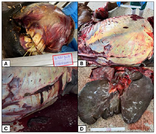
After seven days of hospitalization, the patient died, and an autopsy was performed immediately. Pathological findings included congested oral mucosa with pale areas, as well as ulcerations on the maxillary and mandibular lips (Figure 1A), icteric subcutaneous tissue (Figure 1B), free thoracic fluid with a cloudy, hemorrhagic appearance, and free abdominal fluid that was cloudy, ranging from icteric to hemorrhagic, with fibrin and clots (Figure 1C).
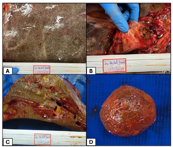
The liver was enlarged (Figure 1D), with an evident lobular pattern and brownish coloration (Figure 2A), with firm parenchyma on cut, with enlarged bile ducts, draining pasty content with formation of firm yellowish lumps (Figure 2C). The common bile duct region was enlarged (Figure 2B) and on cut with a yellowish to brownish calculus (Figure 2D), measuring 5.0x3.0 cm, with a pasty, friable surface, obstructing the intrahepatic ducts.
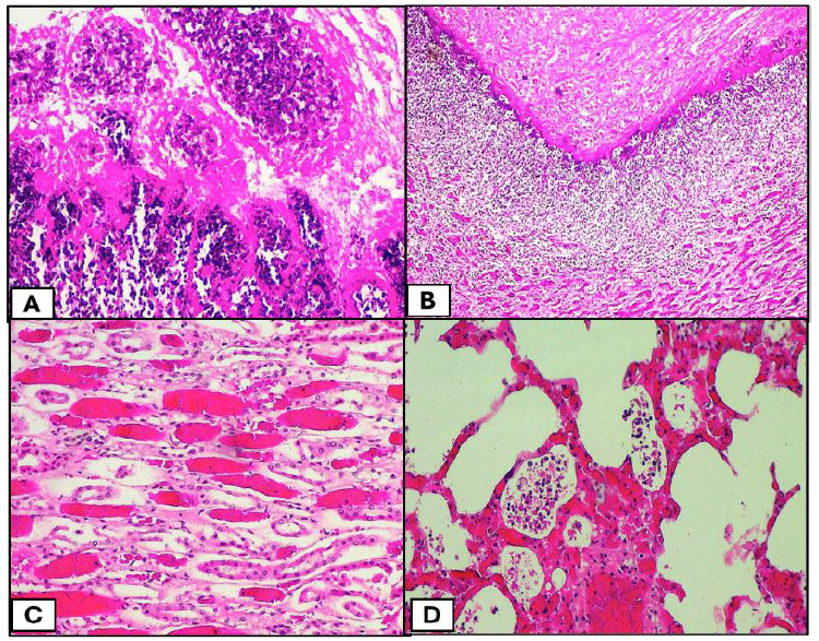
Fragments of liver, kidney, and lung parenchyma were fixed in 10% buffered formalin for subsequent histopathological analysis using hematoxylin and eosin staining. Microscopically, multiple foci of necrosis were observed in the liver, surrounded by an intense inflammatory infiltrate composed of neutrophils and macrophages, with extensive areas of surrounding fibrosis (Figure 3A). Intense ductal proliferation, biliary cholestasis, and bacterial colonies were also noted in areas of necrosis (Figure 3B). The kidneys showed moderate and diffuse vacuolar degeneration of the renal tubules, with intense congestion (Figure 3C). The lungs showed areas of emphysema and intense and diffuse congestion (Figure 3D).
Photographic images of a horse necropsy, demonstrating congested oral mucosa with pale areas and lip ulceration (A); jaundice of subcutaneous tissue (B); cloudy abdominal free fluid, ranging from icteric to hemorrhagic, with fibrin and clots (C) and an enlarged liver (D).
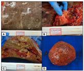
Photographic images of a horse necropsy, demonstrating a liver with an evident lobular pattern and brownish coloration (A); an enlarged common bile duct region (B); presence of lumps in cholestatic vessels (C) and a calculus that was present in the common bile duct region (D).
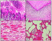
Organ fragments from a horse with choledocholithiasis associated with hepatolithiasis. (A) Liver showing multiple foci of necrosis surrounded by an intense inflammatory infiltrate consisting of neutrophils and macrophages, in addition to extensive areas of periportal fibrosis. Obj. 10x, HE. (B) Marked ductal proliferation, biliary cholestasis, and the presence of bacterial colonies in areas of necrosis. Obj. 4x, HE. (C) Kidney showing diffuse vacuolar degeneration of the renal tubules and intense congestion. Obj. 20x, HE. (D) Lung showing areas of emphysema and diffuse congestion. Staining: hematoxylin and eosin. Obj. 20x, HE.
DISCUSSION
The clinicopathological, histopathological, ultrasonographic, and necroscopic findings of the current horse reported with choledocholithiasis and hepatolithiasis, with consequent chronic bacterial suppurative cholangiohepatitis and hepatic encephalopathy, corroborated exactly those described in other studies in the scientific literature (Santos et al., 2007; Reed et al., 2021). In contrast, regarding clinical signs, Johnston et al. (1989) and Peak and Divers (2000) described that, in cases of choledocholithiasis, colic is frequent, concomitant with fever, jaundice, weight loss, and hepatic encephalopathy, symptoms detected in the patient in the present report, except for fever.
As there is no predisposition by breed or sex, the animal's age is the only relevant risk factor for the development of choledocholithiasis in horses, with middle-aged to elderly animals being the most predisposed (Peek and Divers, 2000; Santos et al., 2007; Reed et al., 2021), thus disagreeing with the described patient who was eight years old. However, this age coincides with the statements by Johnston et al. (1989) that the average age correlated with the condition is nine years. On the other hand, there are reports of young animals affected between the ages of two and six years (Van Der Luer and Kroneman, 1982; Silva et al., 2014; Linn-Peirano et al., 2023).
Regarding laboratory findings related to choledocholithiasis, with the exception of CK and lactate, which remained within the reference values for the species in all evaluations, the other biological markers were persistently and markedly increased, reinforcing the descriptions of Traub et al. (1982), Silva et al. (2014), and Bianchi et al. (2017) that they are directly linked to biliary obstruction and hepatocyte injury. The persistent increase in liver enzymes reflects chronic cholestasis but does not allow differentiation between inflammatory, infectious, or obstructive causes. Therefore, diagnostic confirmation often occurs only post mortem, as in the case described, reinforcing the need for more sensitive complementary investigations, such as liver biopsies or cholangiograms (Reed et al., 2021).
Early clinical diagnosis is hampered by the nonspecificity of signs and limitations of imaging methods, as ultrasound may not detect smaller stones or those located deep in the intrahepatic ducts (Bianchi et al., 2017).
The observed therapeutic failure, despite the adoption of intensive clinical support, highlights the progressive nature and poor prognosis of this condition, especially when associated with suppurative cholangiohepatitis and extensive fibrosis (Peek and Divers, 2000).
The necroscopic findings are mainly located on the hepatobiliary system, in which the liver presents a brownish to greenish capsular surface, firmness to the cut, hepatomegaly, evident lobular pattern, presence of hepatoliths and choledocholiths, dilated bile ducts, in addition to jaundice found in the carcass (Holland et al., 1991; Linn-Peirano et al., 2023) which are compatible with those found in the case discussed. However, Santos et al. (2007) and Bianchi et al. (2017) documented the presence of macroscopic atrophy of the right lateral hepatic lobe in size, but such alteration was not found in the animal in this study.
CONCLUSIONS
Choledocholithiasis in horses, although rare, should be considered in the differential diagnosis of jaundice, liver alterations, and neurological manifestations consistent with hepatic encephalopathy. The reported case highlights the diagnostic and therapeutic complexity of this condition, highlighting that, even with early implementation of supportive measures and antimicrobial treatment, the prognosis remains guarded, especially when advanced liver fibrosis and associated suppurative cholangiohepatitis are present. Post mortem confirmation reinforces the importance of necropsy as an indispensable tool for definitive diagnosis, since clinical and laboratory signs are nonspecific and may overlap with other liver diseases. Therefore, the description of cases such as this one contributes significantly to improving knowledge about the pathophysiology, clinical evolution, and outcomes of choledocholithiasis and hepatolithiasis in horses, providing valuable information for early diagnosis and more assertive therapeutic management in future cases.
REFERENCES
- BIANCHI, M.V.; MELLO, L.S.; LORENZETT, M.P. et al. Secondary biliary hepatic fibrosis caused by cholelithiasis in two horses. J. Equine Vet. Sci., v.56, p.52-55, 2017.
- CARR, E.A.; CARON, J.P.; PERONI, J. Endoscopic diagnosis of choledocholithiasis of the common bile duct in a horse. Equine Vet. Educ., v.16, p.68-71, 2004.
- GERROS, T.C.; MCGUIRK, S.M.; BILLER, D.S. et al. Choledocholithiasis attributable to a foreign body in a horse. J. Am. Vet. Med. Assoc., v. 202, p.301-303, 1993.
- HOLLAND, P.S.; SCHMITZ, D.G.; READ, W.K. Hepatolithiasis in an Arabian Mare. Equine Vet. J., v.23, p.229-232, 1991.
- JOHNSTON, J.K.; DIVERS, T.J.; REEF, V.B.; ACLAND, H. Cholelithiasis in horses: ten cases (1982-1986). J. Am. Vet. Med. Assoc., v.194, p.405-409, 1989.
- LINN-PEIRANO, S.C.; HEPWORTH-WARREN, K.; KINSELLA, H. et al. Ingesta-associated choledocholithiasis in horses: 2 cases and literature review. J. Vet. Diagn. Invest., v.35, p.418-425, 2023.
- PEEK, S.F.; DIVERS, T.J. Medical treatment of cholangiohepatitis and cholelithiasis in mature horses: 9 cases (1991-1998). Equine Vet. J., v.32, p.301-306, 2000.
- REED, S.M.; BAYLY, W.M.; SELLON, D.C. Medicina interna equina. 4.ed. Rio de Janeiro: Guanabara Koogan, 2021. 897p.
- REEF, V.B.; JOHNSTON, J.K.; DIVERS, T.J.; ACLAND, H. Ultrasonographic findings in horses with cholelithiasis: eight cases (1985-1987). J. Am. Vet. Med. Assoc., v.196, p.1836-1840, 1990.
- SANTOS, R.L.; OLIVEIRA, T.F.B.; OLIVEIRA, T.S. et al. Cholelithiasis with atrophy of the right lateral hepatic lobe in a horse. Cienc. Rural, v.37, p.586-589, 2007.
- SCHNEIDER, D.A. Cholestasis and biliary calculi in horses. Compend. Contin. Educ. Pract. Vet., v.19, p.744-754, 1997.
- SILVA, V.C.M.; MAGALHÃES, J.; ECCO, R. et al. Pathological findings of cholelithiasis in two horses. Braz. J. Vet. Pathol., v.7, p.35-37, 2014.
- TRAUB, J.L.; RANTANEN, N.; REED, S.; SCHECTER, L. Cholelithiasis in four horses. J. Am. Vet. Med. Assoc., v.181, p.59-62, 1982.
- VAN DER LUER, R.J.T.; KRONEMAN, J. Three cases of cholelithiasis and biliary fibrosis in the horse. Equine Vet. J., v.14, p.251-253, 1982.
Data-in-article.