Tracheal obstruction due to neuroendocrine carcinoma: case report in a dog
ABSTRACT
Anamnesis. Female, mixed-breed, intact, 13 years and six months old, weighing 7.3kg. Consult motive: acute respiratory distress. Clinical, radiographic, and endoscopic findings. The patient refused to walk, was in an orthopneic position, and presented respiratory distress characterized by inspiratory dyspnea, audible stridor, syncope, cyanosis and muscle weakness. Radiological findings revealed a soft tissue density, with a rounded appearance in the cervical trachea, occupying almost the entire lumen and measuring 1.1cmx1.19cm. Endoscopy confirmed the presence of a rounded, deep pink mass, apparently mucosal, occupying approximately 95% of the lumen. Surgical approach. Video-endoscopy was used for its removal, using a 5mm Maryland ligasure vessel-sealing forceps. Definitive diagnosis. Histopathology revealed presence of neuroendocrine carcinoma. Conclusions: This is one of the most aggressive tumors in humans, and in veterinary medicine there are few publications on its presentation at the level of the trachea in canines, which makes its dissemination important.
Keywords:
tracheal obstruction; carcinomas; neuroendocrine carcinoma (Source: DeCS/MeSH)
RESUMO
Histórico: cadela sem raça definida, de 13 anos, inteira, pesando 7,3kg. Motivo da consulta: desconforto respiratório agudo. Achados clínicos, radiográficos e endoscópicos: a paciente recusava-se a deambular, encontrava-se em posição ortopneica e apresentava desconforto respiratório caracterizado por dispneia inspiratória, estridor audível, síncope, cianose e fraqueza muscular. O exame radiológico revelou uma densidade de forma arredondada, com densidade de tecidos moles na traqueia cervical ocupando quase todo o lúmen e medindo 1,1cm x 1,19cm. A endoscopia confirmou a presença de um nódulo rosa-escuro, aparentemente mucoso, ocupando aproximadamente 95% do lúmen. Abordagem cirúrgica. A neoplasia foi removida por videoendoscopia com pinça de selamento vascular tipo Maryland de 5mm. Diagnóstico definitivo: a histopatologia revelou a presença de carcinoma neuroendócrino. Conclusões: trata-se de um dos tumores mais agressivos relatados em humanos, havendo poucas publicações na medicina veterinária sobre sua apresentação na traqueia canina, tornando sua disseminação importante.
Palavras-chave:
obstrução traqueal; carcinomas; carcinoma neuroendócrino
INTRODUCTION
Neuroendocrine tumors (NET) are a heterogeneous group of neoplasms originating in neuroendocrine cells, which produce monoamines, peptides, and other biologically active substances widely distributed throughout the body. The study of NETs has a history dating back more than a century. In 1888, Lubarsch was one of the pioneers in describing multiple tumors in the ileum during an autopsy; shortly thereafter, in 1907, Oberndorfer coined the term carcinoid to describe a group of tumors with less aggressive behavior compared to adenocarcinomas. In the 1950s, the first reports of characteristic clinical manifestations associated with carcinoid tumors emerged, which were later named carcinoid syndrome (CS) (Fierro M.L.F. et al. 2024, Carrillo D. et al. 2021).
Carcinoid syndrome is caused by the excessive secretion of serotonin and other biologically active compounds by the tumor, including tachykinin and histamine (Bednarczuk T. et al. 2022).
Carcinoid tumors are tumors of the neuroendocrine cells of the digestive tract, although they can also appear in the pancreas, lungs, bile ducts, and liver. Due to their origin in endocrine cells, many produce bioactive products such as amines or peptides (Feldman M. et al. 2015, Mondragón J. 2016).
Neuroendocrine tumors constitute a heterogeneous spectrum of diseases, so there is no ideal marker for their immunohistochemical diagnosis. One of the most widely used is chromogranin A (CgA), a protein of the granin family secreted by neuroendocrine cells together with other hormones and neurotransmitters, whose secretion can persist even when the neuroendocrine cell undergoes neoplastic transformation (Carrillo D. et al. 2021, Bednarczuk T et al. 2022, Feldman M et al. 2015, Clement D et al. 2020).
Primary tumors of the trachea are extremely rare. As a result, there are no large series studies. The truth is that, based on published data, tracheal tumors account for less than 0.1% of all neoplasms, and for every malignant tumor of the trachea there are 20 cases of laryngeal cancer and 160 to 180 cases of lung cancer (Ferrer E. 2017, Bataller L. et al. 2017).
In humans, tracheal carcinomas are rare, with fewer than 20 cases reported in Chinese literature and fewer than 15 cases reported in English literature (Bataller L. et al. 2017, Mukherji A. et al. 2011, Brown M. 2003).
The incidence of tracheal tumors in companion animals is unknown, but in human medicine, the reported annual incidence ranges from 0.142 to 0.27 per 100,000 people (Piirainen K. et al. 2018, Honings J. et al. 2017, Zachary A. et al. 2020, Torres M. et al. 2020). It is understood that tracheal tumors can often be underestimated for months or years, especially in the case of carcinomas due to their asymptomatic and slow-growing characteristic (Rendon M.E. 2011).
Tracheal tumors reported in companion animals include adenocarcinoma, carcinoma, extramedullary plasmacytoma, leiomyoma, fibrosarcoma, mast cell tumor, rhabdomyosarcoma, and squamous cell carcinoma (Piirainen K. et al. 2018, Honings J. et al. 2017, Torres M. et al. 2020).
A study of eight cases in cats between 1989 and 2014 concluded that tracheal tumors often present with nonspecific upper respiratory signs such as dyspnea, wheezing, exercise intolerance, and coughing (Bataller L. 2017, Honings J. et al. 2017, Kanemoto H. et al. 2023, Howard J. et al. 2017).
Dogs with obstructive tracheal tumors often present clinical signs of inspiratory dyspnea, panting, coughing, auscultatory wheezing, or collapse. A tracheal mass can often be demonstrated on radiographs, as the air in the tracheal lumen provides good contrast. Computed tomography (CT) provides detailed information on the size, exact location, and depth of invasion, and airway endoscopy helps us determine the degree of obstruction and appearance of the masses and allows fine-needle aspiration or biopsies of the masses for cytological and histopathological diagnosis (Mukherji A. et al. 2011, Piirainen K. et al. 2018, Torres M. et al. 2020, Sherani K. et al. 2015).
External neck or cervical ultrasound is used as a complement to identify possible cervical metastases in combination with other studies such as computed tomography and to determine lesions in lymph nodes, thyroid glands, parathyroid glands, esophagus, and to perform staging to help determine therapeutic plans for patients (Blom R. et al. 2012), human studies proposed that neck ultrasound was the method of choice for monitoring patients with head and neck carcinoma metastases, as it was an economical, simple, and viable method compared to computed tomography or magnetic resonance imaging for the detection of these metastases (19) (Jiang H. et al. 2021).
In humans, benign and malignant primary tumors of the trachea are usually treated in most cases with tracheal resection, endoscopic resection using laser surgery, and radiotherapy (Torres M. et al. 2020). It has been reported that surgery can cure benign and low-grade malignant tumors, achieve long-term survival in tracheal carcinomas, provide pathological confirmation of complete tumor removal, and permanently relieve airway obstruction (Mukherji A. et al. 2011). It should be noted that this case report is of a tracheal neuroendocrine carcinoma that was removed by airway endoscopy and recurred two months after surgery.
Medical history. A 13.6-year-old mixed-breed female dog named Briseida, weighing 7.3 kg, was brought to the Mascoclínica Veterinary Clinic in Cartagena, Colombia, with severe dyspnea, wheezing, and coughing that had been present for several months.
Clinical examination findings. An emergency physical examination was performed, revealing a patient who refused to walk, with orthopneic position, respiratory distress characterized by inspiratory dyspnea, audible stridor, syncope, muscle weakness, and cyanosis.
Differential diagnosis. Collapse and/or laryngeal paralysis, tracheal foreign body, and tracheal tumor were suspected.
Laboratory tests. Paraclinical tests were performed on blood count, ALT, AST, GGT, albumin, globulin, BUN, urea, and creatinine, with results within normal ranges.
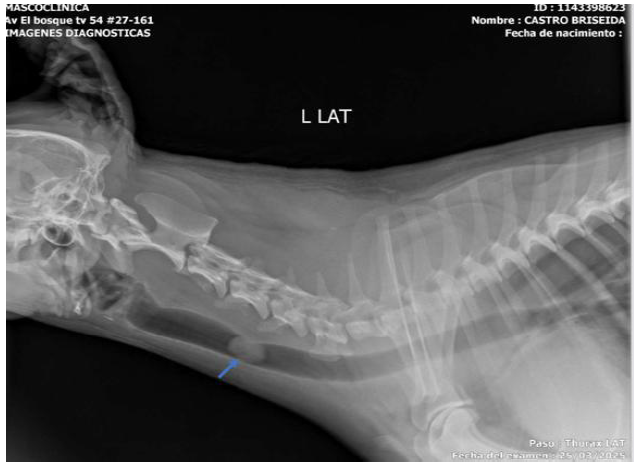
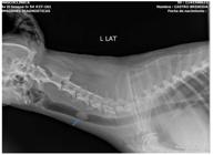
The right lateral cervical digital radiographic study shows a neoformation with increased soft tissue radiopacity, rounded in shape, occupying almost the entire tracheal lumen (Figure 1). The ventral-dorsal view shows a change in density in the vertebral body due to the sum of densities corresponding to nodular density, added to the density of the vertebral body. Therefore, the lateral view was considered diagnostic.
Neck X-ray with right lateral projection in a mixed-breed dog, showing a neoformation with increased soft tissue radiopacity, rounded in shape, measuring 1.1cm high x 1.19cm long and occupying almost the entire lumen of the cervical trachea in its middle portion.
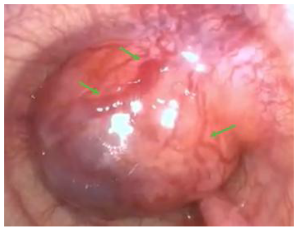
Airway endoscopy revealed a rounded, non-ulcerated, bright pink, vascularized neoplasm, apparently implanted in the mucosa, with a broad pedicle extending into the lumen. In addition, dark areas were observed on their free surface (Figure 2).
Anesthetic protocol. For pre-anesthesia, a combination of acepromazine (0.3mg/kg body weight) + tramadol (4mg/kg body weight) was administered intravenously. Anesthesia was achieved with the intravenous administration of a bolus consisting of ketamine (7 mg/kg) + propofol (6mg/kg) at an effective dose.
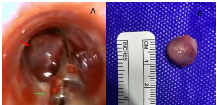
Description of the endoscopic removal technique. An airway endoscopy is performed, accompanied by a 5mm Maryland ligasure vessel sealing clamp, which is used to clamp, hemostasis, and cut the base of the nodule for its removal (Figures 3A and 3B).
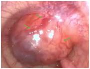
Upper respiratory tract endoscopy revealed a rounded, non-ulcerated, bright pink, vascularized neo-formation that occupied almost the entire lumen of the cervical trachea in its middle portion (green arrows) in a 13-year-old, intact, mixed-breed dog weighing 7.3kg.
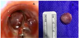
Clamping and cutting of the endotracheal mass (red arrow) with a 5mm Maryland ligasure® vessel sealing clip (green arrow) guided with a flexible videoendoscope (figure 3A) for subsequent removal and measurement (Figure 3B), in a 13-year-old, intact, mixed-breed dog weighing 7.3kg.
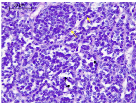
Definitive diagnosis (histopathology and immunohistochemistry). Microscopic findings of the tumor (Figure 4) in the histological sections analyzed show a poorly differentiated infiltrating epithelial neoplasm. It is distributed underlying even stratified and respiratory epithelium. Low polyhedral to cubic epithelial cells are observed, distributed in tubules or acini containing remnants of sparse eosinophilic material and sometimes erythrocytes, occasionally forming solid trabeculae supported by a thin fibrovascular stroma. The neoplastic cells have sparse amphophilic cytoplasm and ovoid nuclei with moderate anisokaryosis and hyperchromatism, mild karyomegaly, an inconspicuous nucleolus and a count of up to 27 mitoses in 2.37mm2, resulting in carcinoma.
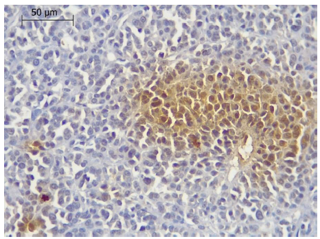
Immunohistochemistry was performed to determine and specify the cell lineage, using antibodies for chromogranin A, cytokeratin 5, and TTF-1 markers, with a negative reaction observed in proliferating cells for cytokeratin 5, and a faint to moderate positive reaction for the chromogranin A marker in groups of proliferating cells, yielding a diagnosis suggestive of neuroendocrine tumor (Figure 5).
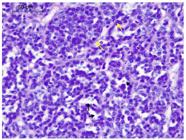
The histological study of the mass extracted from the tracheal lumen of a 13-year-old mixed-breed dog revealed solid trabeculae delimited by a sparse collagen stroma (yellow arrows) and tubules (black arrows). The proliferating cells have ovoid nuclei with karyomegaly, anisokaryosis, and moderate hyperchromatism. H&E staining. 400X.
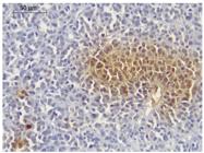
The immunohistochemical study (IHC) chromogranin A marker shows faint to moderate positive reactivity in groups of proliferating cells, from the mass extracted from the lumen of the cervical trachea of a 13-year-old mixed-breed dog. 400X.
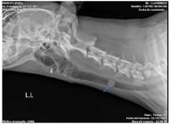
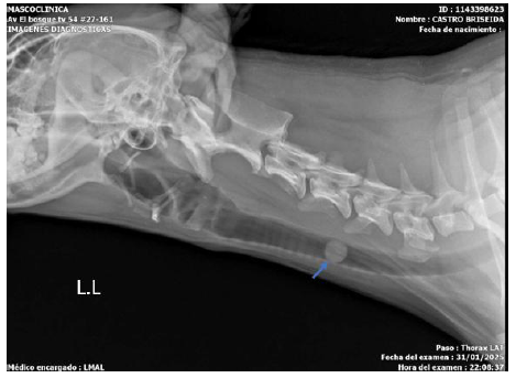
In the days following the procedure, the patient remained stable with no respiratory manifestations. Antimicrobial treatment was administered with enrofloxacin 5mg/kg/24 h/8 days, and for postoperative pain, meloxicam 0.2mg/kg/24 h/3 days accompanied by Traumeel, one tablet orally every 8h/15 days. A radiographic check-up (Figure 6) was performed two months after the procedure, revealing recurrence with almost complete obstruction of the tracheal lumen. The owners decided not to continue with any treatment, and the patient died five days after the check-up, the cause of death being respiratory asphyxia.
Control X-ray of the neck with right lateral projection in a mixed-breed dog, two months after removal of the tracheal mass, showing recurrence (blue arrow).
ETHICAL ASPECTS
This research was not submitted to the Ethics Committee on Animal Use.
DISCUSSION
This case describes a tracheal obstruction in a geriatric dog caused by a neuroendocrine carcinoma, which is an aggressive neoplasm rarely found in this anatomical structure according to a review of the veterinary literature. In humans, its frequency, regardless of the organ, represents less than 0.1% of all neoplasms (Mukherji A. et al. 2011, Zachary A. et al. 2020, Rendon M.E. 2011, Kanemoto H. et al. 2023). Clinically, the respiratory signs observed in the patient (dyspnea, stridor, cyanosis, and syncope) are consistent with the typical symptoms of an obstructive tracheal mass, as described in previous studies in dogs and cats (Mukherji A. et al. 2011, Piirainen K.et al. 2018, Torres M. et al. 2020, Sherani K. et al. 2015). The cause of the obstruction was diagnosed with radiographic studies and airway endoscopy, which revealed the presence of a nodule occupying 95% of the tracheal lumen.
The extraction technique used was endoscopy with a vessel sealing clamp, which allowed the nodule to be removed precisely and with minimal trauma. This technique has been described as effective by Mukherji et al. (2011). Since they used chemoradiation after extraction of tracheal carcinoma in a young man Mukherji A. et al. (2011), this did not coincide with the results of the case, as recurrence was evident two months after the intervention, and no chemoradiation or subsequent oncological treatment was performed because the financial resources for treatment were not available.
The definitive diagnosis was reached by histopathology and immunohistochemistry, finding positivity for chromogranin A, which is a marker for neuroendocrine tumors, and negativity for cytokeratin 5 helped rule out other types of carcinomas. The expression of CgA supports its origin in neuroendocrine cells, as also described in human medicine (Carrillo D. et al. 2021, Bednarczuk T. et al. 2022, Mondragón J. y Uribe J. 2016, Clement D. et al. 2016). In veterinary medicine, reported tracheal neoplasms include adenocarcinomas, leiomyomas, fibrosarcomas, among others (Piirainen K,et al. 2018, Zachary A. et al. 2020, Howard J. et al. 2017) However, neuroendocrine tumors are not commonly found, making a relevant contribution to the global veterinary scientific literature.
In conclusion, tracheal neuroendocrine carcinoma is a rare condition in veterinary medicine, but it is highly aggressive, making timely diagnosis essential. The combination of radiographic techniques, airway endoscopy, histopathological analysis, and immunohistochemistry was crucial for a comprehensive approach to the case. Endoscopic resection is a valuable tool, although not definitive, due to the possibility of recurrence. For a more favorable outcome, tracheal resection with anastomosis is recommended for complete removal of tracheal nodules, masses, or tumors. It is recommended that this type of neoplasm be included in the differential diagnosis of respiratory obstruction in geriatric patients to optimize prognosis through early intervention and support with oncological treatment.
REFERENCES
- BATALLER, L.; TAMBORINI, A.; L'EPLATTENIER, H. et al. Successful treatment of tracheal lymphoma in a Siamese cat. JFMS Open Rep., v.3, n.2, 2017.
- BEDNARCZUK, T.; ZEMCZAK, A.; BOLANOWSKI, M. et al. Neuroendocrine neoplasms of the small intestine and the appendix - update of the diagnostic and therapeutic guidelines (recommended by the Polish Network of Neuroendocrine Tumours). Endokrynol. Pol., v.73, p.549-583, 2022.
- BLOM, R.; VLIEGEN, R.; SCHREURS, W. et al. External ultrasonography of the neck does not add diagnostic value to integrated positron emission tomography-computed tomography (Pet-Ct) scanning in the diagnosis of cervical lymph node metastases in patients with esophageal carcinoma. Dis. Esophagus, v.25, p.555-559, 2012.
- BROWN M. ROGERS KS. Primary tracheal tumors indogs and cats. Compendium, v.25, p.854-860 2023.
- CARRILLO, D.; MUÑOZ, M.; RETAMAL, I. et al. Tumores neuroendocrinos: un desafío transversal. Rev. Med. Chile, v.149, p.888-898, 2021.
- CLEMENT, D.; RAMAGE, J.; SRIRAJASKANTHAN, R. Update on pathophysiology, treatment, and complications of carcinoid syndrome. J. Oncol., v.2020, p.8341426, 2020.
- FELDMAN, M.; FRIEDMAN, L.S.; BRANDT, L.J. Neuroendocrine tumors. In: ______. Sleisenger and Fordtran’s gastrointestinal and liver disease. 10.ed. [Amsterdam]: Elsevier, 2015. v.1, chap.33, p.531-534.
- FERRER, E.; SERRUTO, B. Tumor mucoepidermoide de la tráquea: reporte de caso. Rev. Colomb. Neumol., v.29, n.2, 2017.
- FIERRO, M.L.F.; CÓRDOBA CARREÑO, C.P.; CUÉLLAR-CUÉLLAR, A.A. et al. Consenso colombiano de tumores neuroendocrinos intestinales y síndrome carcinoide. Rev. Colomb. Endocrinol. Diabetes Metab., v.11, p. e896, 2024.
- HONINGS, J.; VAN DIJCK, J.; VERHAGEN, A. et al. Incidence and treatment of tracheal cancer: a nationwide study in the Netherlands. Ann. Surg. Concol., v.4, p.968-976, 2007.
- HOWARD, J.; FISHER, J.; TOLBERT, M. Invasive tracheal neoplasia in eight cats: descriptive cases and reviewof the current literatura. JFMS Open Rep., v.3, p.2055116917690074, 2017.
- JIANG, H.; TAN, Q.; HE, F. et al. Ultrasound in patient with treted head and neck carcinomas: A retrospective análisis for effectiveness of follow-up care. Medicine, v.100, p. e25496, 2021.
- KANEMOTO, H.; FUJIWARA-IGARASHI, A.; KOBAYASHI, T; HARADA, K.; ICHIMATA, M. et al. Retrospective study of feline tracheal mass lesions. J. Feline Med. Surg., v.25, n.5, 2023.
- MONDRAGÓN, J.; URIBE, J. Medicina Interna- Sindrome Carcinoide. Rev. Méd. Costa Rica Centroam., v.73, p.59-63, 2016.
- MUKHERJI, A.; VERMA, P.; SINHA, D.; SHARMA, M.; PAL, M. Carcinoid of the Trachea in a Young Adult Male Responsive to Chemo-Radiation. Indian J. Otolaryngol. Head Neck Surg., v.63, Suppl.1, p. S13-S15, 2011.
- PIIRAINEN, K.; LAPPALAINEN, A.; MÖLSÄ, S. Intraluminal tracheal lipoma as a rare cause of dyspnoea in a dog. Acta Vet. Scand., v.60, p.59, 2018.
- RENDON, M.E. Síndrome carcinoide manejo perioperatorio. Rev. Mex. Anestesiol. Oncol., v.34, p. S72-S77, 2011.
- SHERANI, K.; VAKIL, A.; DODHIA, C.; FEIN, A. Malignant tracheal tumors: a review of current diagnostic and management strategies. Curr. Opin Pulm. Med., v.21, p.322-326, 2015.
- TORRES, M.; ZAMORA, Y; RODRÍGUEZ, J.; CALAÑA, L. Resección traqueal y anastomosis: tratamiento de displasia osteocondral intratraqueal en un canino. Rev. Salud Anim., v.42, n.1, 2020.
- ZACHARY, A.; MILLER, D.; PADGETT, S.; TERREROS, A.; PEARCE, E. Tracheal squamous cell carcinoma treated with tracheal resection and anastomosis in a cat. Case Rep. Vet. Med., v.2020, p.8818660, 2020.
Uninformed - data use not informed; no research data were generated or used.

 Source: Arnache J. and Salinas B.
 Source: Hernández J.
 Source: Hernández J.