Correlation between blood and peritoneal fluid glucose and lactate concentrations with intestinal lesion etiology, survival, and surgical indications in horses with colic
ABSTRACT
Equine colic syndrome is a severe disease with poor prognosis and is associated with hyperglycemia and hyperlactatemia. This study aimed to evaluate blood and peritoneal glucose and lactate concentrations in horses with colic and their relationship with intestinal injury etiology, survival, and treatment choice. Blood and peritoneal fluid samples were collected at the admission of 85 horses. Hyperglycemia was observed in 43.5% (n=38) of the horses, of which 21 (55%) were submitted to surgery. Blood and peritoneal fluid glucose and lactate concentrations were increased in horses treated surgically and increased in those dead and with ischemic injuries. The blood (rs=0.47) and peritoneal fluid (rs=0.27) concentrations of glucose and lactate were correlated, which is a differential of this study. The results reinforce the applicability of lactate assessment as an indicator of survival prognosis and ischemic intestinal injuries. The concentration of glucose and lactate in the blood or peritoneal fluid has clinical application and can be used separately if it is impossible to collect one of them. However, such measurements should be associated with other diagnostic tests in horse colic. Moreover, hyperglycemia is related to recommendation of surgical treatment.
Keywords:
acute abdomen; hyperglycemia; lactatemia; peritoneal fluid; prognostic
RESUMO
A síndrome cólica equina é uma doença grave associada à hiperglicemia e à hiperlactatemia com prognóstico desfavorável. O objetivo deste estudo foi avaliar as concentrações sanguíneas e peritoneais de glicose e lactato em equinos com cólica e sua relação com a etiologia da lesão intestinal, a sobrevivência e o tratamento. Amostras de sangue e líquido peritoneal foram coletadas na admissão de 85 cavalos. Hiperglicemia foi observada em 43,5% (n=38) dos cavalos, dos quais 21 (55%) foram submetidos à cirurgia. As concentrações de glicose e lactato no sangue e no líquido peritoneal foram maiores em cavalos tratados cirurgicamente e também naqueles que não sobreviveram e apresentaram lesões isquêmicas. As concentrações de glicose e lactato no sangue (rs=0,47) e no líquido peritoneal (rs=0,27) apresentaram correlação, o que é um diferencial deste estudo. Esse resultado reforça a aplicabilidade da avaliação de lactato como indicador de prognóstico de sobrevivência em lesões intestinais isquêmicas. A concentração de glicose e lactato no sangue ou no líquido peritoneal tem aplicação clínica e pode ser utilizada separadamente caso seja impossível a coleta de um deles. Entretanto, tais mensurações devem ser associadas a outros exames diagnósticos na cólica equina. Além disso, a hiperglicemia está relacionada à indicação de tratamento cirúrgico.
Palavras-chave:
abdômen agudo; hiperglicemia; lactatemia; líquido peritoneal; prognóstico
INTRODUCTION
The assessment of blood glucose levels is routinely used during hospitalization of critically ill human patients, being hyperglycemia associated with poor prognosis and mortality (Aramendi et al., 2017). Situations of increased cortisol due to stress (Hinchcliff et al., 2005), administration of steroidal anti-inflammatory drugs (Mizen et al., 2017), use of alpha-2 agonists drugs (Ambrósio et al., 2012) and in systemic inflammatory response syndrome (Tóth et al., 2008) explain hyperglycemia in horses. Recent studies have evaluated the use of glycemia as a predictor of the prognosis of survival in horses. Hyperglycemia was associated with mortality and poor prognosis in neonatal foals (Hug et al., 2013), horses with colitis (Urayama et al., 2018), and horses with colic (Hollis et al., 2007; Hassel et al., 2009).
Elevated lactate concentrations in blood and peritoneal fluid are observed in horses presenting intestinal hypoxic conditions, as well as torsions, strangulations, volvulus, or any other disease with decreased blood flow (Shearer et al., 2018). These conditions are associated with a high mortality rate and poor prognosis (Boom et al., 2010). The increased lactic acid in the peritoneal fluid occurs by cells in hypoxia and presents a strong correlation between blood and peritoneal lactate concentrations (Latson et al., 2005). The associated measurement of plasma and peritoneal lactate concentrations has a better prognostic value than isolated measurements (Delesalle et al., 2007).
This study aimed to evaluate plasma and peritoneal glucose and lactate concentrations at the admission of horses with colic and their relationship with the intestinal injury etiology (ischemic or not ischemic), survival, and treatment (medical or surgical). In addition, evaluate the correlation between the variables. This study is different because it assesses the correlation between glucose and lactate.
ETHICAL ASPECTS
The research was submitted to the Ethics Committee on Animal Use of the São Paulo State University, and approved under the number 84/2016
MATERIAL AND METHODS
This study included 85 horses admitted with colic between October of 2013 and December of 2016. The admitted horses were subjected to the emergency colic protocol, including medical history, physical examination (heart and respiratory rate, rectal temperature, mucosa coloration, capillary refill time, intestinal motility auscultation, and rectal examination), and abdominal ultrasound. Blood samples were collected during the initial assessment to evaluate complete blood cell count and physicochemical analysis (total plasma protein and fibrinogen), and the peritoneal fluid for evaluation macroscopic, physicochemical, and cytological analysis. Blood and peritoneal fluid samples were also collected to evaluate glucose and lactate concentrations. Emergency procedures such as nasogastric intubation, venous access, and cecum drainage were performed according to the necessity. Not all animals had results from all evaluations. For example, it was not possible to collect peritoneal fluid from some animals, while for others it was not possible to collect blood.
The horses were clinical or surgically treated according to the case. The criteria for surgical indication were pain refractory to analgesia, transrectal palpation findings, peritoneal fluid indicative of ischemic intestinal lesion, and ultrasonographic findings. For posterior analysis, the data related to diagnosis, intestinal injury etiology (ischemic or not ischemic), survival, treatment (surgical or clinical), age, weight, sex, and breed were recorded.
Blood samples were collected by jugular venipuncture in vacuum tubes with ethylenediaminetetraacetic acid (EDTA) and sodium fluoride at admission for complete blood count and glucose and lactate determination, respectively.
The peritoneal fluid was collected aseptically at admission using a 40x12mm hypodermic needle from the ventral midline region of the abdomen. Samples were collected in tubes with EDTA, without anticoagulant, and sodium fluoride, the latter intended to evaluate glucose and lactate concentrations. Samples with EDTA and without anticoagulant were used for macroscopic, physicochemical, and cytological analysis and processed immediately by conventional methods. Briefly, the macroscopic analysis included color and turbidity; the physicochemical assessed fibrinogen, pH, and total protein; the cytology analyzed the types and number of cells (Weiser, 2015).
Blood and peritoneal fluid samples with sodium fluoride were immediately centrifuged at ~2,400g for 10 minutes. The supernatant aliquots were stored at -80ºC until analysis.
The plasma and peritoneal glucose and lactate concentrations were determined by the electroenzymatic method (YSI 2300 STAT PLUS™ Glucose & L-Lactate Analyzer, YSI Life Sciences, Yellow Springs, OH, USA). Values of glucose above ≥130.869mg/dL in the blood (Hollis et al., 2007) and >113.4mg/dL in the peritoneal fluid (Romero et al., 2011) were considered increased, as well as lactate values above ≥2mmol/L in the blood and >0.6mmol/L in the peritoneal fluid (Latson et al., 2005).
The data of glucose and lactate concentrations from the blood and the peritoneal fluid were compared between the horses alive and dead, treated clinically and surgically, and horses with ischemic and non-ischemic intestinal lesions. Moreover, glucose and lactate from the blood and peritoneal fluid were compared. Variables were assessed for normality using the Kolmogorov-Smirnov test, and the normality failed. Then, the data were analyzed by the Mann-Whitney test since it did not show a normal distribution, even after they were log-transformed.
A one-sample Wilcoxon test compared the blood glucose concentrations of survival vs. dead horses with a reference value of 130.869mg/dL (Hollis et al., 2007).
The glucose and lactate of blood and peritoneal fluid were correlated using the Spearman test. Samples from 85 horses were added to the correlation statistics between blood lactate and glucose concentrations, and samples from 63 horses were added to the correlation statistics between peritoneal fluid lactate and glucose. The correlation was considered as very weak (0-0.19), as weak (0.2-0.39), as moderate (0.40-0.59), as strong (0.6-0.79), and as very strong (0.8-1) (Doria Filho, 1999). Statistical analysis was performed using the Sigma Stat version 3.5 (Systat Software Inc., 2007), and the results were considered significant when the P<0.05. The variables were presented as the median, minimum, and maximum.
RESULTS
From 85 horses included into study, 47 (55.3%) were females and 38 (44.7%) males, with median of 6 years, mean ± standard deviation ages of 7.5±5.8 years (1.5 months - 28 years) and weighing with median of 411Kg and mean ± standard deviation 397.2±109.1kg (45-571kg). The breed distribution in population was 43 (50.5%) Quarter horses, 13 (15.3%) mixed breeds, 8 (9.4%) Mangalarga, 2 (2.4%) Brazilian Equestrian, 2 (2.4%) Arabians, 2 (2.4%) Lusitanos, 2 (2.4%) ponies and 13 (15.2%) other breeds.
Regarding the treatment, 53 (62.4%) were clinically treated and 32 (37.6%) surgically. Forty and nine (56.2%) horses were discharged and considered survivors, and 36 (42.4%) died. Among these 17 were subjected to euthanasia due to the severity of the condition or by the owner’s choice. According to the diagnoses, 71 horses presented non-ischemic lesions in the intestine (83.5%) and 14 ischemic lesions (16.5%).
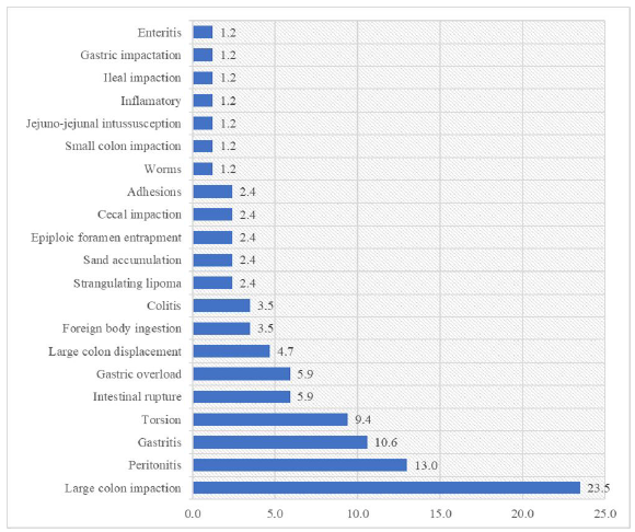
The diseases that affected horses with the colic syndrome were described in Fig. 1, and the principal diagnosis was large colon impaction (23.5%).
The results of the lactate and glucose concentrations in blood and peritoneal fluid according to intestinal injury etiology (ischemic or non-ischemic), survival, and treatment (medical or surgical) are shown in Tab. 1. Blood and peritoneal fluid glucose concentrations were increased in horses treated surgically, and blood and peritoneal fluid lactate concentrations were increased in horses treated surgically, dead, and with ischemic injuries.
There was no difference between blood and peritoneal fluid for glucose and lactate. The median was 122.58mg/dL (minimum and maximum 45.9 and 482,4mg/dL ) and 133.38 mg/dL (minimum and maximum 1.98 and 554.4mg/dL), median [minimum and maximum] respectively) for glucose, and 2.75mmol/L (minimum and maximum 0.76 and 27.00mmol/L) and 3.41mmol/L (minimum and maximum 0.77 and 30.90mmol/L) for lactate concentrations, respectively.
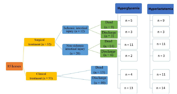
Colic cases distribution according to treatment and survival and the relation with hyperglycemia and hyperlactatemia are presented in Fig. 2.
There was no difference between glucose concentration of survival or dead horses and reference value, and above values of reference were found in 40.4% and 54.3% survival and dead horses, respectively.
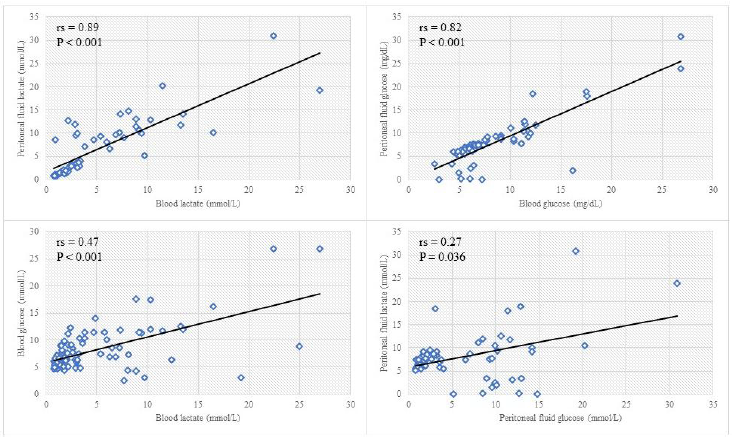
Spearman's correlation results are presented in Fig. 3. There was a very strong positive correlation between blood and peritoneal fluid lactate and blood and peritoneal fluid glucose. However, a moderate and weak positive correlation between blood lactate and glucose, and peritoneal fluid lactate and glucose, respectively, were observed.
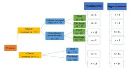
Cases distribution according to treatment (clinical or surgical) and survival related with hyperglycemia and hyperlactatemia in the horses with colic (n=85).
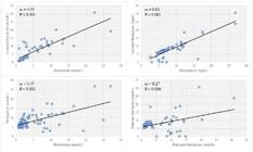
Spearman correlation between variables. A. Blood vs peritoneal fluid lactate. B. Blood vs peritoneal glucose. C. Blood glucose vs blood lactate. D. Peritoneal fluid glucose vs peritoneal fluid lactate (n=85).
DISCUSSION
In our study, the glucose concentrations from survival and diseased horses were not different concerning the reference value for the species (7.2705 mmol/L, Hollis et al., 2007). The results, therefore, suggest that hyperglycemia is expected in the colic case since ~50% of horses presented glycemia >7.2705mmol/L. The absence of long-term survival time records may have been a limitation of our study and contributed to the results obtained.
In horses, hyperglycemia was associated with decreased survival in colic cases (Hollis et al., 2007; Hassel et al., 2009). It’s already described in the literature that hyperglycemia occurs secondary to the release of cortisol in stressful situations (Hinchcliff et al., 2005), administration of steroidal anti-inflammatory drugs (Mizen et al., 2017), use of alpha-2 agonists drugs (Ambrósio et al., 2012) and in systemic inflammatory response syndrome in horses (Tóth et al., 2008). However, a precise and well-described link between hyperglycemia and mortality in colic cases has not been fully elucidated (Hinchcliff et al., 2005).
Although blood glucose was similar for discharged and dead horses, from the overall rate of hyperglycemic horses (43.5% of all animals, 38/85), a total of 51% died (20/38). These results corroborated other studies, in which ~50% of horses with colic presented hyperglycemia (50.2% for Hollis et al., 2007; 45% for Hassel et al., 2009). Similarly, the mortality was described in ~50% of horses with hyperglycemia (56% for Hassel et al., 2009). There is an association between hyperglycemia and death in humans, and it is related to the oxidative stress chain stimulation and endothelial dysfunction (Egi and Bellomo, 2009). Such connection could explain the high mortality rate in horses with hyperglycemia since colic cases involve pain, stress, inflammation, and other factors (Wilkins, 2018). The association of our results with previous studies reinforces the utilization of blood glucose levels as a tool to establish the prognosis in horses with colic.
When caring for a horse with colic several procedures can be performed to arrive at a diagnosis. Physical exam, abdominal auscultation, rectal palpation, nasogastric tube, abdominal ultrasonography, peritoneal fluid collection for example. All are important for defining the prognosis and treatment (Cook and Hassel, 2014).
Blood glucose concentrations above 10 mmol/L are considered extreme hyperglycemia in horses (Hollis et al., 2007) and affect 16.7% of patients with colic (Hassel et al., 2009). In our study, 24.7% (21/85) of horses presented extreme hyperglycemia, and 57% (12/21) did not survive. Of 21 horses with extreme hyperglycemia during admission, 62% were considered severe cases, emphasizing the relationship between blood glucose and the severity of colic cases. If extreme hyperglycemia occurs in severe cases, then the blood glucose level during admission of horses with colic, associated with clinical history, laboratory tests, and imaging exams, can support surgical treatment decision. Furthermore, we observed that in the admission, horses that required surgical treatment (32/85) presented greater blood glucose levels (15/32 showing extreme hyperglycemia) than patients that underwent clinical treatment only.
Furthermore, from the 85 horses evaluated in our study, 37 presented hyperglycemias, and 9 of these were diagnosed with ischemic intestinal lesions, suggesting no clear association between hyperglycemia and ischemic intestinal lesions. However, in the literature, hyperglycemia during admission was previously associated with ischemic intestinal lesions surgically treated with intestinal segment resection (Hollis et al., 2007). Indeed, this is related to disease severity, increase in permeability of the intestinal wall, leading to bacterial translocation and endotoxin release into the blood, resulting in sepsis (Hollis et al., 2007). Possibly, due to the small number of patients with hyperglycemia and ischemic lesions observed in our study (9/85), such a relationship lacked statistical power.
In addition, some factors not related to colic in horses, such as medication and handling, play an essential role in the elevation of blood glucose. Frequently, horses with colic signs receive α2-agonists that can increase the blood glucose by stimulating pancreas receptors producing a hyperglycemic effect for 50 minutes after administration (Thurmon et al., 1982). Despite the unusual, corticosteroids may be used in colic cases, and dexamethasone is frequently administered, stimulating gluconeogenesis in the liver and inhibiting glucose action in the muscles (Mizen et al., 2017). A similar response occurs in stressful situations, as the pain, manipulation, or transport can promote cortisol release (Hinchcliff et al., 2005). These aspects should be highlighted as possible causes of hyperglycemia in horses and considered one limitation since the drugs administered previously to admission and during veterinary care were not recorded.
It's essential to describe that peritoneal fluid glucose concentrations are higher than blood in healthy horses; however, in horses with colic, no difference is observed between both parameters (Brownlow, 1979), which corroborates with our findings.
An exception can be made for horses with ischemic injuries that progress to irreversible infarction/necrosis (Parry, 1987). Moreover, there was a very strong positive correlation between the plasma and peritoneal glucose concentrations that were previously described (Latson et al., 2005). This fact can be explained by the small glucose molecule size that readily diffuses in the tissue through membranes, and its increase in the blood will reflect in peritoneal fluid concentrations (Bonczynski et al., 2003; Aguirre and Abensur, 2014). In our study, the blood and peritoneal fluid glucose concentrations were also high in horses subjected to surgical treatment, which presented the most severe cases of colic. Only 5 (~16%) horses had peritonitis in these cases, justifying the increased glucose consumption by a bacterial infection (Bonczynski et al., 2003), corroborating with the findings of Alonso et al. (2020).
In intestinal ischemia, an anaerobic condition, the lactate concentrations increase in blood and peritoneal fluid (Zilva, 1978). Therefore, lactate concentrations can be used to identify ischemic lesions and become an auxiliary method in determining diseases prognosis in various species (Bonczynski et al., 2003). Indeed, in our study, the increased lactate concentrations of blood and peritoneal fluid were observed in the severe cases in which horses had ischemic intestinal lesions, which were surgically treated or resulted in the patient's death. In colic cases, the ischemic intestinal lesions induce anaerobic glycolysis with lactate as a sub-product, diffusing from intestinal cells to peritoneal fluid and blood (Parry, 1987), explaining the very strong positive correlation between blood and peritoneal fluid lactate observed in our study.
Previous studies demonstrated that lactate concentration in the peritoneal fluid is more sensitive to early recognizing intestinal ischemia and prognostic compared to blood lactate (Latson et al., 2005). Peritoneal fluid lactate >4mmol/L is related to ischemic intestinal lesions with 91% specificity and 27% sensibility (Peloso and Cohen, 2012). Indeed, we observed high peritoneal lactate concentration in horses with ischemic intestinal lesions (median 11.9mmol/L), suggesting a delay in the diffusion from peritoneal lactate to the blood, demonstrating the importance of the parameter measurement for the diagnosis and prognosis within 1 to 6 hours from the admission (Peloso and Cohen, 2012).
Furthermore, a poor prognosis in colic cases was related to increased peritoneal lactate (Latson et al., 2005), corroborating our results since the peritoneal lactate concentration was higher in horses that died or were surgically treated. Therefore, the lactate concentrations in blood and peritoneal fluid can assist the treatment choice in horses with colic.
In our study, there was a correlation between lactate and glucose concentrations both in blood and peritoneal fluid. The lactate concentration in rats in the presence of ischemic intestinal lesions increases rapidly in the peritoneal fluid. Through the permeability of the peritoneal membrane, lactate also increases its concentration in the blood, but at lower concentration when compared to the peritoneal fluid. This shows the high sensitivity of lactate as a biochemical parameter for the diagnosis of ischemic intestinal lesions (Liao et al., 1995). The concentration of glucose increases in the blood due to the elevation of cortisol, pain, excitement or transport of the horse and can also increase in the peritoneal fluid for the same reasons (Latson et al., 2005) since the transport of glucose through the peritoneal membrane is easy. (Aguirre and Abensur, 2014). The use of biochemical parameters as aids in prognostic decision is related both to their concentration and to their location and release mechanism (Liao et al., 1995).
CONCLUSIONS
Hyperglycemia was associated with surgical indication and decreased survival, and extreme hyperglycemia can be a factor of poor prognosis. Peritoneal glucose concentration was identified as a tool to support treatment choice. The lactate concentrations in blood and peritoneal fluid were associated with ischemic intestinal lesions, surgical treatment, and unfavorable prognosis. The blood and peritoneal fluid concentrations of glucose and lactate are more important when associated with other diagnostic parameters in horses with colic. Therefore, they are not reliable as single indicators for the intestinal lesion type, treatment decision, and prognosis.
REFERENCES
- AGUIRRE, A.R.; ABENSUR, H. Physiology of fluid and solute transport across the peritoneal membrane. Braz. J. Nephrol., v.36, p.74-77, 2014.
- ALONSO, J.M.; ESPER, C.S.; PANTOJA, J.C.F. et al. Accuracy of differences in blood and peritoneal glucose to differentiate between septic and non-septic peritonitis in horses. Res. Vet. Sci., v.132, p.237-242, 2020.
- AMBRÓSIO, A.M.; CASAES, A.G.; IDA, K.K. et al. Differences in blood glucose increase between horses receiving xylazine and detomidine for surgical and non-surgical clinical procedures. Braz. J. Vet. Res. Anim. Sci., v.49, p.493-499, 2012.
- ARAMENDI, I.; BURGHI, G.; MANZANARES, W. Dysglycemia in the critically ill patient: current evidence and future perspectives. Rev. Bras. Ter. Intensiva, v.29, p.364-372, 2017.
- BONCZYNSKI, J.J.; LUDWIG, L.L.; BARTON, L.J. Comparison of peritoneal fluid and peripheral blood pH, bicarbonate, glucose, and lactate concentration as a diagnostic tool for septic peritonitis in dogs and cats. Vet. Surg., v.32, p.161-166, 2003.
- BOOM, R.V.D.; BUTLER, C.M.; OOSTERBAAN, M.S.V.O. The usability of peritoneal lactate concentration as a prognostic marker in horses with severe colic admitted to a veterinary teaching hospital. Equine Vet. Educ., v.22, p.420-425, 2010.
- BROWNLOW, M.A. Abdominal paracentesis in the horse clinical evaluation. 1979. Thesis (Master of Veterinary Sciences) - University of Sydney, AUS.
- COOK, V.L.; HASSEL, D.M. Evaluation of the colic in horses: decision for referral. Vet. Clin. North Am. Equine Pract., v.30, p.383-98, 2014.
- DELESALLE, C.; DEWULF, J.; LEFEBVRE, R.A. et al. Determination of lactate concentrations in blood plasma and peritoneal fluid in horses with colic by an accusport analyzer. J. Vet. Intern. Med. v.21, p.293-301, 2007.
- DORIA FILHO, U. Introdução à bioestatística: para simples mortais. 2.ed. São Paulo: Negócio, 1999. 152p.
- EGI, M.; BELLOMO, R. Reducing glycemic variability in intensive care unit patients: a new therapeutic target? J. Diabetes Sci. Technol., v.3, p.1302-1308, 2009.
- HASSEL, D.M.; HILL, A.E.; RORABECK, R.A. Association between hyperglycemia and survival in 228 horses with acute gastrointestinal disease. J. Vet. Intern. Med., v.23, p.1261-1265, 2009.
- HINCHCLIFF, K.W.; RUSH, B.R.; FARRIS, J.W. Evaluation of plasma catecholamine and serum cortisol concentrations in horses with colic. J. Am. Vet. Med. Assoc., v.227, p.276-280, 2005.
- HOLLIS, A.R.; BOSTON, R.C.; CORLEY, K.T.T. Blood glucose in horses with acute abdominal disease. J. Vet. Intern. Med., v.21, p.1099-1103, 2007.
- HUG, S.A.; RIOND, B.; SCHWARZWALD, C.C. Evaluation of a continuous glucose monitoring system compared with an in-house standard laboratory assay and a handheld point-of-care glucometer in critically ill neonatal foals. J. Vet. Emerg. Crit. Care, v.23, p.408-415, 2013.
- LATSON, K.M.; NIETO, J.E.; BELDOMENICO, P.M.; SNYDER, J.R. Evaluation of peritoneal fluid lactate as a marker of intestinal ischaemia in equine colic. Equine Vet. J., v.37, p.342-346, 2005.
- LIAO, X.P.; SHE, Y.X.; SHI, C.R.; LI, M. Changes in body fluid markers in intestinal ischemia. J. Pediatr. Surg., v.30, p.1412-1415, 1995.
- MIZEN, K.; WOODMAN, J.; BOYSEN, S.R. et al. Effect of dexamethasone on resting blood lactate concentrations in horses. J. Vet. Intern. Med., v.31, p.164-169, 2017.
- PARRY, B. W. Use of clinical pathology in evaluation of horses with colic. Vet. Clin. North Am. Equine Pract., v.3, p.529-542, 1987.
- PELOSO, J.G., COHEN, N.D. Use of serial measurements of peritoneal fluid lactate concentration to identify strangulating intestinal lesions in referred horses with signs of colic. J. Am. Vet. Med. Assoc. v.240, p.1208-1217, 2012.
- ROMERO, A.E.; NIETO, J.E.; DECHANT, J.E. et al. Effects of aerobic and anaerobic fluid collection on biochemical analysis of peritoneal fluid in healthy horses and horses with colic. Vet. Surg., v.40, p.40-45, 2011.
- SHEARER, T.R.; NORBY, B.; CARR, E.A. Peritoneal fluid lactate evaluation in horses with nonstrangulating versus strangulating small intestinal disease. J. Equine Vet. Sci., v.61, p.18-21, 2018.
- TÓTH, F.; FRANK, N.; ELLIOTT, S.B. Effects of an intravenous endotoxin challenge on glucose and insulin dynamics in horses. Am. J. Vet. Res., v.69, p.82-88, 2008.
- THURMON, J.C.; NEFF-DAVIS, C.; DAVIS, L.E. et al. Xylazine hydrochloride-induced hyperglycemia and hypoinsulinemia in thoroughbred horses. J. Vet. Pharmacol. Ther., v.5, p.241-245, 1982.
- URAYAMA, S.; ARIMA, D.; FUMIAKI, M. Blood glucose is unlikely to be a prognostic biomarker in acute colitis with systemic inflammatory response syndrome in Thoroughbred racehorses. J. Equine Sci., v.29, p.15-19, 2018.
- WEISER, G. Coleta e processamento da amostra e análise das opções de serviços laboratoriais. In: THRALL, M.A.; ALLISON, R.W.; CAMPBELL, T.W. Hematologia e bioquímica clínica veterinária. 2.ed. São Paulo: Roca, 2015. p.87-99.
- WILKINS, P.A. What’s in a word? The need for SIRS and sepsis definitions in equine medicine and surgery. Equine Vet. J., v.50, p.7-9, 2018.
- ZILVA, J.F. The origin of the acidosis in hyperlactatemia. Ann. Clin. Biochem., v.15, p.40-43, 1978.
The research data are available upon request.